Application of ultrasound in periodontics: Part II
- PMID: 20142946
- PMCID: PMC2813560
- DOI: 10.4103/0972-124X.44096
Application of ultrasound in periodontics: Part II
Abstract
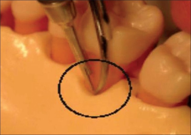
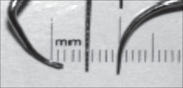
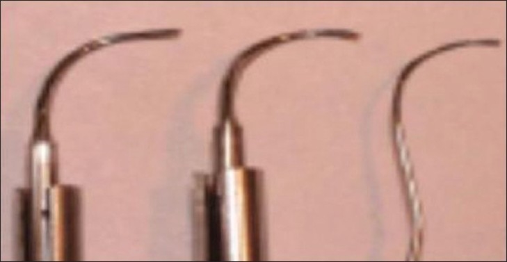
Ultrasound offers great potential in development of a noninvasive periodontal assessment tool that would offer great yield real time information, regarding clinical features such as pocket depth, attachment level, tissue thickness, histological change, calculus, bone morphology, as well as evaluation of tooth structure for fracture cracks. In therapeutics, ultrasonic instrumentation is proven effective and efficient in treating periodontal disease. When used properly, ultrasound-based instrument is kind to the soft tissues, require less healing time, and are less tiring for the operator. Microultrasonic instruments have been developed with the aim of improving root-surface debridement. The dye/paper method of mapping ultrasound fields demonstrated cavitational activity in an ultrasonic cleaning bath. Piezosurgery resulted in more favorable osseous repair and remodeling in comparison with carbide and diamond burs. The effect of ultrasound is not limited to fracture healing, but that bone healing after osteotomy or osteodistraction could be stimulated as well.
Keywords: LIPUS; microstreaming; microultrasonics; piezosurgery; ultrasonic cleaner; ultrasound probe.
Conflict of interest statement
Figures
Similar articles
-
Evaluation of root surface microtopography following the use of four instrumentation systems by confocal microscopy and scanning electron microscopy: an in vitro study.J Periodontal Res. 2012 Oct;47(5):608-15. doi: 10.1111/j.1600-0765.2012.01473.x. Epub 2012 Apr 12. J Periodontal Res. 2012. PMID: 22494068 Clinical Trial.
-
Clinical comparison of instrumentation systems for periodontal debridement: A randomized clinical trial.Int J Dent Hyg. 2022 May;20(2):328-338. doi: 10.1111/idh.12520. Epub 2021 Jul 1. Int J Dent Hyg. 2022. PMID: 34018671 Clinical Trial.
-
Root instrumentation. Power-driven versus manual scalers, which one?Dent Clin North Am. 1998 Apr;42(2):229-44. Dent Clin North Am. 1998. PMID: 9597335 Review.
-
Histological and profilometric evaluation of the root surface after instrumentation with a new piezoelectric device - ex vivo study.Int J Dent Hyg. 2015 May;13(2):138-44. doi: 10.1111/idh.12091. Epub 2014 Jul 3. Int J Dent Hyg. 2015. PMID: 24995862 Clinical Trial.
-
Rationale of mechanical plaque control.J Clin Periodontol. 1996 Mar;23(3 Pt 2):263-7. doi: 10.1111/j.1600-051x.1996.tb02086.x. J Clin Periodontol. 1996. PMID: 8707987 Review.
Cited by
-
Assessing the effect of piezoelectric ultrasonic scaler tip wear on root surface roughness under influence of various working parameters: A profilometric and atomic force microscopic study.J Indian Soc Periodontol. 2023 Nov-Dec;27(6):583-589. doi: 10.4103/jisp.jisp_416_22. Epub 2024 Jan 24. J Indian Soc Periodontol. 2023. PMID: 38434510 Free PMC article.
-
New developments in tools for periodontal diagnosis.Int Dent J. 2012 Apr;62(2):57-64. doi: 10.1111/j.1875-595X.2011.00099.x. Int Dent J. 2012. PMID: 22420472 Free PMC article. Review.
-
The effectiveness of low-level laser therapy and low-intensity pulsed ultrasound in reducing pain induced by orthodontic separation: a randomized controlled trial.BMC Oral Health. 2024 Feb 2;24(1):166. doi: 10.1186/s12903-024-03926-2. BMC Oral Health. 2024. PMID: 38308275 Free PMC article. Clinical Trial.
-
Assessment of periodontal dehiscence and fenestration using ultrasonography and cone-beam computed tomography: an in vitro study.Clin Oral Investig. 2024 Nov 28;28(12):665. doi: 10.1007/s00784-024-06011-8. Clin Oral Investig. 2024. PMID: 39604758
-
Biostimulatory Effects of Low-Intensity Pulsed Ultrasound on Rate of Orthodontic Tooth Movement and Associated Pain, Applied at 3-Week Intervals: A Split-Mouth Study.Pain Res Manag. 2021 May 5;2021:6624723. doi: 10.1155/2021/6624723. eCollection 2021. Pain Res Manag. 2021. PMID: 34035871 Free PMC article.
References
-
- Lefkowitz W. Ultrasound in dentistry. J Proshet Dent. 1958;1:135–6.
-
- Zinner DD. Recent ultrasonic dental studies, including periodontia, without the use of an abrasive. J Dent Res. 1955;34:748–9.
-
- Carr M. Ultrasonics; access. Special Supplemental Issue. 1999 May-Jun;:2–8.
-
- McCombs GB, Hinders M. The potential of the ultrasonic probe. Dimensions Dent Hyg. 2006;4:16–8.
-
- Spranger H. Ultrasonic diagnosis of marginal periodontal diseases. Int Dent J. 1971;21:442–55. - PubMed
LinkOut - more resources
Full Text Sources
Other Literature Sources