. 2010 Jan 11:10:1.
doi: 10.1186/1475-2867-10-1.

ASPM-associated stem cell proliferation is involved in malignant progression of gliomas and constitutes an attractive therapeutic target

Affiliations

ASPM-associated stem cell proliferation is involved in malignant progression of gliomas and constitutes an attractive therapeutic target

Sandra-Nadia Ngwabyt Bikeye et al. Cancer Cell Int. .

Erratum in

Abstract

Background: ASPM (Abnormal Spindle-like Microcephaly associated) over-expression was recently implicated in the development of malignant gliomas.

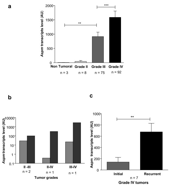
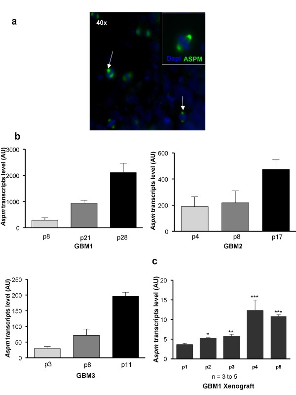
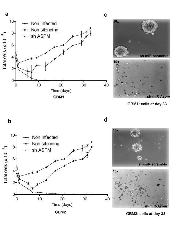
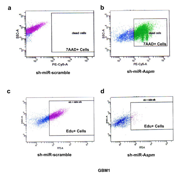
Results: To better characterize the involvement of ASPM in gliomas, we investigated the mRNA expression in 175 samples, including 8 WHO Grade II, 75 WHO Grade III and 92 WHO Grade IV tumors. Aspm expression was strongly correlated with tumor grade and increased at recurrence when compared to the initial lesion, whatever the initial grade of the primary tumor. ASPM expression also increased over serial passages in gliomaspheres in vitro and in mouse xenografts in vivo. Lentivirus-mediated shRNA silencing of ASPM resulted in dramatic proliferation arrest and cell death in two different gliomasphere models.

Conclusion: These data suggest that ASPM is involved in the malignant progression of gliomas, possibly through expansion of a cancer stem cell compartment, and is an attractive therapeutic target in glioblastoma multiforme.


Figures

Figure 1
Figure 1
Aspm expression correlates with glioma grade and increases at recurrence. (a) Aspm is up-regulated in gliomas compared to non-tumor brain and increases with the tumor grade;(b) ASPM expression increases between the initial tumor and recurrence at a higher grade; (c) ASPM expression also increases at recurrence when the initial tumor is already a Grade IV GBM. The scale bar represents the standard error of the mean (SEM).
Figure 2
Figure 2
ASPM expression in gliomaspheres increases with successive passages in vitro and in vivo. (a) Metaphase staining of ASPM protein at both poles of the spindle. ASPM protein (green) is detected in gliomaspheres (arrow). Nuclei are stained with DAPI (blue); (b) ASPM expression increases with successive passages (p) in gliomaspheres issued from GBM 1, 2 and 3. Passages were performed in vitro every 8 to 12 days; (c) GBM1 cells were subcutaneously engrafted into nude mice and Aspm expression was measured over four passages. Aspm expression increased progressively in xenograft tumor (mean +/- SEM; n = 3 to 5 mice for each point. In vivo passages were performed every 8-16 weeks.
Figure 3
Figure 3
ASPM knockdown in glioblastoma gliomaspheres. Cell kinetics of GBM1 (a) and GBM2 (b) gliomaspheres. The data were generated from three independent experiments. Gliomaspheres treated with sh-miR-Aspm do not grow as compared to non-transduced gliomaspheres and gliomaspheres transduced with non-silencing sh-miR-RNA, which start growing again after a transient drop; (c, d) at day 33, there is extensive cell death in cell populations of GBM1 and GBM2 expressing the ASPM-silencing sh-miR-RNA as compared to the cells expressing a non-targeted sh-miR-RNA.
Figure 4
Figure 4
Cell death and proliferation inhibition induced by ASPM silencing, evaluated by 7-Actinomycin D (7-AAD) labeling and 5- Ethyl -2'- Deoxyuridine (EdU) incorporation. Compared to control (a), transduction of GBM1 with sh-miR-Aspm results in massive cell death (b) and a marked decrease in proliferation of surviving cells, with only 25% of the viable cells being EdU+ (d), compared to 64% for the controls (c).

References

    1. Galli R, Binda E, Orfanelli U, Cipelletti B, Gritti A, De Vitis S, Fiocco R, Foroni C, Dimeco F, Vescovi A. Isolation and characterization of tumorigenic, stem-like neural precursors from human glioblastoma. Cancer Res. 2004;64(19):7011–7021. doi: 10.1158/0008-5472.CAN-04-1364. - DOI - PubMed
    1. Singh SK, Hawkins C, Clarke ID, Squire JA, Bayani J, Hide T, Henkelman RM, Cusimano MD, Dirks PB. Identification of human brain tumour initiating cells. Nature. 2004;432(7015):396–401. doi: 10.1038/nature03128. - DOI - PubMed
    1. Fish JL, Kosodo Y, Enard W, Paabo S, Huttner WB. Aspm specifically maintains symmetric proliferative divisions of neuroepithelial cells. Proc Natl Acad Sci USA. 2006;103(27):10438–10443. doi: 10.1073/pnas.0604066103. - DOI - PMC - PubMed
    1. Bond J, Roberts E, Mochida GH, Hampshire DJ, Scott S, Askham JM, Springell K, Mahadevan M, Crow YJ, Markham AF. ASPM is a major determinant of cerebral cortical size. Nat Genet. 2002;32(2):316–320. doi: 10.1038/ng995. - DOI - PubMed
    1. Kouprina N, Pavlicek A, Collins NK, Nakano M, Noskov VN, Ohzeki J, Mochida GH, Risinger JI, Goldsmith P, Gunsior M. The microcephaly ASPM gene is expressed in proliferating tissues and encodes for a mitotic spindle protein. Hum Mol Genet. 2005;14(15):2155–2165. doi: 10.1093/hmg/ddi220. - DOI - PubMed

LinkOut - more resources