Review
. 2010 Nov;18(3-4):367-76.
doi: 10.1007/s12640-010-9154-6. Epub 2010 Feb 9.

Dopamine system dysregulation by the ventral subiculum as the common pathophysiological basis for schizophrenia psychosis, psychostimulant abuse, and stress

Affiliations
Review

Dopamine system dysregulation by the ventral subiculum as the common pathophysiological basis for schizophrenia psychosis, psychostimulant abuse, and stress

Anthony A Grace. Neurotox Res. 2010 Nov.

Abstract

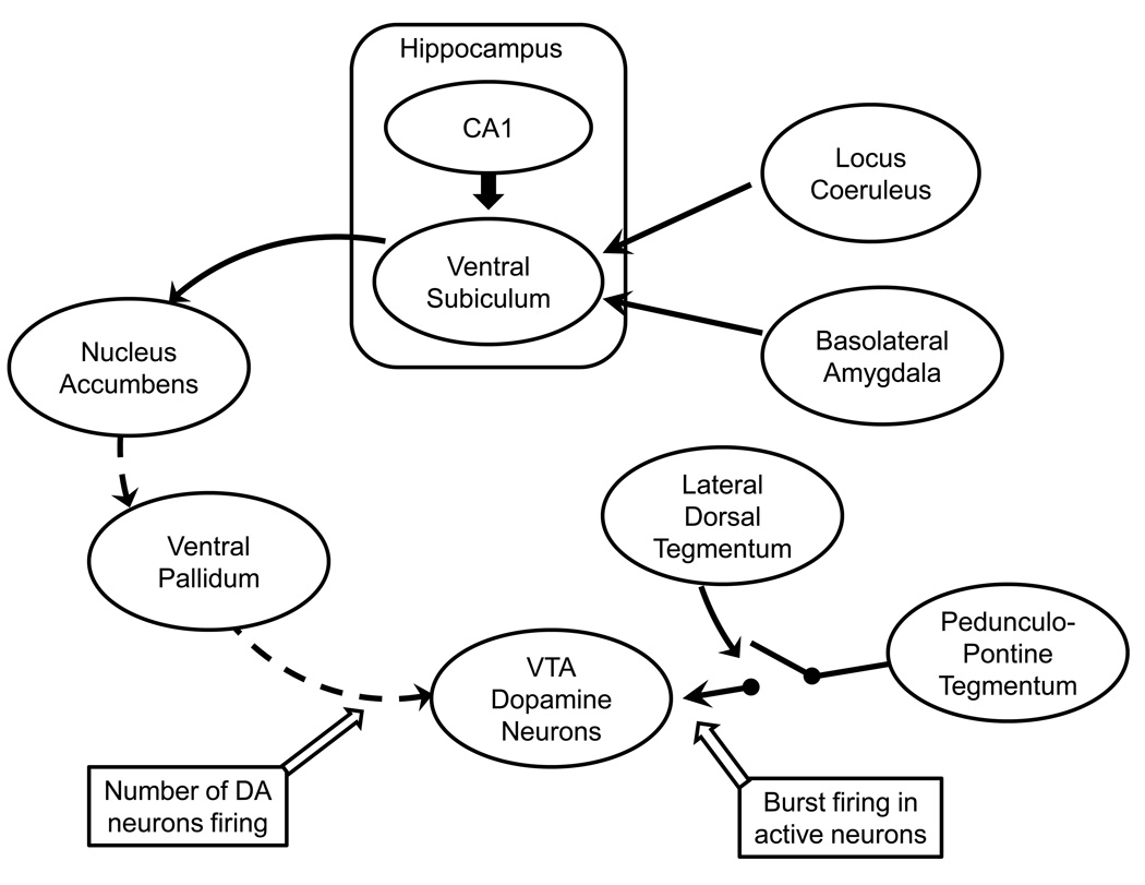
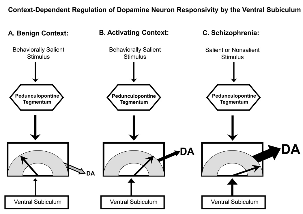
The dopamine system is under multiple forms of regulation, and in turn provides effective modulation of system responses. Dopamine neurons are known to exist in several states of activity. The population activity, or the proportion of dopamine neurons firing spontaneously, is controlled by the ventral subiculum of the hippocampus. In contrast, burst firing, which is proposed to be the behaviorally salient output of the dopamine system, is driven by the brainstem pedunculopontine tegmentum (PPTg). When an animal is exposed to a behaviorally salient stimulus, the PPTg elicits a burst of action potentials in the dopamine neurons. However, this bursting only occurs in the portion of the dopamine neuron population that is firing spontaneously. This proportion is regulated by the ventral subiculum. Therefore, the ventral subiculum provides the gain, or the amplification factor, for the behaviorally salient stimulus. The ventral subiculum itself is proposed to carry information related to the environmental context. Thus, the ventral subiculum will adjust the responsivity of the dopamine system based on the needs of the organism and the characteristics of the environment. However, this finely tuned system can be disrupted in disease states. In schizophrenia, a disruption of interneuronal regulation of the ventral subiculum is proposed to lead to an overdrive of the dopamine system, rendering the system in a constant hypervigilant state. Moreover, amphetamine sensitization and stressors also appear to cause an abnormal dopaminergic drive. Such an interaction could underlie the risk factors of drug abuse and stress in the precipitation of a psychotic event. On the other hand, this could point to the ventral subiculum as an effective site of therapeutic intervention in the treatment or even the prevention of schizophrenia.


Figures

Figure 1
Figure 1
Diagram illustrating the interconnections of the brain regions regulating dopamine neuron firing. Dopamine neurons in the ventral tegmental area (VTA) are differentially regulated by two projection systems. Spatial location is integrated with limbic and stress-related information from the basolateral amygdala (emotion and stress) and noradrenergic locus coeruleus (attention and stress) within the ventral subiculum, providing a contextual representation of the behavioral contingencies. The ventral subiculum then drives nucleus accumbens neuron activity which, in turn, inhibits the ventral pallidum. The ventral pallidum provides a powerful inhibition to the VTA dopamine neurons, causing a portion to be inhibited and non-firing. By controlling the ventral pallidum, this system determines the number of VTA dopamine neurons that are firing spontaneously. In contrast, the pedunculopontine tegmentum provides a glutamatergic drive over the dopamine neurons that causes only the spontaneously active neurons (determined by the ventral pallidum) to burst fire in response to a phasic stimulus. The ability of the pedunculopontine tegmentum to drive burst firing is gated by the lateral dorsal tegmentum; inhibition of the lateral dorsal tegmentum prevents burst activity altogether. These systems interact to provide phasic behaviorally salient dopamine release (via the pedunculopontine tegmentum) in response to a stimulus, with the amplitude of the phasic release dependent on the number of dopamine neurons firing (via the ventral subiculum). Solid lines = excitatory/facilitatory projections; dashed lines = inhibitory projections
Figure 2
Figure 2
Dopamine neuron activity states are regulated by the ventral subiculum of the hippocampus and the pedunculopontine tegmentum. Stimuli that are behaviorally salient activate the pedunculopontine tegmentum, causing glutamate release onto mesolimbic dopamine neurons and leading them to burst fire. The amplitude of this phasic dopamine signal is dependent on the number of dopamine neurons that the pedunculopontine can activate, since it can only cause burst firing in dopamine neurons that are already spontaneously firing. By controlling the population activity (i.e., proportion of dopamine neurons firing spontaneously), the ventral subiculum regulates the gain of the phasic signal. A) In a safe, benign context, novel salient stimuli activate the pedunculopontine tegmentum. However, because of the benign environmental context, the ventral subiculum only allows a small proportion of dopamine neurons to be firing spontaneously. As a result, the dopamine signal is small, garnering little behavioral activation of the subject. B) In an activating context, stimuli would likely have a strong motivating or threatening association, such as when searching for prey (rewarding) or in dangerous situations (threatening). In this case, the ventral subiculum causes a large proportion of dopamine neurons to be spontaneously firing. Now when a salient stimulus activates the pedunculopontine tegmentum, the dopamine signal is large in amplitude. C) In schizophrenia, a dysfunctional and overactive ventral subiculum overdrives the dopamine system, causing essentially all of the dopamine neurons to fire spontaneously. In this condition, even minor activation of the pedunculopontine tegmentum by salient or even nonsalient stimuli still results in massive activation of the dopamine signal. As a result, the subject is forced to attend to every signal it receives as if it were a life-threatening situation, clamoring for attention.
Figure 3
Figure 3
Amphetamine sensitization increases dopamine neuron population activity via activation of the ventral subiculum. A) Acute administration of amphetamine (1.5 mg/kg i.p.) causes a decrease in the population activity of dopamine neurons in the midbrain ventral tegmental area. In contrast, after 5 days repeated amphetamine and 5 days withdrawal, there is a substantial increase in the number of dopamine neurons firing spontaneously. B) Inactivation of the ventral subiculum (cross-hatched) does not produce a strong effect on the population activity in control animals, likely due to its low level of activity in anesthetized rats. In contrast, inactivation of the ventral subiculum in amphetamine sensitized rats restores the increased population activity back to control levels. C) Inactivation of the ventral subiculum with TTX injection does not affect the locomotor response of control rats to amphetamine injection (1.5 mg/kg i.v., vertical dashed line). D) In contrast, in amphetamine-sensitized rats inactivation of the ventral subiculum restores the locomotor response to amphetamine back to control levels. Adapted from (Lodge and Grace, 2008). dPBS = distilled phosphate buffered saline; †Significant group effect of treatment

Similar articles

Cited by

References

    1. Abercrombie ED, Keefe KA, DiFrischia DS, Zigmond MJ. Differential effect of stress on in vivo dopamine release in striatum, nucleus accumbens, and medial frontal cortex. J Neurochem. 1989;52:1655–1658. - PubMed
    1. Amaral DG, Dolorfo C, Alvarez-Royo P. Organization of CA1 projections to the subiculum: a PHA-L analysis in the rat. Hippocampus. 1991;1:415–435. - PubMed
    1. Angrist B, Sathananthan G, Wilk S, Gershon S. Amphetamine psychosis: behavioral and biochemical aspects. Journal of Psychiatric Research. 1974;11:13–23. - PubMed
    1. Angrist B, Thompson H, Shopsin B, Gershon S. Clinical studies with dopamine-receptor stimulants. Psychopharmacologia. 1975;44:273–280. - PubMed
    1. Anstrom KK, Woodward DJ. Restraint increases dopaminergic burst firing in awake rats. Neuropsychopharmacology. 2005;30:1832–1840. - PubMed

MeSH terms