. 2010 Apr;38(4):1288-313.
doi: 10.1007/s10439-010-9949-x. Epub 2010 Feb 9.

Quantification of hemodynamics in abdominal aortic aneurysms during rest and exercise using magnetic resonance imaging and computational fluid dynamics

Affiliations

Quantification of hemodynamics in abdominal aortic aneurysms during rest and exercise using magnetic resonance imaging and computational fluid dynamics

Andrea S Les et al. Ann Biomed Eng. 2010 Apr.

Abstract

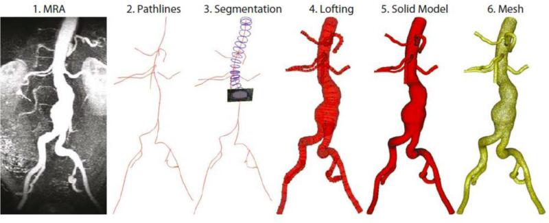
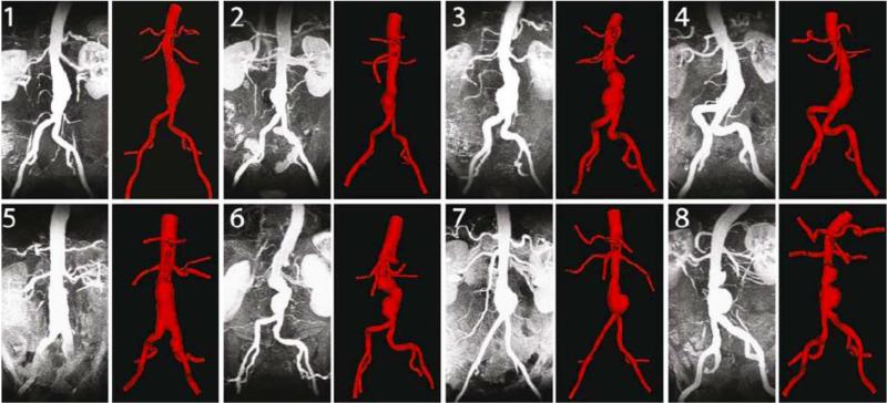
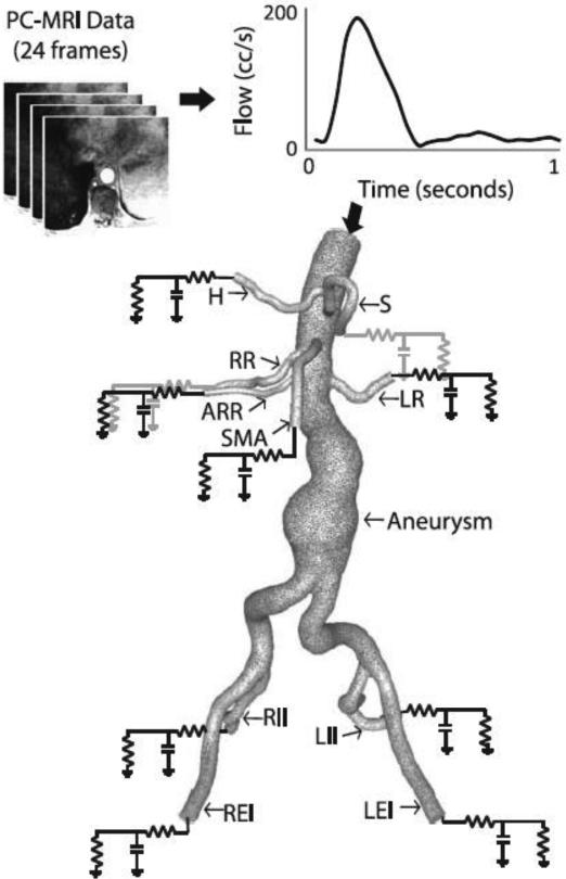
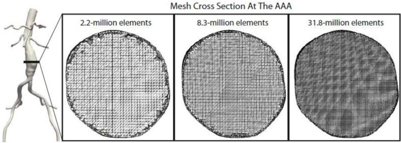
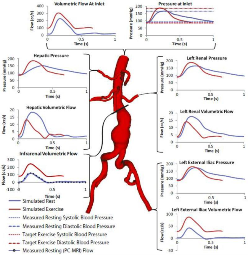
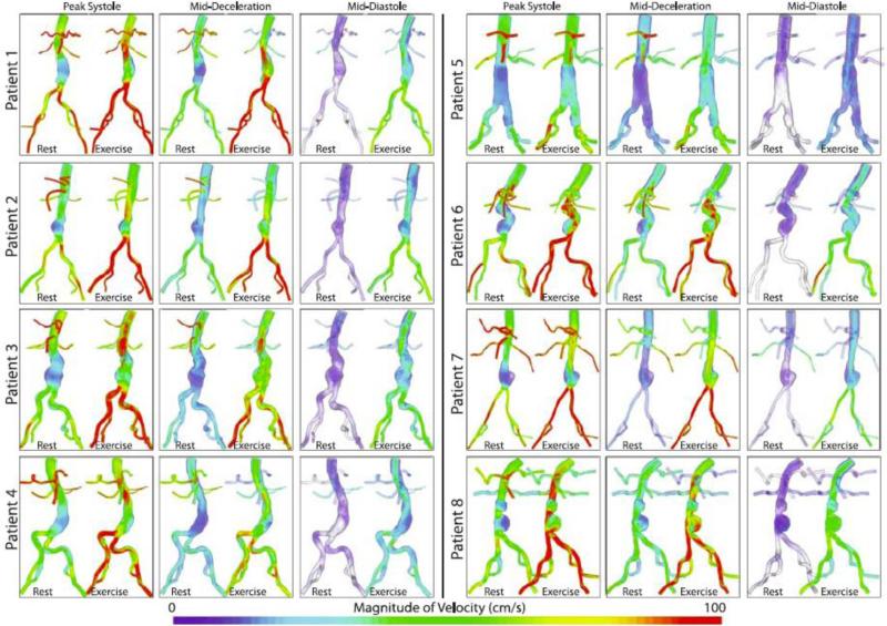
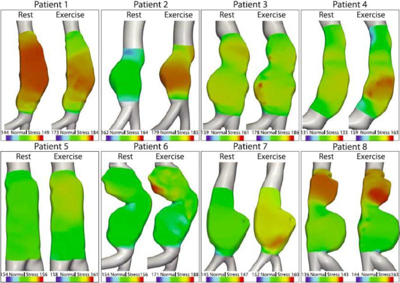
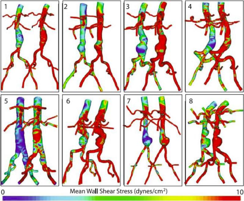
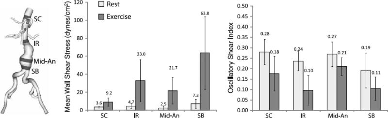
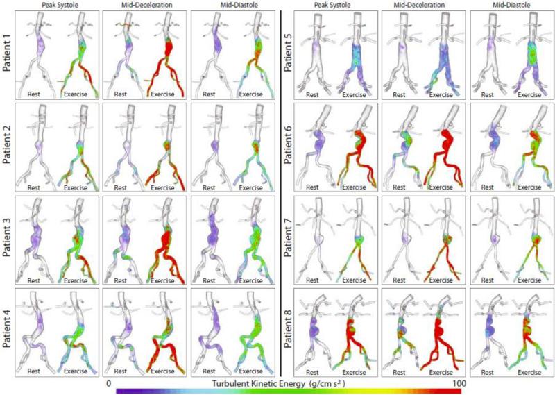
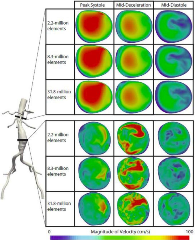
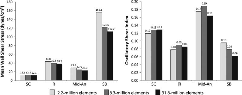
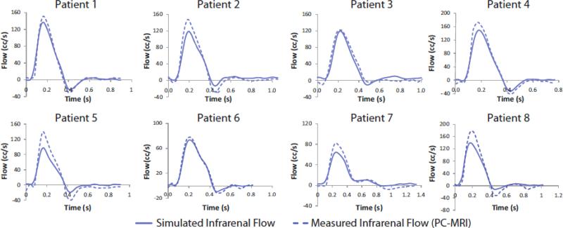
Abdominal aortic aneurysms (AAAs) affect 5-7% of older Americans. We hypothesize that exercise may slow AAA growth by decreasing inflammatory burden, peripheral resistance, and adverse hemodynamic conditions such as low, oscillatory shear stress. In this study, we use magnetic resonance imaging and computational fluid dynamics to describe hemodynamics in eight AAAs during rest and exercise using patient-specific geometric models, flow waveforms, and pressures as well as appropriately resolved finite-element meshes. We report mean wall shear stress (MWSS) and oscillatory shear index (OSI) at four aortic locations (supraceliac, infrarenal, mid-aneurysm, and suprabifurcation) and turbulent kinetic energy over the entire computational domain on meshes containing more than an order of magnitude more elements than previously reported results (mean: 9.0-million elements; SD: 2.3 M; range: 5.7-12.0 M). MWSS was lowest in the aneurysm during rest 2.5 dyn/cm(2) (SD: 2.1; range: 0.9-6.5), and MWSS increased and OSI decreased at all four locations during exercise. Mild turbulence existed at rest, while moderate aneurysmal turbulence was present during exercise. During both rest and exercise, aortic turbulence was virtually zero superior to the AAA for seven out of eight patients. We postulate that the increased MWSS, decreased OSI, and moderate turbulence present during exercise may attenuate AAA growth.


Figures

Figure 1
Figure 1
Building a model from the MRA data (1) entails generating pathlines (2), segmenting along the pathlines (3), lofting the segmentations to create vessels (4), unioning the vessels to create a single, solid model representing the flow domain (5), and then discretizing the model into a finite-element mesh (6).
Figure 2
Figure 2
The maximum intensity projection (MIP) of the MRA is compared to the 3-D computer model for all eight patients.
Figure 3
Figure 3
A patient-specific SC flow waveform measured by phase-contrast MRI (resting conditions) is mapped to the inflow face using a Womersley velocity profile. A patient-specific 3-element Windkessel model with a proximal resistance (Rp), capacitance (C), distal resistance (Rd) boundary condition is used to represent the resistance and compliance of the vasculature downstream of each outlet. The outlet labels are as follows: S=splenic artery, LR=left renal artery, LEI=left external iliac artery, LII=left internal iliac artery, RII=right internal iliac artery, REI=right external iliac artery, SMA=superior mesenteric artery, ARR=accessory right renal artery (Note: accessory renals are not found in the majority of patients), RR=right renal artery, H=hepatic artery. Please note that typically (as in this patient), the splenic and hepatic arteries branch from a common celiac trunk.
Figure 4
Figure 4
Cross sections of the 2.2, 8.3, and 31.8-million finite-element meshes are shown at the mid-aneurysm level for patient 1.
Figure 5
Figure 5
Pressure and flow waveforms at the inlet and selected outlets of patient 3 under rest and exercise conditions. The pressure waveforms at each outlet were calculated by averaging the pressure field over the outlet area. The flow waveforms at each outlet were calculated by integrating the velocities over the outlet area. The flow and pressure waves depicted in this figure show a number of physiologically-realistic features. For instance, for the renal arteries under resting conditions and the abdominal aorta under exercise conditions, the flow waveforms lack backflow and have a relatively high diastolic flow, matching literature measurements and calculations., In addition, not only are the outlet pressures in the physiologic range, the inlet resting maximum and minimum pressures agree well with the measured systolic and diastolic pressure, and the inlet exercise maximum and minimum pressure agree well with the physiologic target systolic and diastolic pressures.
Figure 6
Figure 6
The magnitude of the ensemble average of the velocity field at three points in the cardiac cycle (peak systole, mid-deceleration, and mid-diastole) is plotted using a volume rendering technique for all patients for both rest and exercise. In general, the flow features look more complex during exercise than during rest, for all three reported time points. In particular, during exercise, features such as jets and recirculation zones can be observed, especially during diastole and mid-deceleration.
Figure 7
Figure 7
The distribution of normal stresses acting on the luminal wall of the aneurysms at peak flow (defined as peak systole throughout the paper) is shown for both rest and exercise. Values are presented in mmHg.
Figure 8
Figure 8
Mean wall shear stress (MWSS) for rest (left of each pair) and exercise (right of each pair) is shown for all eight patients. MWSS was quantified from cardiac cycles four through eight.
Figure 9
Figure 9
Oscillatory shear index (OSI) for rest (left of each pair) and exercise (right of each pair) is shown for all eight patients. OSI was quantified from cardiac cycles four through eight.
Figure 10
Figure 10
Mean wall shear stress (left) and oscillatory shear index (right) values were quantified in 1-cm strips at four locations (SC=supraceliac, IR=infrarenal, Mid-An=mid-aneurysm, SB=suprabifurcation) for rest and exercise. The location of the strips for patient 3 are shown at left. The standard deviation for each value are indicated with vertical bars. MWSS and OSI were quantified from cardiac cycles four through eight.
Figure 11
Figure 11
Turbulent kinetic energy (TKE) at three points in the cardiac cycle (peak systole, mid-deceleration, and mid-diastole) are plotted for all patients for both rest and exercise. TKE was quantified from cardiac cycles four through eight.
Figure 12
Figure 12
The instantaneous magnitude of the velocity field for the eighth cardiac cycle of patient 1 under exercise conditions are shown at the mid-aneurysm and suprarenal locations for the 2.2, 8.3, and 31.8-million finite-element meshes at peak systole, mid-deceleration, and mid-diastole. The velocity field changed little between meshes at the suprarenal location, where the TKE was virtually zero, but changed more dramatically at the mid-aneurysm location where TKE was moderate. Mesh independence of the instantaneous velocity fields is not achievable in turbulent regions.
Figure 13
Figure 13
Mean wall shear stress (left) and oscillatory shear index (right) at four locations (SC=supraceliac, IR=infrarenal, Mid-An=mid-aneurysm, SB=suprabifurcation) for three meshes (2.2-million, 8.3-million, and 31.8-million elements) are shown for patient 1 during exercise. MWSS and OSI were quantified from cardiac cycles four through eight.
Figure 14
Figure 14
A comparison between simulated and measured volumetric flow at the infrarenal level during rest for all eight patients is shown above.

Similar articles

Cited by

References

    1. Allardice JT, Allwright GJ, Wafula JM, Wyatt AP. High prevalence of abdominal aortic aneurysm in men with peripheral vascular disease: Screening by ultrasonography. Brit. J. Surg. 1988;75:240–2. - PubMed
    1. Asbury CL, Ruberti JW, Bluth EI, Peattie RA. Experimental investigation of steady flow in rigid models of abdominal aortic aneurysms. Ann Biomed Eng. 1995;23:29–39. - PubMed
    1. Bax L, Bakker CJ, Klein WM, Blanken N, Beutler JJ, Mali WP. Renal blood flow measurements with use of phase-contrast magnetic resonance imaging: Normal values and reproducibility. J. Vasc. Interv. Radiol. 2005;16:807–14. - PubMed
    1. Berguer R, Bull JL, Khanafer K. Refinements in mathematical models to predict aneurysm growth and rupture. Ann. N.Y. Acad. Sci. 2006;1085:110–6. - PubMed
    1. Bluestein D, Niu L, Schoephoerster RT, Dewanjee MK. Steady flow in an aneurysm model: Correlation between fluid dynamics and blood platelet deposition. Journal of Biomechamical Engineering. 1996;118:280–6. - PubMed

Publication types