

Competitiveness for the niche and mutual dependence of the germline and somatic stem cells in the Drosophila testis are regulated by the JAK/STAT signaling
- PMID: 20143337
- PMCID: PMC2894562
- DOI: 10.1002/jcp.22073
Competitiveness for the niche and mutual dependence of the germline and somatic stem cells in the Drosophila testis are regulated by the JAK/STAT signaling
Abstract
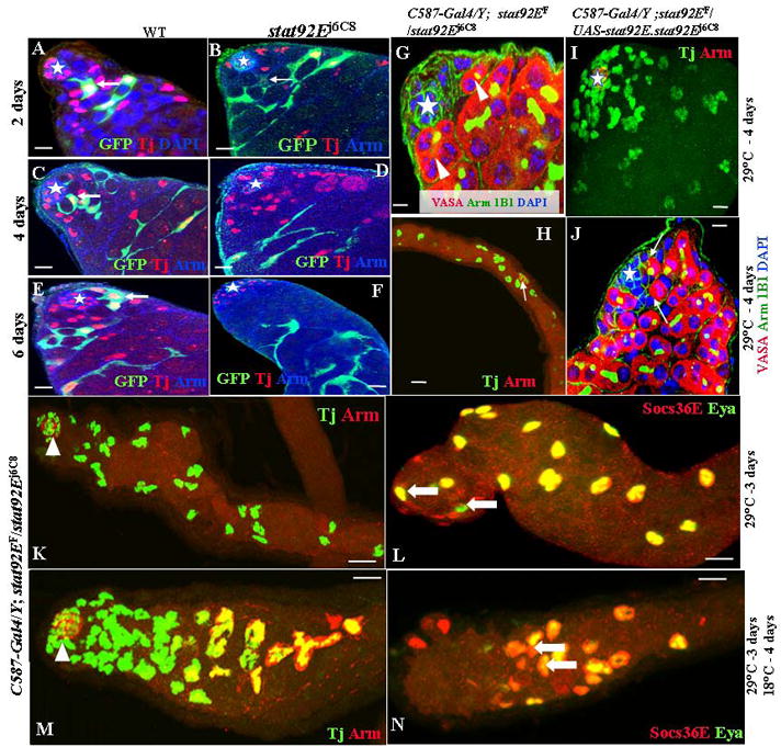
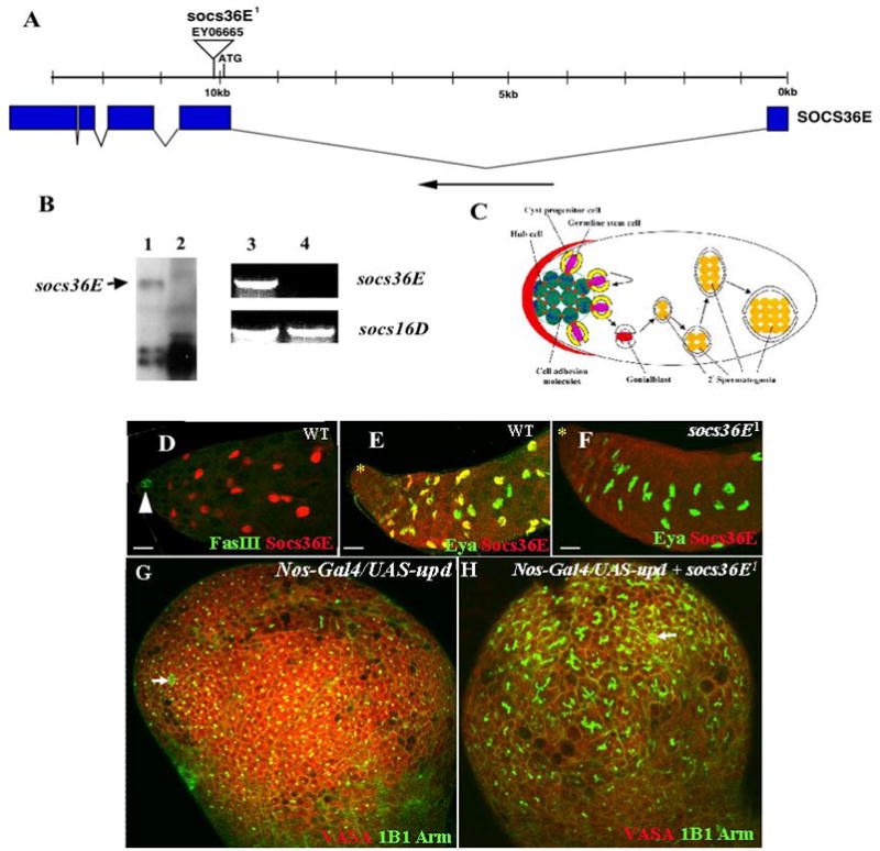
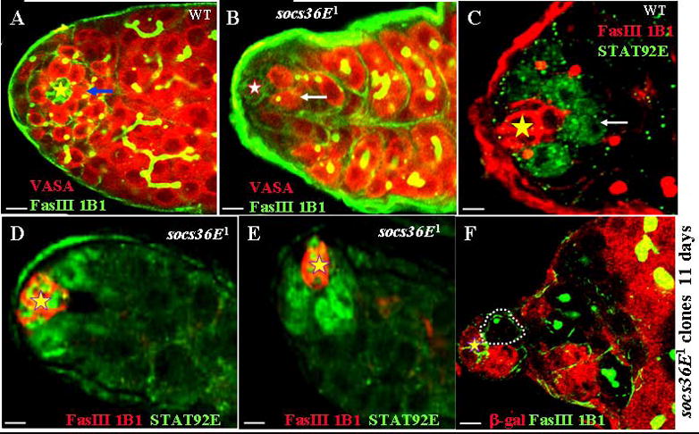
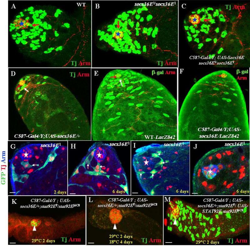
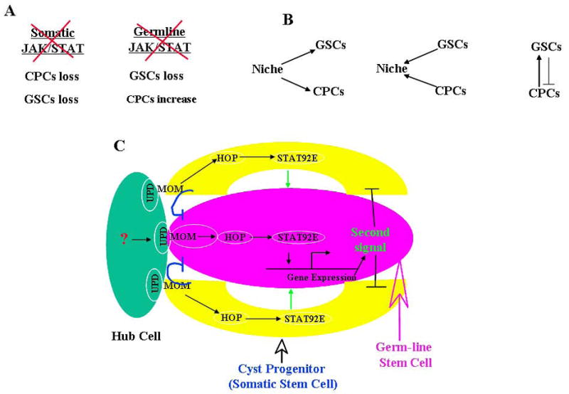
In many tissues, two or more types of stem cells share a niche, and how the stem cells coordinate their self-renewal and differentiation is poorly understood. In the Drosophila testis, germ line stem cells (GSCs) and somatic cyst progenitor cells (CPCs) contact each other and share a niche (the hub). The hub expresses a growth factor unpaired (Upd) that activates the Janus kinase/signal transducer and activator of transcription (JAK/STAT) pathway in GSCs to regulate the stem cell self-renewal. Here, we demonstrate that the JAK/STAT signaling also regulates CPCs self-renewal. We also show that a negative regulator, the suppressor of cytokine signaling 36E (SOCS36E), suppresses JAK/STAT signaling in somatic cells, preventing them from out-competing the GSCs. Furthermore, through selectively manipulating the JAK/STAT signaling level in either CPCs or GSCs, we demonstrate that the somatic JAK/STAT signaling is essential for self-renewal and maintenance of both CPCs and GSCs. These data suggest that a single JAK/STAT signal from the niche orchestrate the competitive and dependent co-existence of GSCs and CPCs in the Drosophila testis niche.
Figures
Similar articles
-
Jak-STAT regulation of cyst stem cell development in the Drosophila testis.Dev Biol. 2012 Dec 1;372(1):5-16. doi: 10.1016/j.ydbio.2012.09.009. Epub 2012 Sep 23. Dev Biol. 2012. PMID: 23010510 Free PMC article.
-
JAK-STAT signal inhibition regulates competition in the Drosophila testis stem cell niche.Science. 2009 Oct 2;326(5949):153-6. doi: 10.1126/science.1176817. Science. 2009. PMID: 19797664 Free PMC article.
-
A regulatory loop of JAK/STAT signalling and its downstream targets represses cell fate conversion and maintains male germline stem cell niche homeostasis.Cell Prolif. 2024 Oct;57(10):e13648. doi: 10.1111/cpr.13648. Epub 2024 Jul 10. Cell Prolif. 2024. PMID: 38987866 Free PMC article.
-
Hedgehog in the Drosophila testis niche: what does it do there?Protein Cell. 2013 Sep;4(9):650-5. doi: 10.1007/s13238-013-3040-y. Epub 2013 Jun 26. Protein Cell. 2013. PMID: 23807635 Free PMC article. Review.
-
Local signaling within stem cell niches: insights from Drosophila.Curr Opin Cell Biol. 2012 Apr;24(2):225-31. doi: 10.1016/j.ceb.2012.01.004. Epub 2012 Jan 30. Curr Opin Cell Biol. 2012. PMID: 22296770 Free PMC article. Review.
Cited by
-
The Drosophila histone methyltransferase SET1 coordinates multiple signaling pathways in regulating male germline stem cell maintenance and differentiation.Development. 2024 Aug 1;151(15):dev202729. doi: 10.1242/dev.202729. Epub 2024 Aug 9. Development. 2024. PMID: 39007366 Free PMC article.
-
Possible therapeutic use of spermatogonial stem cells in the treatment of male infertility: a brief overview.ScientificWorldJournal. 2012;2012:374151. doi: 10.1100/2012/374151. Epub 2012 Mar 12. ScientificWorldJournal. 2012. PMID: 22536138 Free PMC article. Review.
-
The stem cell niche: lessons from the Drosophila testis.Development. 2011 Jul;138(14):2861-9. doi: 10.1242/dev.056242. Development. 2011. PMID: 21693509 Free PMC article. Review.
-
Without children is required for Stat-mediated zfh1 transcription and for germline stem cell differentiation.Development. 2014 Jul;141(13):2602-10. doi: 10.1242/dev.109611. Epub 2014 Jun 5. Development. 2014. PMID: 24903753 Free PMC article.
-
Escort cell encapsulation of Drosophila germline cells is maintained by irre cell recognition module proteins.Biol Open. 2019 Mar 5;8(3):bio039842. doi: 10.1242/bio.039842. Biol Open. 2019. PMID: 30837217 Free PMC article.
References
-
- Arbouzova NI, Zeidler MP. JAK/STAT signalling in Drosophila: insights into conserved regulatory and cellular functions. Development. 2006;133:2605–2616. - PubMed
-
- Baksa K, Parke T, Dobens LL, Dearolf CR. The Drosophila STAT protein, Stat92E, regulates follicle cell differentiation during oogenesis. Dev Biol. 2002;243:166–175. - PubMed
-
- Brand AH, Perrimon N. Targeted gene expression as a means of altering cell fates and generating dominant phenotypes. Development. 1993;118:401–415. - PubMed
-
- Brawley C, Matunis E. Regeneration of male germline stem cells by spermatogonial dedifferentiation in vivo. Science. 2004;304:1331–1334. - PubMed
-
- Bunt SM, Hime GR. Ectopic activation of Dpp signalling in the male Drosophila germline inhibits germ cell differentiation. Genesis. 2004;39:84–93. - PubMed
Publication types
MeSH terms
Substances
Grants and funding
LinkOut - more resources
Full Text Sources
Medical
Molecular Biology Databases
