

Cortical gamma-oscillations modulated by auditory-motor tasks-intracranial recording in patients with epilepsy
- PMID: 20143383
- PMCID: PMC2891961
- DOI: 10.1002/hbm.20963
Cortical gamma-oscillations modulated by auditory-motor tasks-intracranial recording in patients with epilepsy
Abstract
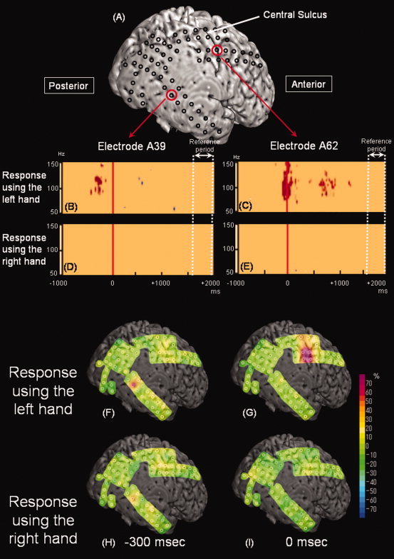
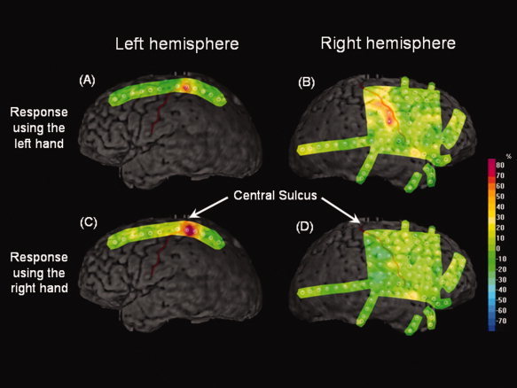
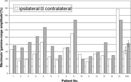
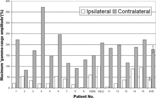
Human activities often involve hand-motor responses following external auditory-verbal commands. It has been believed that hand movements are predominantly driven by the contralateral primary sensorimotor cortex, whereas auditory-verbal information is processed in both superior temporal gyri. It remains unknown whether cortical activation in the superior temporal gyrus during an auditory-motor task is affected by laterality of hand-motor responses. Here, event-related γ-oscillations were intracranially recorded as quantitative measures of cortical activation; we determined how cortical structures were activated by auditory-cued movement using each hand in 15 patients with focal epilepsy. Auditory-verbal stimuli elicited augmentation of γ-oscillations in a posterior portion of the superior temporal gyrus, whereas hand-motor responses elicited γ-augmentation in the pre- and postcentral gyri. The magnitudes of such γ-augmentation in the superior temporal, precentral, and postcentral gyri were significantly larger when the hand contralateral to the recorded hemisphere was required to be used for motor responses, compared with when the ipsilateral hand was. The superior temporal gyrus in each hemisphere might play a greater pivotal role when the contralateral hand needs to be used for motor responses, compared with when the ipsilateral hand does.
© 2010 Wiley-Liss, Inc.
Figures
Similar articles
-
Three- and four-dimensional mapping of speech and language in patients with epilepsy.Brain. 2017 May 1;140(5):1351-1370. doi: 10.1093/brain/awx051. Brain. 2017. PMID: 28334963 Free PMC article.
-
Young patients with focal seizures may have the primary motor area for the hand in the postcentral gyrus.Epilepsy Res. 2007 Sep;76(2-3):131-9. doi: 10.1016/j.eplepsyres.2007.07.007. Epub 2007 Aug 27. Epilepsy Res. 2007. PMID: 17723289 Free PMC article.
-
Cortical gamma oscillations modulated by word association tasks: intracranial recording.Epilepsy Behav. 2010 May;18(1-2):116-8. doi: 10.1016/j.yebeh.2010.03.008. Epub 2010 May 6. Epilepsy Behav. 2010. PMID: 20451464 Free PMC article.
-
Event-related oscillations reflect functional asymmetry in children with attention deficit/hyperactivity disorder.Suppl Clin Neurophysiol. 2013;62:289-301. doi: 10.1016/b978-0-7020-5307-8.00018-1. Suppl Clin Neurophysiol. 2013. PMID: 24053046 Review.
-
The Control of Movements via Motor Gamma Oscillations.Front Hum Neurosci. 2022 Jan 17;15:787157. doi: 10.3389/fnhum.2021.787157. eCollection 2021. Front Hum Neurosci. 2022. PMID: 35111006 Free PMC article. Review.
Cited by
-
Gamma activity modulated by naming of ambiguous and unambiguous images: intracranial recording.Clin Neurophysiol. 2015 Jan;126(1):17-26. doi: 10.1016/j.clinph.2014.03.034. Epub 2014 Apr 18. Clin Neurophysiol. 2015. PMID: 24815577 Free PMC article.
-
Cortical gamma-oscillations modulated by visuomotor tasks: Intracranial recording in patients with epilepsy.Epilepsy Behav. 2010 Jul;18(3):254-61. doi: 10.1016/j.yebeh.2010.02.015. Epub 2010 May 23. Epilepsy Behav. 2010. PMID: 20580900 Free PMC article.
-
Human occipital cortices differentially exert saccadic suppression: Intracranial recording in children.Neuroimage. 2013 Dec;83:224-36. doi: 10.1016/j.neuroimage.2013.06.046. Epub 2013 Jun 20. Neuroimage. 2013. PMID: 23792979 Free PMC article.
-
Evaluating the arcuate fasciculus with combined diffusion-weighted MRI tractography and electrocorticography.Hum Brain Mapp. 2014 May;35(5):2333-47. doi: 10.1002/hbm.22331. Epub 2013 Aug 24. Hum Brain Mapp. 2014. PMID: 23982893 Free PMC article.
-
Combining Gamma With Alpha and Beta Power Modulation for Enhanced Cortical Mapping in Patients With Focal Epilepsy.Front Hum Neurosci. 2020 Dec 21;14:555054. doi: 10.3389/fnhum.2020.555054. eCollection 2020. Front Hum Neurosci. 2020. PMID: 33408621 Free PMC article.
References
-
- Abler B, Roebroeck A, Goebel R, Höse A, Schönfeldt‐Lecuona C, Hole G, Walter H ( 2006): Investigating directed influences between activated brain areas in a motor‐response task using fMRI. Magn Reson Imaging 24: 181–185. - PubMed
-
- Akiyama T, Otsubo H, Ochi A, Galicia EZ, Weiss SK, Donner EJ, Rutka JT, Snead OC III ( 2006): Topographic movie of ictal high‐frequency oscillations on the brain surface using subdural EEG in neocortical epilepsy. Epilepsia 47: 1953–1957. - PubMed
-
- Auranen T ( 2002): Nonparametric statistical analysis of time‐frequency representations of magnetoencephalographic data. Master's Thesis, Helsinki University of Technology, Department of Electrical and Communications Engineering, Espoo, Finland.
