. 2010 Mar 23;49(11):2529-39.
doi: 10.1021/bi9018237.

Peroxynitrite-mediated oxidative modifications of complex II: relevance in myocardial infarction

Affiliations

Peroxynitrite-mediated oxidative modifications of complex II: relevance in myocardial infarction

Liwen Zhang et al. Biochemistry. .

Abstract

Increased O(2)(*-) and NO production is a key mechanism of mitochondrial dysfunction in myocardial ischemia/reperfusion injury. In complex II, oxidative impairment and enhanced tyrosine nitration of the 70 kDa FAD-binding protein occur in the post-ischemic myocardium and are thought to be mediated by peroxynitrite (OONO(-)) in vivo [Chen, Y.-R., et al. (2008) J. Biol. Chem. 283, 27991-28003]. To gain deeper insights into the redox protein thiols involved in OONO(-)-mediated oxidative post-translational modifications relevant in myocardial infarction, we subjected isolated myocardial complex II to in vitro protein nitration with OONO(-). This resulted in site-specific nitration at the 70 kDa polypeptide and impairment of complex II-derived electron transfer activity. Under reducing conditions, the gel band of the 70 kDa polypeptide was subjected to in-gel trypsin/chymotrypsin digestion and then LC-MS/MS analysis. Nitration of Y(56) and Y(142) was previously reported. Further analysis revealed that C(267), C(476), and C(537) are involved in OONO(-)-mediated S-sulfonation. To identify the disulfide formation mediated by OONO(-), nitrated complex II was alkylated with iodoacetamide. In-gel proteolytic digestion and LC-MS/MS analysis were conducted under nonreducing conditions. The MS/MS data were examined with MassMatrix, indicating that three cysteine pairs, C(306)-C(312), C(439)-C(444), and C(288)-C(575), were involved in OONO(-)-mediated disulfide formation. Immuno-spin trapping with an anti-DMPO antibody and subsequent MS was used to define oxidative modification with protein radical formation. An OONO(-)-dependent DMPO adduct was detected, and further LC-MS/MS analysis indicated C(288) and C(655) were involved in DMPO binding. These results offered a complete profile of OONO(-)-mediated oxidative modifications that may be relevant in the disease model of myocardial infarction.


Figures
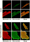
Fig. 1
Fig. 1. Hypoxia/Reoxygenation (H/RO) increases 3-nitrotyrosine staining in the mitochondria of myocytes
Cardiomyocytes were plated on laminin-coated coverslips in 24 well plates. Hypoxic treatment was accomplished by forming a layer of oil on the surface of the glucose-free medium and incubating for 1 h, after which reoxygenation was carried out by incubating in medium with glucose for 2 h. After treatment myocytes were loaded with MitoTracker® Red (250 nM for 15 min), fixed, and stained for nitrotyrosine using anti-nitrotyrosine polyclonal antibody (the secondary antibody was Alexa 488 conjugated). Fluorescence images were acquired with confocal microscopy and were merged in order to determine whether the increase in nitrotyrosine signal colocalized in mitochondria.
Fig. 2
Fig. 2
A and B, The rat heart model of in vivo myocardial ischemia/reperfusion and TCC staining of infarct region in the post-ischemic myocardium. Myocardial tissue homogenates from non-ischemic and infarct (risk) regions were subjected to analysis of complex II activity (in A) and immunoprecipitation with a polyclonal antibody against complex II 70 kDa and subsequently subjected to SDS-PAGE and immunoblotted with anti-3-nitrotyrosine (upper panel) and anti-70 kDa (lower panel) antibodies (in B). Note that 100% of the basal level of enzymatic activity (TTFA sensitive) is 60.3 nmole of DCPIP reduction/min/mg of protein in A. C, Right panel, isolated complex II was subjected to in vitro protein tyrosine nitration. Protein (1 μM, based on heme b) was incubated with various concentrations of OONO (0–80 μM) at 37 °C for 1 h. Excess OONO was removed by uric acid (1 mM). The OONO –treated complex II was subjected to SDS-PAGE and then immunoblotting with anti-3-nitrotyrosine antibody. Left panel, SDS-PAGE of OONO –treated complex II and stained by Coomassie blue. D, the OONO –treated complex II was subjected to analysis of electron transfer activity. 100% of basal level of purified bovine complex II activity is 15.0 μmol of succinate oxidized/min/mg protein.
Fig. 3
Fig. 3
Amino acid sequence of the precursor of the complex II-70 kDa FAD-binding subunit. The regions labeled with bold represent the amino acid residues identified with LC/MS/MS under the reduced conditions in the presence of β-ME (A) and non-reduced conditions in the absence of β-ME (B). In (A), the cysteinyl residues involved in S-sulfonation are highlighted with gray and they are C267, C476, and C537. The cysteinyl residues involved in protein radical formation are underlined and they are C288 and C655. In (B), the cysteinyl residues involved in disulfide linkage are highlighted with gray and they are C306, C312, C439, C444, C288, and C575. The region labeled with a dotted underline is the N-terminal extension (aa 1–43), which acts as an import sequence and does not exist in the mature protein.
Fig. 4
Fig. 4
MS/MS of doubly protonated molecular ion of S-sulfonated peptide (263TYFSC267(SO3) TSAHTSTGDGTAMVTR283) (where Cys267 was sulfonated) of the 70 kDa subunit from peroxynitrite-treated complex II. The sequence-specific ions are labeled as y and b ions on the spectrum. Note that the same spectrum is shown in upper and lower panels.
Fig. 5
Fig. 5
Disulfide bond linkage between C306 and C312 as determined by MS/MS spectrum of tryptic/chymotryptic digests of the 70 kDa subunit of peroxynitrite-treated complex II. The sequence-specific ions are labeled as yn (n=2–7), yn* (yn-2, n=14–15), and bn* (bn-2, n=10–16) on the spectrum. Note that the same spectrum is shown in upper and lower panels.
Fig. 6
Fig. 6
Disulfide bond linkage between C288 and C575 as determined by MS/MS spectrum of tryptic/chymotryptic digests of the 70 kDa subunit of peroxynitrite-treated complex II. Chain A and Chain B represent peptides containing aa 568–577 and aa 287–291, respectively. A disulfide bond linking Chain A with Chain B is indicated in red. The sequence-specific ions are labeled as bnA (n = 2–9) for Chain A, y3B and b3B for Chain B on the spectrum. M indicates Chain A plus Chain B.
Fig. 7
Fig. 7. Left panel
Schematic delineation of immuno-spin trapping of OONO–induced protein radical with anti-DMPO polyclonal antibody. Right panel: Detection of the DMPO adduct of the complex II-derived protein radicals by Western blot using anti-DMPO nitrone adduct polyclonal antibody (lane 3). The reaction mixture contained complex II (dithiothretol treated, 1 μM heme b) and DMPO (100 mM) in PBS. OONO (60 μM) was added to initiate the reaction. The reaction was allowed to incubate for 1 h at 37 °C, terminated by addition of uric acid (0.2 mM) and sample buffer containing 0.4% SDS and 1% β-ME, and then heated at 70 °C for 5 min. Aliquotes of 30 pmol of protein were subjected to SDS-PAGE and Western blot using anti-DMPO polyclonal antibody. Lane 1, OONO was removed from the complete system. Lane 3, uric acid (0.2 mM) was pre-incubated with complex II and DMPO before the reaction was initiated by OONO. Lane 4, DMPO was omitted from the complete system.
Fig. 8
Fig. 8. MS/MS spectra of the doubly protonated molecular ions of the DMPO-binding peptides
A, 284AGLPC288QDLEFVQF296. B, 649TLNETDC655ATVPPAIR663. The sequence-specific ions are labeled as y and b ions on the spectra. The amino acid residues involved in DMPO binding are identified to be C288 and C655.

Similar articles

Cited by

References

    1. Lemos RS, Fernandes AS, Pereira MM, Gomes CM, Teixeira M. Quinol:fumarate oxidoreductases and succinate:quinone oxidoreductases: phylogenetic relationships, metal centres and membrane attachment. Biochim Biophys Acta. 2002;1553:158–170. - PubMed
    1. Chen YR, Chen CL, Pfeiffer DR, Zweier JL. Mitochondrial Complex II in the Post-ischemic Heart: OXIDATIVE INJURY AND THE ROLE OF PROTEIN S-GLUTATHIONYLATION. J Biol Chem. 2007;282:32640–32654. - PubMed
    1. Chen CL, Chen J, Rawale S, Varadharaj S, Kaumaya PP, Zweier JL, Chen YR. Protein tyrosine nitration of the flavin subunit is associated with oxidative modification of mitochondrial complex II in the post-ischemic myocardium. J Biol Chem. 2008;283:27991–28003. - PMC - PubMed
    1. Liu B, Tewari AK, Zhang L, Green-Church KB, Zweier JL, Chen YR, He G. Proteomic analysis of protein tyrosine nitration after ischemia reperfusion injury: Mitochondria as the major target. Biochim Biophys Acta. 2009;1794:476–485. - PMC - PubMed
    1. Zhao X, Chen YR, He G, Zhang A, Druhan LJ, Strauch AR, Zweier JL. Endothelial nitric oxide synthase (NOS3) knockout decreases NOS2 induction, limiting hyperoxygenation and conferring protection in the postischemic heart. Am J Physiol Heart Circ Physiol. 2007;292:H1541–1550. - PubMed

Publication types

MeSH terms