. 2010 Mar 16;49(10):2205-15.
doi: 10.1021/bi902133s.

A conserved mode of protein recognition and binding in a ParD-ParE toxin-antitoxin complex

Affiliations

A conserved mode of protein recognition and binding in a ParD-ParE toxin-antitoxin complex

Kevin M Dalton et al. Biochemistry. .

Abstract

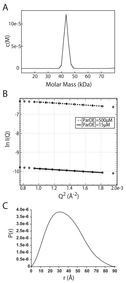
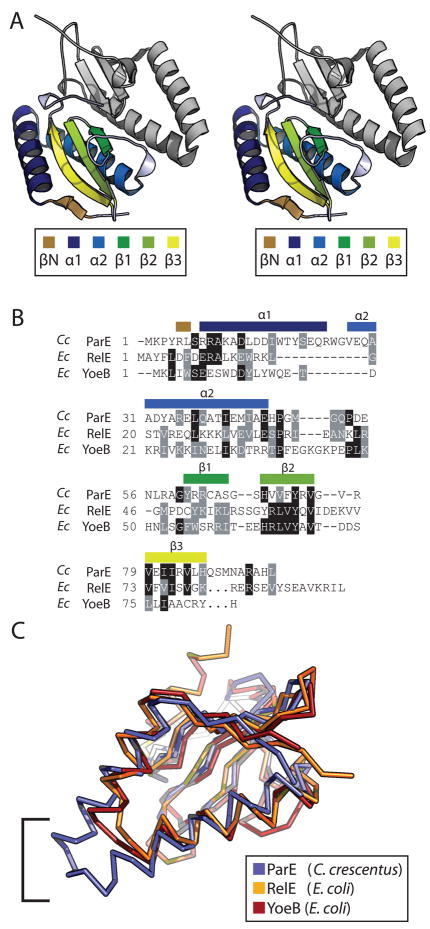
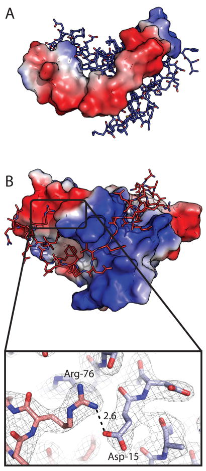
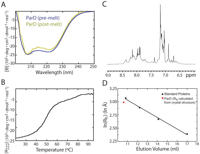
Toxin-antitoxin (TA) systems form a ubiquitous class of prokaryotic proteins with functional roles in plasmid inheritance, environmental stress response, and cell development. ParDE family TA systems are broadly conserved on plasmids and bacterial chromosomes and have been well characterized as genetic elements that promote stable plasmid inheritance. We present a crystal structure of a chromosomally encoded ParD-ParE complex from Caulobacter crescentus at 2.6 A resolution. This TA system forms an alpha(2)beta(2) heterotetramer in the crystal and in solution. The toxin-antitoxin binding interface reveals extensive polar and hydrophobic contacts of ParD antitoxin helices with a conserved recognition and binding groove on the ParE toxin. A cross-species comparison of this complex structure with related toxin structures identified an antitoxin recognition and binding subdomain that is conserved between distantly related members of the RelE/ParE toxin superfamily despite a low level of overall primary sequence identity. We further demonstrate that ParD antitoxin is dimeric, stably folded, and largely helical when not bound to ParE toxin. Thus, the paradigmatic model in which antitoxin undergoes a disorder-to-order transition upon toxin binding does not apply to this chromosomal ParD-ParE TA system.


Figures

Figure 1
Figure 1
2.6 Å structure of the heterotetrameric ParD-ParE complex of C. crescentus. The ParE toxin is rendered in blue and the ParD antitoxin in red. The N-terminal ParD ribbon-helix-helix DNA binding motif (RHH) is labeled. The 4 secondary structural elements (®1, α1, α2, α3) of ParD are labeled in red on both monomers.
Figure 2
Figure 2
A) Sedimentation velocity c(M) plot for the ParD-ParE complex. The predominant species, at 0.6 mg/ml, has a sedimentation coefficient that corresponds to a molecular weight of 43.5 kDa. B) Guinier analysis of small angle X-ray scattering data for the ParD-ParE complex. Fitted plots of protein solution at 500μM and 15μM yielded Rg values of 28.2 ± 0.04 and 27.8 ± 0.5 Å, respectively. C) P(r) plot of ParD-ParE calculated from the crystal structure yields a predicted hydration shell radius of gyration of 27.9 Å, consistent with the experimental SAXS data.
Figure 3
Figure 3
A) Wall-eyed stereo rendering of the ParE toxin dimer. The lower toxin monomer is colored by secondary structure as indicated in the legend. The second monomer is rendered in grey for completeness. B) Structure-based sequence alignment of C. crescentus ParE, and E. coli RelE and YoeB, based on pairwise least-squares fitting of Ca for each structure. Sequence position of secondary structure elements are labeled above the alignment. C) An overlay of the Ca traces for C. crescentus ParE, E. coli RelE, and E. coli YoeB colored as indicated in the legend. The bracket indicates the area of C. crescentus ParE α1 + α2 that does not overlay with its structural homologs due to longer helices.
Figure 4
Figure 4
Surface representations of the C. crescentus ParE toxin monomer bound to the ParD antitoxin. The ParD antitoxin is shown as a stick model rendered in red; only Ala46 through Ala79 are shown. A) Hydrophobic surface plot of ParE based on the Protscale script for PyMOL. Blue indicates hydrophobic surface; white indicates hydrophilic surface. B) The ParE monomer is rendered in grey with yellow indicating residues identified by Anantharaman and Aravind (1) as greater than 80% conserved across the ParE/RelE superfamily. C) Surface representation of E. coli RelE toxin bound to RelB (red). D) E. coli YoeB surface bound to YefM (red).
Figure 5
Figure 5
View of the two hydrophobic subdomains of the ParD-ParE binding groove. ParE is rendered as a hydrophobic surface based on the Protscale script for PyMOL. Blue represent hydrophobic surface while white indicates hydrophilic. The ParD antitoxin backbone is rendered in red with key hydrophobic sidechains rendered as ball and stick models. A) At hydrophobic subdomain 1, the helical antitoxin inserts F67, F69, F72, & I73 into a cavity on the toxin surface. The region N-terminal to this on ParD adopts a random coil conformation stabilized by F63 and I64. B) α2 of ParD buries L48, L51, L55, & I56 against the toxin surface.
Figure 6
Figure 6
A) ParD electrostatic contact potential map (red = acidic, blue = basic) modeled with ParE as ball-and-stick. B) Electrostatic contact potential of ParE with ParD modeled as ball-and stick. ParD contacts an intensely basic binding groove in the toxin surface potential map. Inset: Salt-bridge between ParD R76 and ParE D15. Carbon atoms of ParD are represented in red, carbon atoms of ParE are represented in blue. Simulated annealing omit map was generated by omitting R76 and D15 prior to refinement; map contoured at 1 sigma. The electrostatic potential surfaces were calculated in PyMOL using the generate-vacuum electrostatics function.
Figure 7
Figure 7
Interfacial salt bridges. (A) Ring of salt bridges linking residues R9, D13, R83 of ParE and E57 of ParD. (B) Chain of salt bridges linking residues R58, R74, E79, and R5 of ParE and E65 and E59 of ParD. A water molecule binding to the carboxyl group of ParD E65 and ParE R5 is shown as a red sphere. Side chains are rendered in red for ParD and blue for ParE. Labeled distances are in Å. Simulated annealing omit maps were generated by omitting the labeled residues; maps contoured at 1.0 sigma.
Figure 8
Figure 8
A) Normalized circular dichroism spectrum of ParD. Two spectra are plotted, pre- and post-thermal denaturation. B) Melting curve of ParD over a temperature gradient of 20 to 95°C at 1°C/min. Helical signal at 222nm is plotted on the ordinate. C) The amide proton region of a 1H nuclear magnetic resonance spectrum of 500μM ParD. D) Size exclusion chromatography of ParD on Superdex 75. Elution volume is plotted versus the logarithm of the calculated hydrodynamic radii of ParD (red) and four standards (black) (see Materials and Methods). Standard proteins are indicated with black squares. ParD is indicated with a red asterisk. Linear regression of the gel filtration standard curve is indicated with a black line.
Figure 9
Figure 9
Output of the coiled-coil prediction algorithm, Coils (38), for the ParD antitoxin. ParD antitoxin is rendered as sticks; ParE toxin is rendered as a transparent blue surface. ParD residues with a greater than 80% probability of forming a coiled-coil are shown in white.

Similar articles

Cited by

References

    1. Anantharaman V, Aravind L. New connections in the prokaryotic toxin-antitoxin network: relationship with the eukaryotic nonsense-mediated RNA decay system. Genome Biol. 2003;4:R81. - PMC - PubMed
    1. Gronlund H, Gerdes K. Toxin-antitoxin systems homologous with relBE of Escherichia coli plasmid P307 are ubiquitous in prokaryotes. J Mol Biol. 1999;285:1401–1415. - PubMed
    1. Pandey DP, Gerdes K. Toxin-antitoxin loci are highly abundant in free-living but lost from host-associated prokaryotes. Nucleic Acids Res. 2005;33:966–976. - PMC - PubMed
    1. Gerdes K, Christensen SK, Lobner-Olesen A. Prokaryotic toxin-antitoxin stress response loci. Nat Rev Microbiol. 2005;3:371–382. - PubMed
    1. Buts L, Lah J, Dao-Thi MH, Wyns L, Loris R. Toxin-antitoxin modules as bacterial metabolic stress managers. Trends Biochem Sci. 2005;30:672–679. - PubMed

Publication types

MeSH terms

LinkOut - more resources