. 2009 Dec;7(4):189-96.
doi: 10.1089/lrb.2009.0033.

Non-peptide small molecule regulators of lymphangiogenesis

Affiliations

Non-peptide small molecule regulators of lymphangiogenesis

Changge Fang et al. Lymphat Res Biol. 2009 Dec.

Abstract

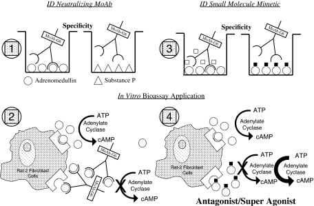
Adrenomedullin (AM) and gastrin releasing peptide (GRP) are neuroendocrine peptides that have been previously implicated as regulators of angiogenesis and lymphangiogenesis. Using an immortalized human dermal microvascular lymphatic endothelial cell line stably transfected with red fluorescent protein (LEC/RFP), we demonstrate the ability of AM and GRP to augment tube formation complexity of this target cell in a dose-dependent manner. Maximum tube density was initiated at 1 nM for both peptides, and as concentrations exceeded 10 nM a decrease in tube formation was noted, hence following a classic rise/fall biological response curve. In addition, we show that appropriate small molecule mimetics to neutralizing monoclonal antibodies of AM or GRP, at 1 microM concentration, can function to either inhibit (antagonist) or enhance (super agonist) peptide-induced tube formation of LEC/RFP. Our small molecule reagents by themselves have no activity, but in the presence of their respective peptides can mediate a positive or negative response, hence the super agonist designation. These compounds represent new regulatory drugs of the lymphatic system with possible patient application in the clinical management of edema and metastatic disease.


Figures

FIG. 1.
FIG. 1.
Carboxy terminal amidation of peptides: a hallmark of peptide biological activity.27–29 Two such amidated peptides, AM and GRP, are proven inducers of neovascularization and have recently been implicated in lymphangiogenesis.21–26, 32–34 A distinct amino acid (AA) motif within the prohormone signals for amidation to take place and consists of a target AA to be amidated (any AA), followed by glycine (only known amide donor AA), followed by a series of basic AAs (arginine or lysine). An initial trypsin-like cleavage event releases the amidation motif from the prohormone, followed by carboxypeptidase processing, giving rise to a glycine-extended intermediate product. This intermediate then serves as the substrate for a unique series of enzyme complexes known as peptitdyl-glycine–amidating monoxygenase that ultimately generates the amide of glycine's penultimate neighbor AA. The prohormone amidation motifs for AM (a tyrosine amide) and GRP (a methionine amide) are shown. The glycine intermediate or free-acid derivatives of both AM and GRP are 100 to 1000 time less potent on a molar basis than their respective amide counter-part depending on the biological assay used (boxed structures).
FIG. 2.
FIG. 2.
Small molecule drug screening approach. Screening strategy design for high throughput analysis using a 96-well microtiter plate format and based on prior NCI technology. Section No. 1 indicates the initial testing of hybridoma products for ligand binding specificity. Here AM (a tyrosine amide) was used as the immunogen and the solid phased target peptide, while substance P (amethionine amide) was used as a negative control peptide. Section No. 2 depicts the bioassay used to identify preselected anti-AM monoclonal antibodies (MoAb) as peptide neutralizing reagents. AM induces Rat-2 fibroblast cells to concert ATP > cAMP and MoAb-G6, identified in Section No. 1, blocks that event. Section No. 3. A secondary binding assay was established involving MoAb-G6/AM interaction and the NCI/Developmental Therapeutics Program (DTP) diversity set of 2000 small molecules family members (representing 140,000 compounds) were screened for their ability to inhibit ligand/antibody complex formation. Section No. 4. Candidate compounds making it through this initial screening were then evaluated in the Rat-2 fibroblast assay for their effects on AM-induced cAMP production. As indicated, small molecule AM antagonist and super agonist compounds were found using this collective screening strategy. An identical collective screening approach was used to identify small molecule regulators of GRP bioactivity with MoAb-2A11 serving as the neutralizing anti-GRP antibody.30,35
FIG. 3.
FIG. 3.
Chemical structure of small molecule AM/GRP antagonists and super agonists. Average formula weight (FW) approximately 300 daltons (NSC 697169 exception with FW 781).
FIG. 4.
FIG. 4.
In vitro tube formation assay. Telomerase immortalized human dermal lymphatic endothelial cells stably transfected with RFP (LEC/RFP) were utilized in this 96-well analysis, seeding density of 19,000 cells/well, and evaluated after 3.5 h incubation at 37°C/5% CO2. A, B, and C represent serum induced tube formation at 0%, 0.5%, and 5% FBS, respectively. A mid-range of 0.5% FBS supplementation was chosen for all follow-up studies related to peptide-induced tube formation. D, E, F, G, and H represent resulting tubes observed when stimulated with AM at 0.01 nM, 0.1 nM, 1.0 nM, 10 nM, and 100 nM concentration. I, J, K, L, and M are the GRP-induced tubes formed when tested at a peptide concentration of 0.01 nM, 0. 1 nM, 1.0 nM, 10 nM, and 100 nM. Note that both peptides induce a biphasic dose response curve (AM having a steeper response curve than GRP) with maximum tube formation generated at a peptide concentration of 1.0 nM.
FIG. 5.
FIG. 5.
Testing small molecule regulators of AM and GRP in the tube formation assay. An identical assay as described in Figure 4 was established to evaluate the ability of small molecule antagonists and agonists to regulate peptide-induced tube formation with LEC/RFP target cells. A, B, and C represent tube formation observed in the presence of 0%, 0.5%, and 5% FBS supplementation, respectively. D, E, and F are representative examples of the lymphatic tube formation generated in the presence of 0.5% FBS +0.1 nM AM, followed by the addition of either 1 μM AM antagonist (NSC 16311) or 1 μM AM super agonist (NSC 697169). G, H, and I are representative responses with 0.5% FBS +0.1 nM GRP followed by the addition of either 1 μM GRP antagonist (NSC 77427) or 1 μM GRP super agonist (NSC 372874).

References

    1. Olszewski W. The lymphatic system in body homeostasis: Physiological conditions. Lymphat Res Biol. 2003;1:11–21. - PubMed
    1. Butler MG. Isogai S. Weinstein BM. Lymphatic development. Birth Defects Res. 2009;87:222–231. - PMC - PubMed
    1. Rocha SF. Adams RH. Molecular differentiation and specialization of vascular beds. Angiogenesis. 2009;12:139–147. - PubMed
    1. Banerji S. Ni J. Wang SX. Clasper S. Su J. Tammi R. Jones M. Jackson DG. LYVE-1, a new homologue of the CD44 glycoprotein, is a lymph-specific receptor for hyaluronan. J Cell Biol. 1999;144:789–801. - PMC - PubMed
    1. Wigle JT. Oliver G. Prox1 function is required for the development of the murine lymphatic system. Cell. 1999;98:769–778. - PubMed

Publication types

MeSH terms