Review
. 2010 Mar 1;50 Suppl 2(S2):S54-65.
doi: 10.1086/648966.

Review of meningococcal group B vaccines

Affiliations
Review

Review of meningococcal group B vaccines

Dan M Granoff. Clin Infect Dis. .

Abstract

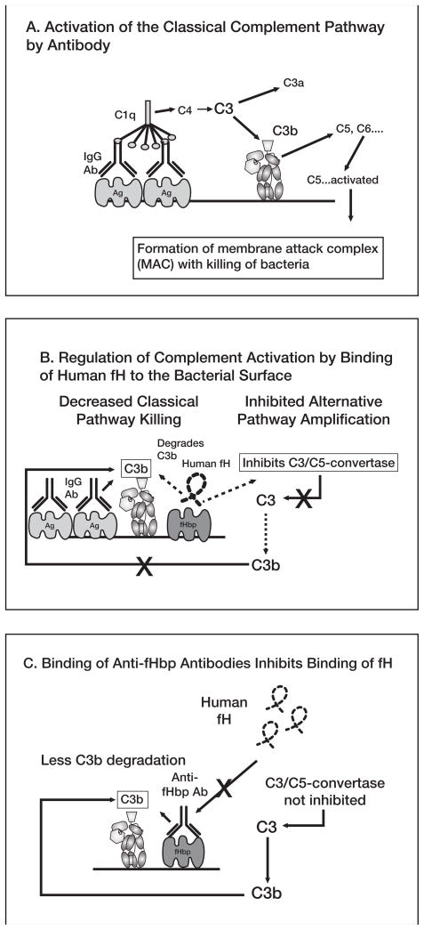
No broadly effective vaccines are available for prevention of group B meningococcal disease, which accounts for >50% of all cases. The group B capsule is an autoantigen and is not a suitable vaccine target. Outer-membrane vesicle vaccines appear to be safe and effective, but serum bactericidal responses in infants are specific for a porin protein, PorA, which is antigenically variable. To broaden protection, outer-membrane vesicle vaccines have been prepared from >1 strain, from mutants with >1 PorA, or from mutants with genetically detoxified endotoxin and overexpressed desirable antigens, such as factor H binding protein. Also, recombinant protein vaccines such as factor H binding protein, given alone or in combination with other antigens, are in late-stage clinical development and may be effective against the majority of group B strains. Thus, the prospects have never been better for developing vaccines for prevention of meningococcal disease, including that caused by group B strains.


Conflict of interest statement

Conflict of interest. The author is principal investigator of laboratory research conducted on behalf of Children’s Hospital Oakland Research Institute that is funded by grants from Novartis Vaccines and Diagnostics, and Sanofi Pasteur. He also holds a paid consultancy from Novartis and is an inventor on patents or patent applications in the area of meningococcal B vaccines.

Figures

Figure 1
Figure 1
Detergent-extracted OMV vaccines. Panel A. Electron micrograph of outer membrane vesicles of N. meningitides. The scale bar is 100 nm and the vesicle diameter is about 50–200 nm (80nm on average). Panel B. Major outer membrane proteins (PorA, PorB, reduction modifiable protein (RmpM) and opacity protein A (OpcA)) as visualized by Coommassie-stained SDS PAGE. Lane 1, molecular mass standards; lane 2, strain NZ98/254; lane 3, strain H44/76. After immunization, the SBA–responses of infants and children are directly predominantly against PorA. Adapted from published data [23]. Reprinted from Vaccine, vol 27, Supplement 2, 2009, Holst J, Martin D, Arnold R, et al. Properties and clinical performance of vaccines containing outer membrane vesicles from Neisseria meningitidis, with permission from Elsevier.
Figure 2
Figure 2
Release of TNF-α after incubation of human PBMCs for 4 hours with OMV vaccines. The OMV concentrations that resulted in a 10-fold increased release of TNF-α concentrations above background are shown on the X intercepts. White circles, native OMV from wildtype strain; gray squares, OMV vaccine prepared from LpxL1 KO mutants; black triangles, detergent-extracted OMV vaccines from corresponding wildtype strains. Adapted from published data [65] with permission from American Society for Microbiology.
Figure 3
Figure 3
Serum bactericidal activity elicited in mice by a native OMV vaccine prepared from a mutant with attenuated endotoxin activity and overexpressed fHbp. N. meningitidis strain designations are shown below the X axis (all expressed fHbp in the variant 1 group). Vaccine groups: recombinant (open bars), multicomponent vaccine containing 2 fusion proteins, GNA 2091-fHbp variant 1 and GNA 2132-GNA 1030, and NadA (See Figure 6); Detergent-OMV, WT (hatched bars), a clinical lot of detergent-treated OMV vaccine from Norway (strain H44/76); native OMV, mutant (gray bars), a native (not treated with detergent) OMV from a mutant strain of H44/76 with attenuated endotoxin (LpxL1KO) and overexpressed fHbp in the variant 1 group. Al(OH)3 (black bars), adjuvant alone (also used for the 3 vaccines). Copyright © 2008 by the Infectious Diseases Society of America. All rights reserved.
Figure 4
Figure 4
A, Activation of classical complement pathway. Binding of 2 optimally spaced IgG molecules to the bacterial surface engages C1q and activates the classical complement pathway, which results in increased deposition of C3b. Bound C3b can serve as an opsonin and can also lead to bacteriolyis by cleavage of C5 and assembly of the C5b-9 membrane attack complex. Not shown are the components of the alternative pathway, which can be activated by the classical pathway and serve as an amplification loop. B, Regulation of complement activation by binding of human factor H (fH) to the bacterial surface. Human fH binds to surface-exposed fHbp. fH accelerates the decay of alternative pathway C3/C5 convertases, which downregulates the positive feedback amplification loop of the alternative pathway. Binding of fH also leads to degradation of C3b by factor I (not shown), which decreases classical pathway activation and amplification by the alternative pathway. C, Binding of antibodies to fHbp activates classical complement pathway bacteriolysis and also inhibits binding of fH to the bacterial surface. With decreased amounts of fH bound to the bacterial surface, there is less downregulation of complement activation and the organism becomes more susceptible to complement-mediated bacteriolysis.
Figure 5
Figure 5
A. Phylogram of 70 unique fHbp amino acid sequences showing division of the proteins into 2 subfamilies, designated as A and B by Fletcher et al. [100]. Subfamily B contains the proteins in the variant 1 group described by Masignani et a. [61]. Subfamily A is subdivided into 2 branches, designated by Masignani et al. as variants 2 and 3, respectively. Each branch represents a distinctive protein sequence. The scale bar represents 5 amino acid differences per 100 amino acids. B. Modular structure. The architecture of fHbp consists of different combinations of five variable segments, designated VA to VD [116]. Each segment is derived from one of two genetic lineages, designated alpha (gray segments) or beta (white segments). All of the distinctive fHbp protein amino acid sequences referred to in Panel A could be assigned to one of six “modular groups”, designated I–VI. Panel B is reprinted from Microbiology 2009;155:2873–83. Copyright ©2009 by the Society for General Microbiology.
Figure 6
Figure 6
Schematic showing three recombinant proteins (five antigens) contained in a multicomponent meningococcal vaccine [96, 119]. Two of the components are fusion proteins (GNA 2132 fused with GNA 1030, and GNA 2091 fused with fHbp. The third component is recombinant NadA. N- and C- refer to the amino- and carboxy terminal portions, respectively, of the proteins. The scale bar represents 100 amino acids (AA). Of the five antigens, fHbp, GNA 2132 and NadA, shown in gray, were reported to be responsible for most of the SBA responses in mice [96]. In the vaccine formulation being investigated in phase 3 clinical trials, the three recombinant proteins are combined with a detergent-extracted OMV vaccine from group B strain NZ98/254.
Figure 7
Figure 7
Summary of the proportion of immunized subjects with titers ≥ 1:4 in SBA (3 strains), or passive protection (PP) in a human blood bacteremia model with group B strains NZ98/254 and S3032 (strain H44/76 was not tested). The bars represent the proportion of pre- (open bars) or postimmunization sera (closed bars) that were positive when tested at a 1:4 dilution in each assay and the respective 95% confidence intervals. Postimmunization passive protective activity was 2.6-to 2.8-fold more frequent then an SBA titer ≥ 1:4. Adapted with permission. Copyright 2009© American Society for Microbiology, Clin Vaccine Immunol, 16:785–91, 2009.

Similar articles

Cited by

References

    1. Harrison LH, Trotter CL, Ramsay ME. Global epidemiology of meningococcal disease. Vaccine. 2009;27:B51–B63. - PubMed
    1. Trotter CL, Chandra M, Cano R, et al. A surveillance network for meningococcal disease in Europe. FEMS Microbiol Rev. 2007;31:27–36. - PubMed
    1. Gray SJ, Trotter CL, Ramsay ME, et al. Epidemiology of meningococcal disease in England and Wales 1993/94 to 2003/04: contribution and experiences of the Meningococcal Reference Unit. J Med Microbiol. 2006;55:887–96. - PubMed
    1. Trotter CL, Andrews NJ, Kaczmarski EB, Miller E, Ramsay ME. Effectiveness of meningococcal serogroup C conjugate vaccine 4 years after introduction. Lancet. 2004;364:365–7. - PubMed
    1. Shepard CW, Rosenstein NE, Fischer M. Neonatal meningococcal disease in the United States, 1990 to 1999. Pediatr Infect Dis J. 2003;22:418–22. - PubMed

Publication types

MeSH terms