

Screening for diabetic retinopathy using computer vision and physiological markers
- PMID: 20144333
- PMCID: PMC2769953
- DOI: 10.1177/193229680900300431
Screening for diabetic retinopathy using computer vision and physiological markers
Abstract
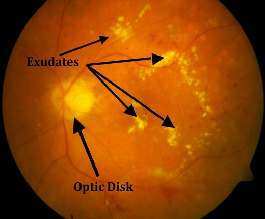
Background: Hyperglycemia and diabetes result in vascular complications, most notably diabetic retinopathy (DR). The prevalence of DR is growing and is a leading cause of blindness and/or visual impairment in developed countries. Current methods of detecting, screening, and monitoring DR are based on subjective human evaluation, which is also slow and time-consuming. As a result, initiation and progress monitoring of DR is clinically hard.
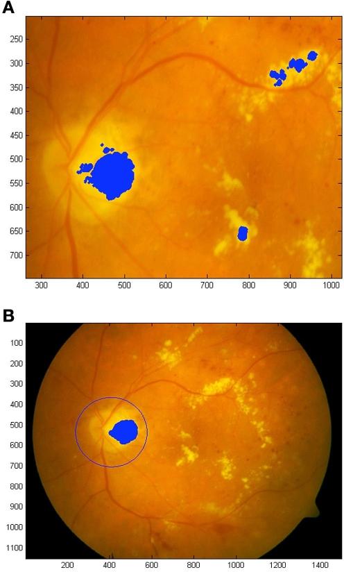
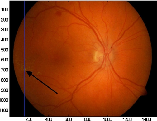
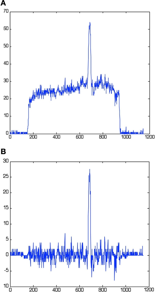
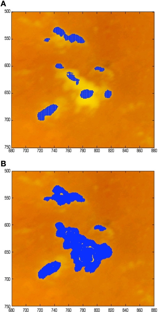
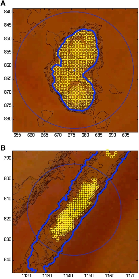
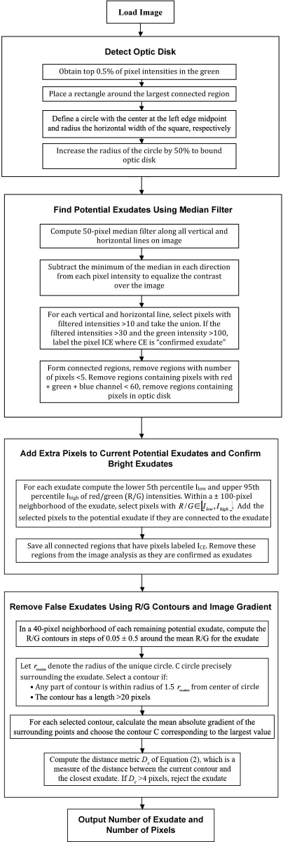
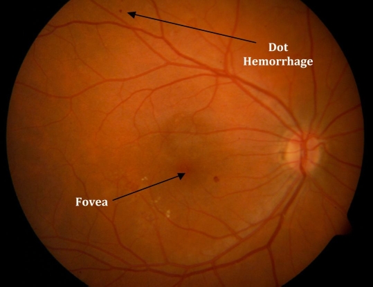
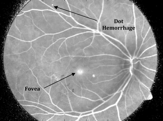
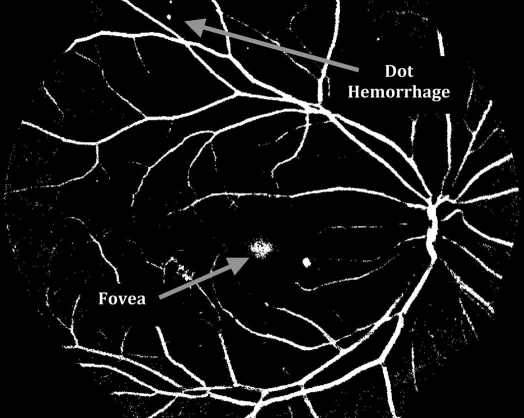
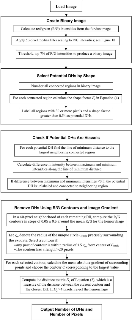
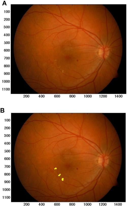
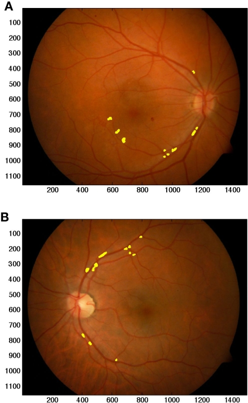
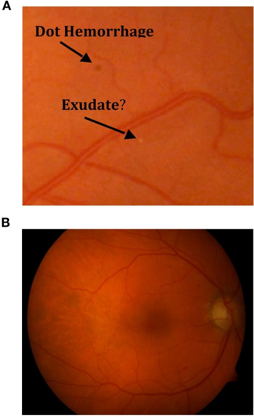
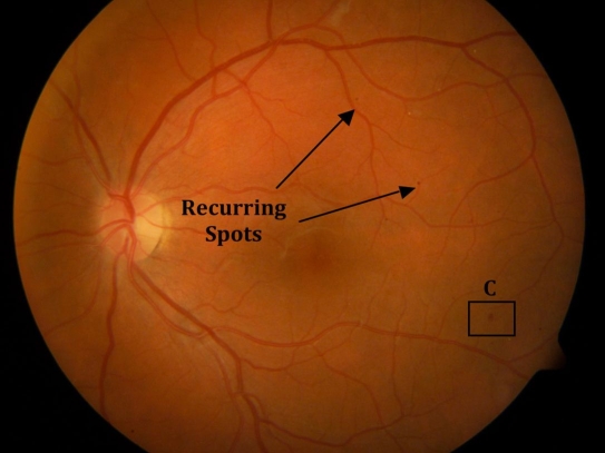
Methods: Computer vision methods are developed to isolate and detect two of the most common DR dysfunctions-dot hemorrhages (DH) and exudates. The algorithms use specific color channels and segmentation methods to separate these DR manifestations from physiological features in digital fundus images. The algorithms are tested on the first 100 images from a published database. The diagnostic outcome and the resulting positive and negative prediction values (PPV and NPV) are reported. The first 50 images are marked with specialist determined ground truth for each individual exudate and/or DH, which are also compared to algorithm identification.
Results: Exudate identification had 96.7% sensitivity and 94.9% specificity for diagnosis (PPV = 97%, NPV = 95%). Dot hemorrhage identification had 98.7% sensitivity and 100% specificity (PPV = 100%, NPV = 96%). Greater than 95% of ground truth identified exudates, and DHs were found by the algorithm in the marked first 50 images, with less than 0.5% false positives.
Conclusions: A direct computer vision approach enabled high-quality identification of exudates and DHs in an independent data set of fundus images. The methods are readily generalizable to other clinical manifestations of DR. The results justify a blinded clinical trial of the system to prove its capability to detect, diagnose, and, over the long term, monitor the state of DR in individuals with diabetes.
Copyright 2009 Diabetes Technology Society.
Figures
Similar articles
-
Evaluation of automated fundus photograph analysis algorithms for detecting microaneurysms, haemorrhages and exudates, and of a computer-assisted diagnostic system for grading diabetic retinopathy.Diabetes Metab. 2010 Jun;36(3):213-20. doi: 10.1016/j.diabet.2010.01.002. Epub 2010 Mar 10. Diabetes Metab. 2010. PMID: 20219404
-
Retinal images benchmark for the detection of diabetic retinopathy and clinically significant macular edema (CSME).Biomed Tech (Berl). 2019 May 27;64(3):297-307. doi: 10.1515/bmt-2018-0098. Biomed Tech (Berl). 2019. PMID: 30055096
-
An Automated Grading System for Detection of Vision-Threatening Referable Diabetic Retinopathy on the Basis of Color Fundus Photographs.Diabetes Care. 2018 Dec;41(12):2509-2516. doi: 10.2337/dc18-0147. Epub 2018 Oct 1. Diabetes Care. 2018. PMID: 30275284
-
Deep learning based computer-aided diagnosis systems for diabetic retinopathy: A survey.Artif Intell Med. 2019 Aug;99:101701. doi: 10.1016/j.artmed.2019.07.009. Epub 2019 Aug 7. Artif Intell Med. 2019. PMID: 31606116 Review.
-
Survey on recent developments in automatic detection of diabetic retinopathy.J Fr Ophtalmol. 2021 Mar;44(3):420-440. doi: 10.1016/j.jfo.2020.08.009. Epub 2021 Jan 30. J Fr Ophtalmol. 2021. PMID: 33526268 Review.
References
-
- Icks A, Trautner C, Haastert B, Berger M, Giani G. Blindness due to diabetes: population based age- and sex- specific incidence rates. Diabet Med. 1997;14(7):571–575. - PubMed
-
- Fong D, Aiello L, Gardner T, King G, Blankenship G, Cavallerano J, Ferris F, 3rd, Klein R. Retinopathy in diabetes. Diabetes Care. 2004;27(Suppl 1):S84–S87. - PubMed
-
- Kempen JH, O'Colmain BJ, Leske MC, Haffner SM, Klein R, Moss SE, Taylor HR, Hamman RF. Eye Diseases Prevalence Research Group. The prevalence of diabetic retinopathy among adults in the United States. Arch Ophthalmol. 2004;122(4):552–563. - PubMed
-
- Wild S, Roglic G, Green A, Sicree R, King H. Global prevalence of diabetes: estimates for the year 2000 and projections for 2030. Diabetes Care. 2004;27(5):1047–1053. - PubMed
-
- Fong D, Aiello L, Gardner T, King G, Blankenship G, Cavallerano J, Ferris FL, 3rd, Klein R. American Diabetes Association. Diabetic retinopathy. Diabetes Care. 2003;26(1):226–229. - PubMed
MeSH terms
LinkOut - more resources
Full Text Sources
Medical
Research Materials
