. 2010 Apr;43(4):422-30.
doi: 10.1016/j.mcn.2010.01.009. Epub 2010 Feb 6.

Regulation of axonal trafficking of cytochrome c oxidase IV mRNA

Affiliations

Regulation of axonal trafficking of cytochrome c oxidase IV mRNA

Armaz Aschrafi et al. Mol Cell Neurosci. 2010 Apr.

Abstract

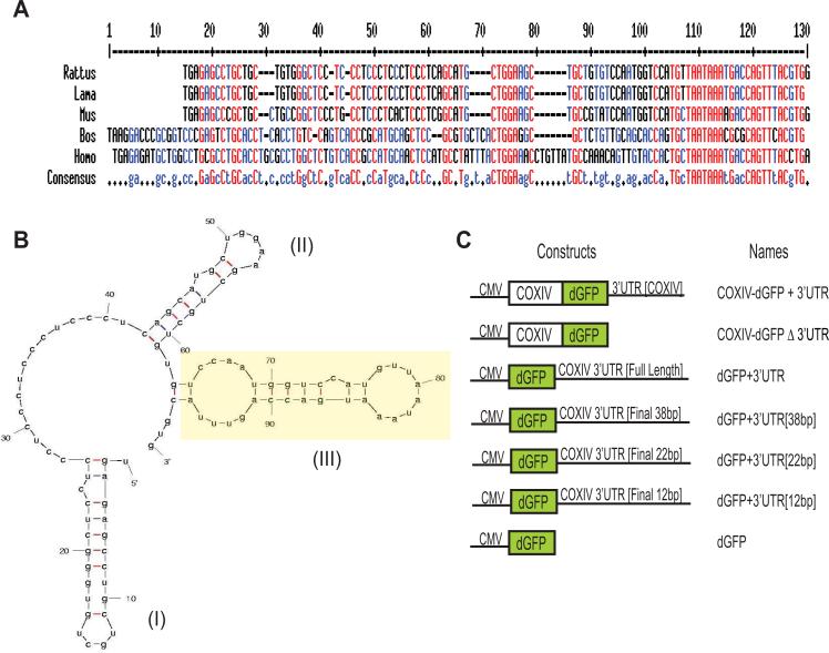
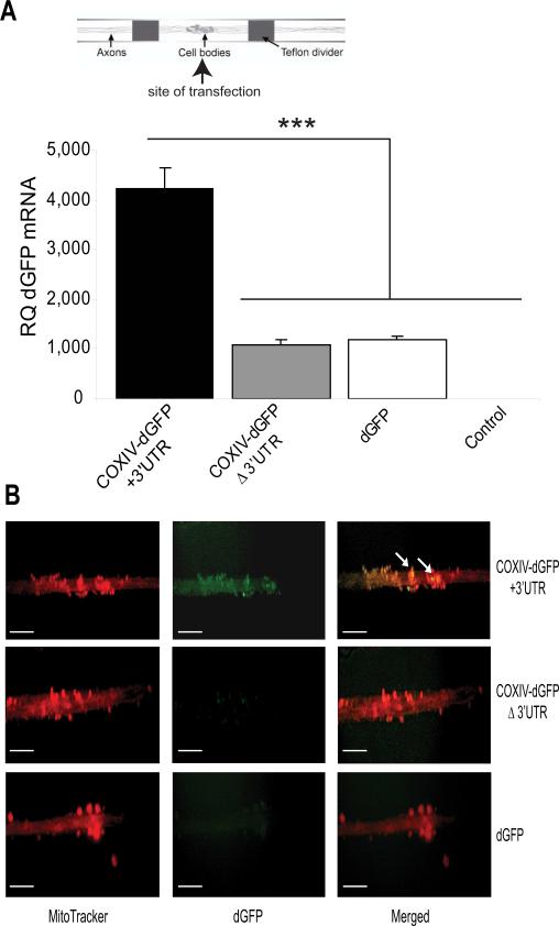
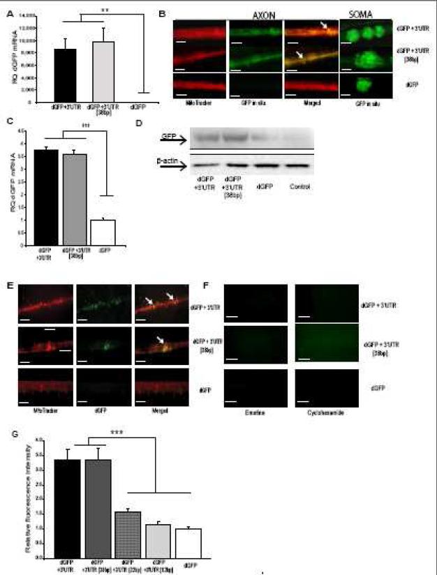
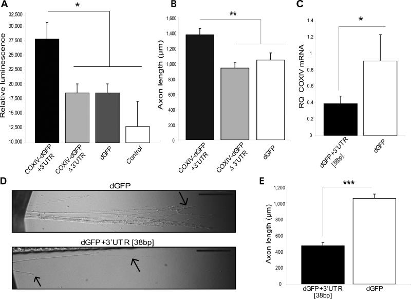
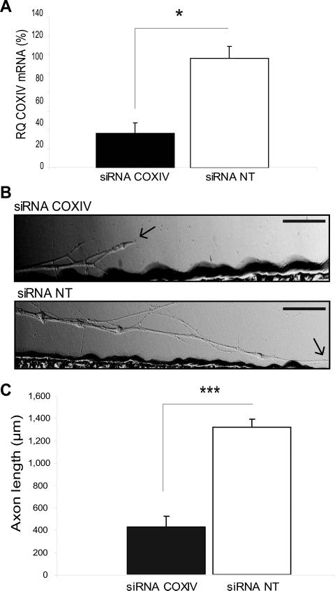
Trafficking and local translation of axonal mRNAs play a critical role in the development and function of this neuronal subcellular structural domain. In this report, we studied cytochrome c oxidase subunit IV (COXIV) mRNA trafficking into distal axons of primary superior cervical ganglia (SCG) neurons, and provided evidence that axonal trafficking and mitochondrial association of the mRNA are mediated by an element located in a 38bp-long, hairpin-loop forming region within the 3'UTR of the transcript. Our results also suggest that suppression of local translation of COXIV mRNA results in significant attenuation of axonal elongation. Taken together, the results provide the first evidence for the existence of a cis-acting axonal transport element within a nuclear-encoding mitochondrial gene, and demonstrate the importance of the axonal trafficking and local translation of nuclear-encoded mitochondrial mRNAs in axonal growth.


Figures

Fig. 1
Fig. 1
The 3'UTR of mammalian COXIV mRNA is highly conserved. (A) Sequence alignment of the 3'UTR of COXIV mRNAs from five mammalian species shows a high degree of sequence conservation. The red and the blue shading represent nucleotides conserved among all five and in four out of the five species, respectively. Each 3’UTR sequence begins with the stop codon. (B) Structure of the COXIV 3’UTR, as determined by secondary structure prediction analysis (Mfold). Yellow highlighted hairpin-loop segment (III) facilitates mRNA transport into distal axons. (C) Overview of constructs used in this study. Plasmid constructs consisting of the COXIV ORF followed by the destabilized GFP (dGFP) cDNA that contain the COXIV 3’UTR [+3’UTR] or the SV40 3′UTR [Δ3’UTR], are diagrammed. In addition, schematic representation of reporter gene plasmids carrying the dGFP cDNA followed by either the full length 3’UTR of rat COXIV, the final 38 bp, 22 bp or 12 bp of the COXIV 3’UTR, respectively, is shown. Expression of all fusion gene products was driven by the human cytomegalovirus promoter (CMV).
Fig. 2
Fig. 2
COXIV mRNA 3’UTR contains an axonal targeting element. (A) SCG cultures were transfected with the chimeric COXIV-dGFP constructs (2 μg) in the center compartment of the Campenot chambers, as shown. Using GFP-specific primers, quantitation of COXIV-dGFP mRNA levels in the distal axons of SCG neurons were determined by qRT-PCR 24 h after plasmid DNA or sham-transfection (control). GFP mRNA levels are expressed relative to β-actin mRNA. Error bars represent the SEM for n = 3 samples. ***, p < 0.0001. (B) COXIV-dGFP+3’UTR puncta colocalize with the mitochondrial marker MitoTracker in the distal SCG axons, indicating local translation of the COXIV-dGFP mRNA and mitochondrial association of the translation product. Arrowheads depict examples of areas of co-localization. The scale bars represent 100 μm.
Fig. 3
Fig. 3
The final 38 bp within the COXIV mRNA 3’UTR defines axonal localization, mitochondrial targeting, and dGFP local translation. (A) SCG cultures (6 DIV) were transfected with the dGFP constructs (2 μg) in the center compartment as shown in Fig. 2. Quantitation of GFP mRNA levels in the distal axons of SCG neurons was determined by qRT-PCR 24 h after DNA transfection. GFP mRNA levels are expressed relative to β-actin mRNA. Error bars represent the SEM for n = 3 samples (dGFP constructs versus sham-transfected controls). Student's t test, **p < 0.001. (B) SCG neuronal cell bodies were transfected as in (A). Distal axons and cell bodies were processed for in situ hybridization with a FITC-labeled LNA probe specific for GFP. Higher-magnification images illustrate co-localization of dGFP mRNAs carrying the entire COXIV 3’UTR or the final 38bp of this 3’UTR with axonal mitochondria, but complete absence of FITC fluorescence in axons of dGFP-control transfected neurons. Representative examples from one of four independent experiments are shown. FITC-labeled, scramble, LNA probe served as negative control, and produced only faint fluorescence under identical hybridization and wash conditions (not shown). Scale bar, 100 μm (C) SCG cultures (6 DIV) were transfected with the dGFP constructs as described in (A). Total RNA was prepared in the presence of 3 mM MgCl2 from an enriched mitochondrial fraction in distal axons following the protocols described in (Russo et al., 2006). Quantitation of dGFP mRNA levels from total mitochondrial RNA was determined by qRT-PCR 24 h after DNA transfection. dGFP mRNA levels are expressed relative to COXII mRNA. Error bars represent the SEM for n = 3 samples (dGFP constructs versus sham-transfected controls). Student's t test, ***p < 0.0001. (D) Protein lysates (5 μg) were prepared from distal axons of SCG neurons that were transfected as in (A), and analyzed by western blot using a monoclonal GFP antibody. Levels of β-actin protein served as an internal control. (E) dGFP encoded by transcripts containing either the entire COXIV 3’UTR or the final 38 bp segment are visualized using fluorescence microscopy as puncta that colocalize with the mitochondrial marker MitoTracker in the distal SCG axons, indicating mitochondrial association of the dGFP translation product. In contrast, dGFP encoded by transcripts devoid of the COXIV 3’UTR sequence were not detected in axons. Arrowheads depict areas of colocalization. The scale bars represent 100 μm. (F) Fluorescent image analysis of distal axons treated with protein synthesis inhibitors. Application of emetine or cyclohexamide for 3-4 h caused complete disappearance of the dGFP fluorescent signal in the axon. The scale bars represent 100 μm. (G) A 38 bp fragment within the COXIV 3’UTR represents the minimal axonal transport element of the mRNA. SCG cultures (6 DIV) were transfected with dGFP vectors (2 μg) that contained the full 3’UTR (103 bp), or final sub-fragments in length of 38 bp, 22 bp, and 12 bp of the COXIV 3’UTR, respectively, in the center compartment. Axonal GFP levels were visualized by fluorescence microscopy, and quantified using Image J. Fluorescence levels were normalized to the fluorescence intensity of axons from non-transfected neurons. Data are averages ±SEM from the measurement of 35-45 axons. The experiment was repeated three times with similar results. ANOVA, ***, p < 0.0001.
Fig. 4
Fig. 4
Axonal trafficking and protein synthesis of COXIV mRNA is important for axonal respiration and elongation. (A) A marked increase in axonal ATP levels was observed upon local translation of COXIV-dGFP carrying the COXIV 3’UTR but not the SV40 3’UTR. ATP levels were assessed using CellTiter-Glo luminescent cell viability assay from Promega as previously described (Aschrafi et al., 2007). Values were plotted in arbitrary luminescence units. Data represent mean ±SEM; One-Way ANOVA, *, p<0.01. (B) Axons locally translating COXIV-dGFP mRNA containing the 3’UTR elongate more rapidly when grown in compartmented cultures than axons translating the construct lacking the 3’UTR or dGFP mRNA alone. Axon length is shown for primary SCG neurons that were transfected with the COXIV-dGFP constructs or dGFP 24 h after initial plating in compartmented dishes. Length of distal axons located in the side compartments was measured 2-3 d after the transfection of neurons located in the central compartment. Data are averages ±SEM from measurement of 35-45 axons. ANOVA, **, p < 0.001. (C) Over-expression of dGFP mRNA containing the final 38 bp segment of the COXIV 3’UTR reduces abundance of endogenous COXIV mRNA levels in distal axons. SCG cultures were transfected with dGFP-3’UTR [38 bp], or dGFP (1 μg each) in the center compartment as shown in Fig. 2. Quantitation levels of endogenous COXIV mRNA in the distal axons of SCG neurons were determined by qRT-PCR 24 h after DNA transfection. COXIV mRNA levels are expressed relative to β-actin mRNA. Error bars represent the SEM for n = 3 samples. *, p < 0.05. (D) Overexpression of dGFP mRNA containing the 38bp 3’UTR attenuates neurite outgrowth. SCG neurons were cultured for 1 d and were subsequently transfected with dGFP-3’UTR [38 bp], or dGFP constructs (2 μg each). Arrows indicate location of axonal distal or terminal regions. Scale bars: 10 μm. (E) Neurite extension was measured after 2 d in the lateral sides of compartmentalized culture. Data are averages ±SEM from measurement of 30-40 axons. The experiment was repeated three times with similar results. Student's t test, ***, p < 0.0001.
Fig. 5
Fig. 5
siRNA-mediated knockdown of local COXIV levels attenuates neurite elongation in sympathetic neurons. (A) siRNA oligonucleotides (25 nM) targeted against rat COXIV mRNA were introduced into distal axons of SCG neurons by lipofection and COXIV mRNA quantitated 24 h later by qRT-PCR. Quantitation showed a significant reduction in COXIV mRNA levels after the introduction of siRNA (*, p < 0.05, Student's t test). (B) SCGs were cultured for 3 days and neurites located in the lateral compartments of compartmentalized cultures were transfected with siRNA (25 nM) targeted against rat COXIV mRNA, or transfected with non-targeted control siRNA (25 nM). Arrows indicate location of growth cones. Scale bars: 100 μm. (C) Neurite extension was measured 24 h after transfection. Data are averages ±SEM from measurement of 12-24 axons. The experiment was repeated three times with similar results. Student's t test, ***, p < 0.0001.

Similar articles

Cited by

References

    1. Amiri M, Hollenbeck PJ. Mitochondrial biogenesis in the axons of vertebrate peripheral neurons. Dev. Neurobiol. 2008;68:1348–1361. - PMC - PubMed
    1. Aronov S, Aranda G, Behar L, Ginzburg I. Axonal tau mRNA localization coincides with tau protein in living neuronal cells and depends on axonal targeting signal. J. Neurosci. 2001;21:6577–6587. - PMC - PubMed
    1. Aschrafi A, Schwechter AD, Mameza MG, Natera-Naranjo O, Gioio AE, Kaplan BB. MicroRNA-338 regulates local cytochrome c oxidase IV mRNA levels and oxidative phosphorylation in the axons of sympathetic neurons. J. Neurosci. 2008;28:12581–12590. - PMC - PubMed
    1. Bassell G, Singer RH. mRNA and cytoskeletal filaments. Curr. Opin. Cell Biol. 1997;9:109–115. - PubMed
    1. Bernstein BW, Bamburg JR. Actin-ATP hydrolysis is a major energy drain for neurons. J. Neurosci. 2003;23:1–6. - PMC - PubMed

Publication types

MeSH terms