

Review
doi: 10.1016/j.stem.2010.01.011.
Stem cells and the niche: a dynamic duo
Affiliations
- PMID: 20144784
- PMCID: PMC3012646
- DOI: 10.1016/j.stem.2010.01.011
Item in Clipboard
Review
Stem cells and the niche: a dynamic duo
Cell Stem Cell.
.
Abstract
Stem cell niches are dynamic microenvironments that balance stem cell activity to maintain tissue homeostasis and repair throughout the lifetime of an organism. The development of strategies to monitor and perturb niche components has provided insight into the responsive nature of the niche and offers a framework to uncover how disruption of normal stem cell niche function may contribute to aging and disease onset and progression. Additional work in the identification of genetic factors that regulate the formation, activity, and size of stem cell niches will facilitate incorporation of the niche into stem cell-based therapies and regenerative medicine.
Copyright 2010 Elsevier Inc. All rights reserved.
Figures
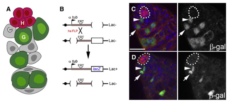
(A) Schematic of the stem cell niche in the Drosophila testis. Germline (G) and somatic cyst (C) stem cells are in direct physical contact with hub cells (H). (B) Lineage tracing analysis utilizing an inducible recombination strategy that permanently labels a random mitotic cell and its daughter due to reconstitution of the tubulin promoter upstream of the lacZ gene. (C) Labeled germline stem cell (green, arrowhead) adjacent to the hub (outline). Note its immediate daughter (arrow) is displaced from the hub. (D) Labeled cyst stem cell (green, arrowhead) adjacent to the hub (outline). Note its immediate daughter (arrow) is displaced from the hub. Immunofluorescence images of testes stained for E-cadherin (red), β-galactosidase (green), and DAPI (blue). Lineage tracing performed as described in (Voog et al., 2008). Scale bar, 20 μm.
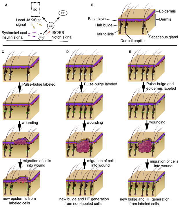
(A) Local signals mediate stem cell response to injury in the Drosophila midgut. Notch signaling regulates intestinal stem cell (ISC) and enteroblast (EB) cell-fate decisions. In response to injury, localized JAK-STAT signaling from enterocytes (EC) promotes proliferation of ISCs and differentiation of EBs. EBs are capable of generating ECs or enteroendocrine (ee) cells. Systemic and localized insulin signaling also influences recovery. (B) Schematic of mammalian epidermis. Epidermal stem cells reside in the basal layer (red) of the epidermis in contact with the basement membrane. Multipotent stem cells (white) reside in the bulge region of the outer root sheath of the hair follicle. (C) Bulge stem cells can repair the epidermis upon injury. Lineage tracing strategy pulse-labels bulge stem cells (white) with an observable marker (green). Upon injury to epidermis, labeled bulge stem cells migrate and aid in transient repair of the epidermis by contributing to basal cell layer. (D) Non-bulge stem cells contribute to de novo HF and bulge regeneration. Lineage tracing strategy pulse labels bulge stem cells; however, during wound repair, new hair follicles (HF) are generated that lack labeled bulge stem cells. (E) Interfollicular epidermal cells contribute to de novo HF and bulge regeneration. Lineage tracing strategy pulse-labels bulge and epidermal stem cells. During wound repair, new bulge and HFs are generated that contain labeled cells.
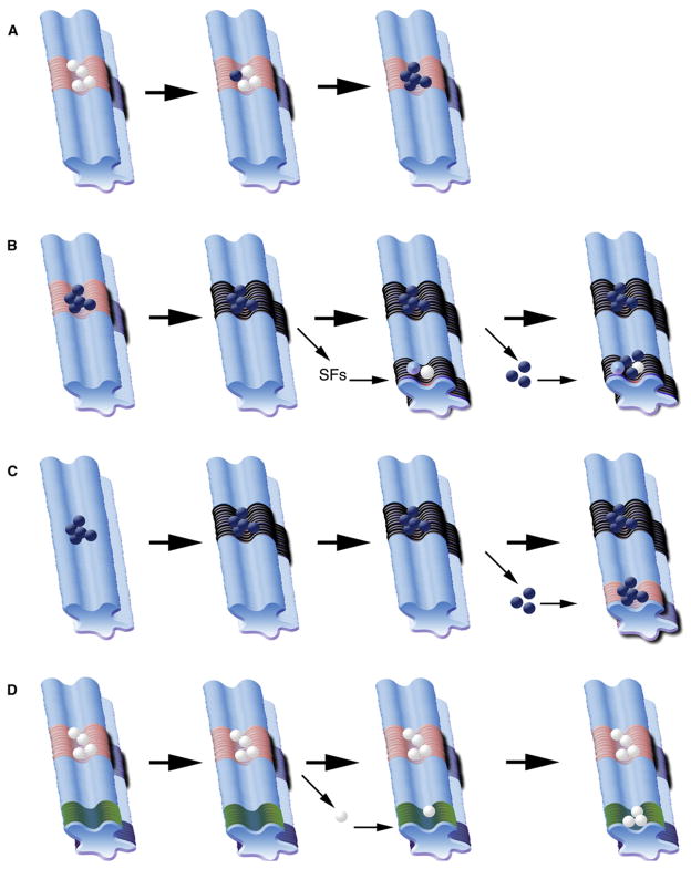
(A) In an endogenous niche, stem cells (white) are in contact with niche support cells (pink region) near associated cells (light blue region). Cancer cells (navy blue) may outcompete stem cells for access to niche-derived signals, resulting in expansion of cancer cells and loss of endogenous stem cells. (B) Cancer cells (navy blue) that occupy an endogenous niche (pink region) may transform or recruit surrounding support cells (black region). Distant premetastatic or malignant niches induced by signaling factors (SF) from primary tumor can recruit endogenous progenitor cells (white) or nonprogenitor cells (light blue) to aid in metastasis (navy blue spheres). (C) Cancer cells may contribute to transformation of local environment (black region); metastatic cancer cells may home to distant endogenous niches (pink region). (D) In a “latent niche” model, competent cells (white) from an endogenous niche (pink region) become physically displaced through developmental or injury induced mechanisms, and distinct signaling cells (green region) act as a niche to influence ectopic proliferation.
References
-
- Adams GB, Chabner KT, Alley IR, Olson DP, Szczepiorkowski ZM, Poznansky MC, Kos CH, Pollak MR, Brown EM, Scadden DT. Stem cell engraftment at the endosteal niche is specified by the calcium-sensing receptor. Nature. 2006;439:599–603. - PubMed
-
- Amsel S, Dell ES. Response of the preosteoblast and stem cell of rat bone marrow to a lethal dose of x-irradiation or cyclophosphamide. Cell Tissue Kinet. 1971;4:255–261. - PubMed
-
- Arai F, Hirao A, Ohmura M, Sato H, Matsuoka S, Takubo K, Ito K, Koh GY, Suda T. Tie2/angiopoietin-1 signaling regulates hematopoietic stem cell quiescence in the bone marrow niche. Cell. 2004;118:149–161. - PubMed
-
- Austin J, Kimble J. glp-1 is required in the germ line for regulation of the decision between mitosis and meiosis in C. elegans. Cell. 1987;51:589–599. - PubMed
Publication types
MeSH terms
Grants and funding
LinkOut - more resources
Full Text Sources
Other Literature Sources
Medical
