

doi: 10.1016/j.neuropsychologia.2010.02.004.
Epub 2010 Feb 7.
An animal model of recognition memory and medial temporal lobe amnesia: history and current issues
Affiliations
- PMID: 20144894
- PMCID: PMC2975590
- DOI: 10.1016/j.neuropsychologia.2010.02.004
Item in Clipboard
An animal model of recognition memory and medial temporal lobe amnesia: history and current issues
Neuropsychologia.
2010 Jul.
Abstract
The medial temporal lobe includes a system of anatomically connected structures that are essential for declarative memory (conscious memory for facts and events). A prominent form of declarative memory is recognition memory (the ability to identify a recently encountered item as familiar). Recognition memory has been frequently assessed in humans and in the experimental animal. This article traces the successful development of an animal model of human medial temporal lobe amnesia, which eventually identified the structures in the medial temporal lobe important for memory. Attention is given to two prominent behavioral paradigms (delayed nonmatching to sample and tests of spontaneous novelty preference).
Figures
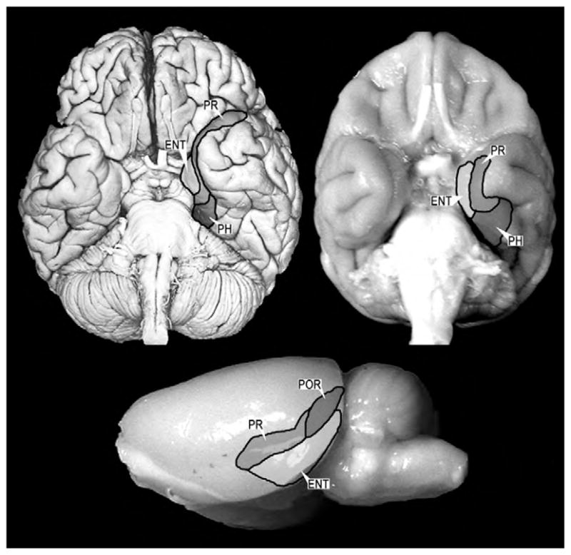
Ventral view of a human brain (upper left), a monkey brain (upper right) and a lateral view of a rat brain (lower center). The major cortical components of the medial temporal lobe are highlighted and outlined. The organization and connections of these structures are highly conserved across these species. Abbreviations: PR: perirhinal cortex, PH: parahippocampal cortex, ENT: entorhinal cortex, POR: postrhinal cortex (referred to as parahippocampal cortex in primates).
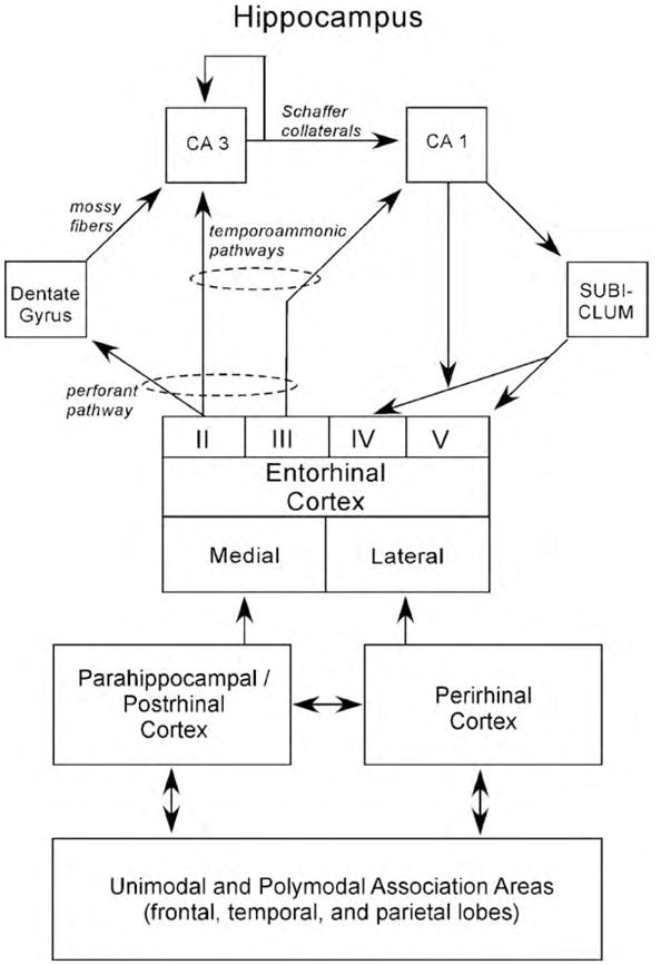
A schematic view of the medial temporal lobe memory system: The hippocampus, defined here as the dentate gyrus (DG), CA3, and CA1, is anatomically situated to receive highly processed information from widespread neocortical regions through three temporal cortical areas: the entorhinal, perirhinal, and parahippocampal cortices (in the rat the postrhinal cortex is used in place of the parahippocampal cortex), and through other direct projections from extra-temporal areas. The main pathway for the transmission of sensory information to the hippocampus is the entorhinal cortex. Layer II of this structure provides the major input to the hippocampus. This unidirectional projection, forming part of the perforant pathway, provides a substantial input to the DG, which, in turn, provides the major input to CA3 via the mossy fiber projection. There is also a smaller unidirectional projection to CA3 from layer II of the entorhinal cortex. CA3 provides the major input to CA1 via the Schaffer collateral/commissural pathway, but there is a substantial recurrent associational projection back to the CA3 field. CA1 also receives a direct temporoammonic projection from layer III of the entorhinal cortex (as does the subiculum, not shown). Both Schaffer collateral and temporoammonic projections to CA1 are unidirectional. CA1 primarily projects to the subiculum, but also sends a projection to entorhinal cortex layer V. The subiculum sends a prominent projection primarily to the entorhinal cortex layers IV and V (see Witter & Amaral, 2004 for review). The figure shows a simplified view of the way in which information enters the hippocampus from the superficial layers of the entorhinal cortex and then flows in a largely unidirectional, feed-forward, clockwise direction to ultimately return predominantly to the deep layers of entorhinal cortex. The perirhinal and parahippocampal/postrhinal cortex account for a substantial portion of the cortical input to the entorhinal cortex. The parahippocampal/postrhinal cortex preferentially projects to medial entorhinal cortex, and the perirhinal cortex preferentially projects to the lateral entorhinal cortex. These structures in turn receive projections from unimodal and polymodal areas in the frontal, temporal, and parietal lobes.
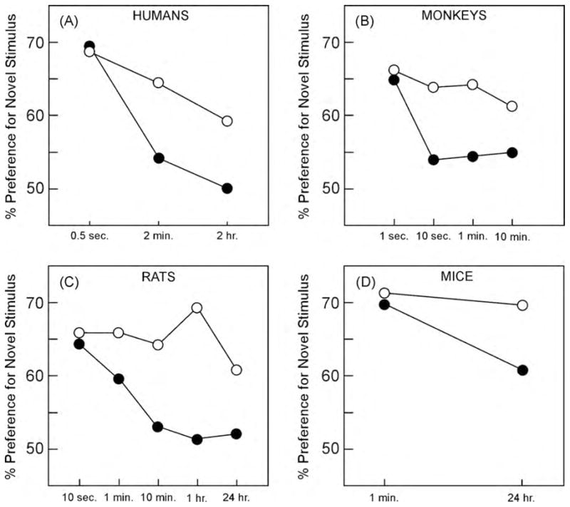
Performance of humans, monkeys, rats, and mice on the VPC/NOR task. In all panels the white circles depict control groups, and the black circles depict groups with hippocampal damage or disruption. (A) Human participants (data from McKee & Squire, 1993). (B) Monkeys (data from Zola et al., 2000). (C) Rats (data from Clark et al., 2000). (D) Mice (data from Hammond et al., 2004). All four studies revealed delay-dependent impairment after hippocampal damage or disruption.

Performance on the NOR task on 4 different days. On each day there was a familiarization and test phase by sham operated animals (CON, n = 47) and animals with hippocampal lesions (H, n = 44). Performance was scored over 30 s of cumulative object exploration. The figure shows the 4-day mean for the hippocampal lesion group (black bar) and the sham group (white bar). Both groups performed above chance (chance = 50%). Group difference is indicated by an asterisk (p < 0.05).
References
-
- Aggleton JP. One-trial object recognition by rats. Quarterly Journal of Experimental Psychology. 1985;37B:279–294.
-
- Aggleton JP, Brown MW. Interleaving brain systems for episodic and recognition memory. Trends in Cognitive Science. 2006;10:455–463. - PubMed
-
- Aggleton JP, Hunt PR, Rawlins JNP. The effects of hippocampal lesions upon spatial and non-spatial tests of working memory. Behavioral Brain Research. 1986;19:133–146. - PubMed
-
- Ainge JA, Heron-Maxwell C, Theofilas P, Wright P, de Hoz L, Wood ER. The role of the hippocampus in object recognition in rats: examination of the influence of task parameters and lesion size. Behavioral Brain Research. 2006;167:183–195. - PubMed
Publication types
MeSH terms
Grants and funding
LinkOut - more resources
Full Text Sources
