. 2010 Jun;38(10):3222-32.
doi: 10.1093/nar/gkq056. Epub 2010 Feb 9.

Binding of NF-kappaB p65 subunit to the promoter elements is involved in LPS-induced transactivation of miRNA genes in human biliary epithelial cells

Affiliations

Binding of NF-kappaB p65 subunit to the promoter elements is involved in LPS-induced transactivation of miRNA genes in human biliary epithelial cells

Rui Zhou et al. Nucleic Acids Res. 2010 Jun.

Abstract

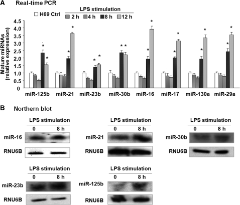
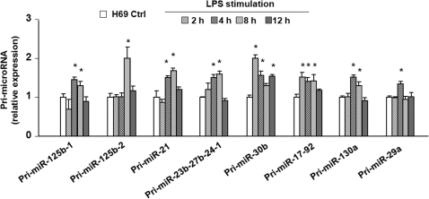
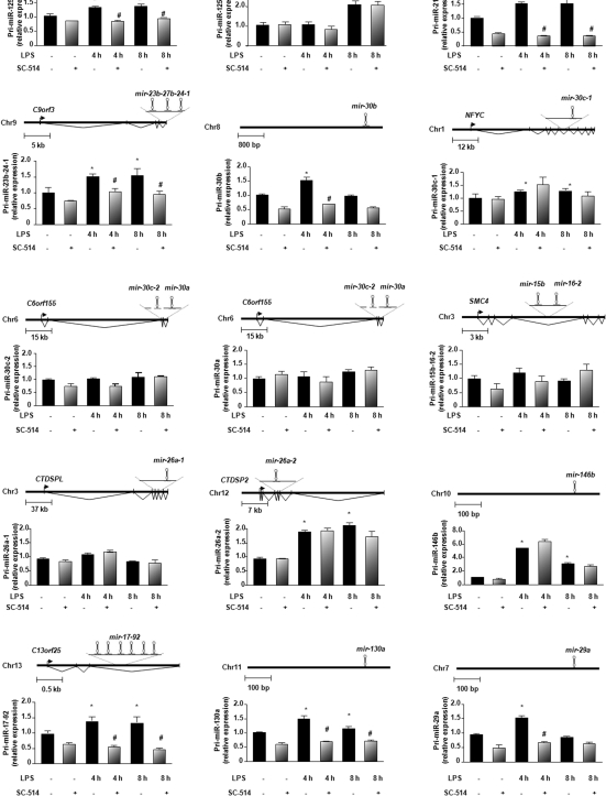
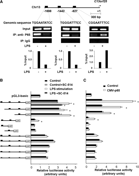
The majority of human miRNA genes is transcribed by polymerase II and can be classified as class II genes similar to protein-coding genes. Whereas current research on miRNAs has focused on the physiological and pathological functions, the molecular mechanisms underlying their transcriptional regulation are largely unknown. We recently reported that lipopolysaccharide (LPS) alters mature miRNA expression profile in human biliary epithelial cells. In this study, we tested the role of transcription factor NF-kappaB in LPS-induced transcription of select miRNA genes. Of the majority of LPS-up-regulated mature miRNAs in cultured human biliary epithelial cells, potential NF-kappaB binding sites were identified in the putative promoter elements of their corresponding genes. Inhibition of NF-kappaB activation by SC-514, an IKK2 inhibitor, blocked LPS-induced up-regulation of a subset of pri-miRNAs, including pri-miR-17-92, pri-miR-125b-1, pri-miR-21, pri-miR-23b-27b-24-1, pri-miR-30b, pri-miR-130a and pri-miR-29a. Moreover, direct binding of NF-kappaB p65 subunit to the promoter elements of mir-17-92, mir-125b-1, mir-21, mir-23b-27b-24-1, mir-30b and mir-130a genes was identified by chromatin immunoprecipitation analysis and confirmed by the luciferase reporter assay. Thus, a subset of miRNA genes is regulated in human biliary epithelial cells through NF-kappaB activation induced by LPS, suggesting a role of the NF-kappaB pathway in the transcriptional regulation of miRNA genes.


Figures

Figure 1.
Figure 1.
Altered expression of selected miRNAs identified by real-time PCR and northern blot following LPS stimulation in H69 cells. (A) Alterations of selected miRNA expression in H69 cells after exposure to LPS for various periods of time as assessed by real-time PCR. The amount of mature miRNAs was obtained by normalizing to the level of snRNA RNU6B in the samples. Data are expressed as the amount of mature miRNAs in LPS-stimulated samples relative to the control non-stimulated samples and representative of three independent experiments. (B) Alterations of selected miRNA expression in cells after exposure to LPS for 8 h as determined by northern blot. snRNA RNU6B was used as a control to ensure equal loading. Representative northern blots (LPS-stimulated cells versus non-stimulated control) from three independent experiments are shown. *P < 0.05 t-test versus non-stimulated cells.
Figure 2.
Figure 2.
Differential expression of primary transcripts of selected LPS-up-regulated mature miRNAs in H69 cells. H69 cells were exposed to LPS for 2–12 h and primary transcripts (pri-miRNAs) of select miRNAs were quantified by real-time PCR. The amount of pri-miRNAs was obtained by normalizing to the level of GAPDH in the samples. Data are expressed as the amount of pri-miRNAs in LPS-stimulated samples relative to the control non-stimulated samples and representative of three independent experiments. *P < 0.05 t-test versus the non-stimulated cells.
Figure 3.
Figure 3.
NF-κB dependent miRNAs identified among a subset of LPS-up-regulated mature miRNAs in biliary epithelial cells. Data are presented as the relative expression level of each pri-miRNA in H69 cells following LPS stimulation for 4 and 8 h in the presence or absence of SC-514 as assessed by real-time PCR. A schematic diagram shows the structure of each miRNA gene. The amount of pri-miRNAs was obtained by normalizing to the level of GAPDH in the samples. Data are expressed as the amount of pri-miRNAs in the stimulated samples relative to the control non-stimulated samples and representative of three independent experiments. *P < 0.05 t-test versus the non-stimulated cells; #P < 0.05 t-test versus LPS-stimulated cells.
Figure 4.
Figure 4.
Promoter binding of p65 transactivates mir-17-92 gene in H69 cells following LPS stimulation. (A) LPS induced the promoter element binding of p65 to C13orf25 gene. The schematic diagram shows three potential binding sites in the putative promoter element of to C13orf25 gene. ChIP analysis revealed increased binding of p65 to the binding site at −827, but not at −1442 and −1698, of C13orf25 promoter element in cells following LPS stimulation. Representative ChIP gels are shown in the upper panel and densitometry analysis in the lower panel. (B) H69 cells were transfected with various luciferase reporter constructs spanning the potential p65 binding sites of the C13orf25 promoter. The transfected cells were exposed to LPS in the presence or absence of SC-514. Luciferase activity was measured and presented as the ratio of the activity of the test construct with the control luciferase reporter construct. Reporter constructs containing different nucleotide genomic regions with the putative p65 binding sites were also utilized for the analysis as indicated. H69 cells were co-transfected with the pCMV-p65 to overexpress p65 and the luciferase reporter constructs for 24 h followed by measurement of luciferase activity. *P < 0.05 t-test versus the non-stimulated cells or empty pCMV vector control; #P < 0.05 t-test versus LPS-stimulated cells.

References

    1. Cai X, Hagedorn CH, Cullen BR. Human microRNAs are processed from capped, polyadenylated transcripts that can also function as mRNAs. RNA. 2004;10:1957–1966. - PMC - PubMed
    1. Lee Y, Kim M, Han J, Yeom KH, Lee S, Baek SH, Kim VN. MicroRNA genes are transcribed by RNA polymerase II. EMBO. J. 2004;23:4051–4060. - PMC - PubMed
    1. Winter J, Jung S, Keller S, Gregory RI, Diederichs S. Many roads to maturity: microRNA biogenesis pathways and their regulation. Nat. Cell. Biol. 2009;11:228–234. - PubMed
    1. Eulalio A, Huntzinger E, Izaurralde E. Getting to the root of miRNA-mediated gene silencing. Cell. 2008;132:9–14. - PubMed
    1. Asirvatham AJ, Gregorie CJ, Hu Z, Magner WJ, Tomasi TB. MicroRNA targets in immune genes and the Dicer/Argonaute and ARE machinery components. Mol. Immunol. 2008;45:1995–2006. - PMC - PubMed

Publication types