. 2010 Mar 1;123(Pt 5):795-805.
doi: 10.1242/jcs.050682. Epub 2010 Feb 9.

Control of mitotic and meiotic centriole duplication by the Plk4-related kinase ZYG-1

Affiliations

Control of mitotic and meiotic centriole duplication by the Plk4-related kinase ZYG-1

Nathaniel Peters et al. J Cell Sci. .

Abstract

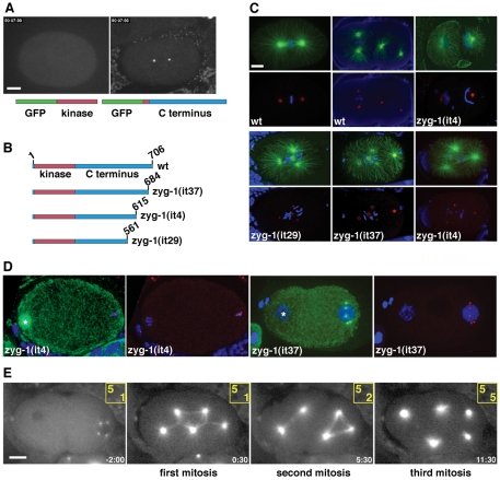
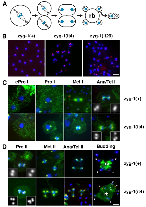
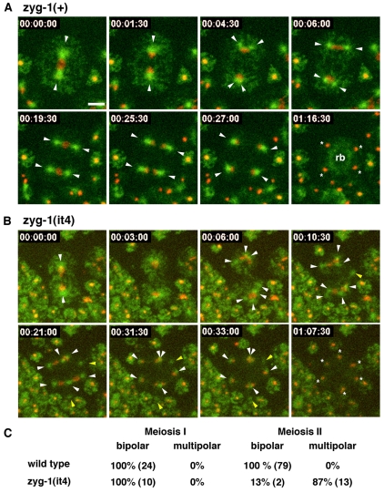
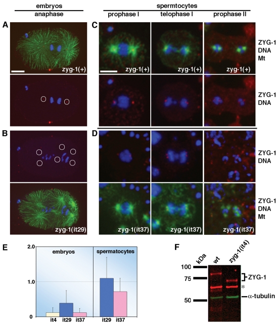
Centriole duplication is of crucial importance during both mitotic and male meiotic divisions, but it is currently not known whether this process is regulated differently during the two modes of division. In Caenorhabditis elegans, the kinase ZYG-1 plays an essential role in both mitotic and meiotic centriole duplication. We have found that the C-terminus of ZYG-1 is necessary and sufficient for targeting to centrosomes and is important for differentiating mitotic and meiotic centriole duplication. Small truncations of the C-terminus dramatically lower the level of ZYG-1 at mitotic centrosomes but have little effect on the level of ZYG-1 at meiotic centrosomes. Interestingly, truncation of ZYG-1 blocks centrosome duplication in the mitotic cycle but leads to centrosome amplification in the meiotic cycle. Meiotic centriole amplification appears to result from the overduplication of centrioles during meiosis I and leads to the formation of multipolar meiosis II spindles. The extra centrioles also disrupt spermatogenesis by inducing the formation of supernumerary fertilization-competent spermatids that contain abnormal numbers of chromosomes and centrioles. Our data reveal differences in the regulation of mitotic and meiotic centrosome duplication, particularly with regard to ZYG-1 activity, and reveal an important role for centrosomes in spermatid formation.


Figures

Fig. 1.
Fig. 1.
Elements within the C-terminus of ZYG-1 are important for localization to the centrosome and maintaining proper centrosome number. (A) GFP fused to the ZYG-1 C-terminus localizes to the centrosome (right), whereas ZYG-1 fused to the kinase domain is cytoplasmic (left). Scale bar: 10 μm. (B) Presumptive structure of the wild-type ZYG-1 protein and the three class II mutants. Due to mutation of a 3′ splice site, the zyg-1(it29) allele probaby produces a mixture of properly and improperly spliced messages. (C) Class II embryos stained for microtubules (green), DNA (blue) and centrosomes (red). Embryos are shown with (top) and without (bottom) microtubules. In the top set of panels, centrosomes are stained for SPD-2. The zyg-1(it4) embryo has failed to duplicate the sperm centrioles and as a result assembles monopolar spindles at the two-cell stage. In the bottom set of panels, the zyg-1(it29) embryo is stained for SPD-2, the zyg-1(it37) embryo is stained for SPD-5, and the zyg-1(it4) embryo is stained for SAS-4. Scale bar: 10 μm. (D) Young class II mutant embryos showing extra centrosomes exclusively associated with the male pronucleus. Centrosomes are stained for SPD-2. In the zyg-1(it4) embryo, the female meiotic spindle is visible (asterisk) but is not associated with centrosomes. In the zyg-1(it37) embryo, the female pronucleus (asterisk) lacks centrosomes. (E) Frames from a recording of zyg-1(it29) embryo expressing GFP-tubulin. Though the embryo inherits five centrosomes, they do not duplicate during the embryonic divisions. Numbers in yellow indicate the number of centrosomes (top) and cells (bottom) at each point in the recording. Time (minutes:seconds) is relative to first metaphase. Scale bar: 10 μm (D,E).
Fig. 2.
Fig. 2.
The constancy of zygotic centrosome number is disrupted in class II zyg-1 mutants. For each of the class II alleles, the distribution of centrosome number (MTOCs, microtubule-organizing centers) in live and fixed one-cell embryos was determined. For each allele, the percentage of embryos with one, two, or more than two centrosomes is given in the inset. Although not shown, wild-type one-cell embryos always have two centrosomes.
Fig. 3.
Fig. 3.
The meiotic centrosome cycle is abnormal in zyg-1 class II mutants. (A) Schematic of C. elegans spermatogenesis showing the arrangement of chromosomes and centrioles during meiosis I (MI), meiosis II (MII), spermatid formation, and in sperm. Spermatids form in the vicinity of spindle poles through budding, which produces a residual body (rb) that is later degraded. (B) Wild-type sperm contain a nucleus of uniform size and a single focus of SPD-2 (red), which corresponds to a centriole pair. Class II mutant sperm are heterogeneous with respect to DNA content and the number and intensity of SPD-2 foci. Scale bar: 5 μm. (C,D) zyg-1(+); him-8(e1489) and zyg-1(it4); him-8(e1489) spermatocytes at various stages of meiosis I and meiosis II. Spermatocytes are stained for microtubules (green), DNA (blue) and SPD-2 (red). Scale bar: 5 μm. (C) Meiosis I is mostly normal in zyg-1(it4) spermatocytes. Extra centrosomes first become apparent in anaphase or telophase I when more than two SPD-2 foci are detected at the poles of the spindle. (D) In zyg-1(it4) spermatocytes, meiosis II often occurs in the presence of too many centrosomes and DNA segregates on multipolar spindles. The extra centrosomes give rise to abnormal budding patterns in which more than four spermatids (asterisks) are formed from a single residual body. Notice that more than one SPD-2 focus is apparent in some of the budding mutant spermatids.
Fig. 4.
Fig. 4.
Selected frames from recordings of zyg-1(+) and zyg-1(it4) spermatocytes expressing GFP-tubulin and mCherry-histone (Movie 5). Arrowheads indicated centrosomes and asterisks indicate spermatids. (A) A zyg-1(+) primary spermatocyte undergoing two meiotic divisions with bipolar spindles. At 00:04:30, each pole of the meiosis I spindle splits to form two centrosomes. At 00:27:00, meiosis is complete and each haploid nucleus is associated with a single aster. At 01:16:30, the four spermatids have finished budding. The asters have disassembled and the tubulin has relocated to the residual body (rb). (B) A zyg-1(it4) spermatocyte assembles multipolar spindles during meiosis II. In the first frame, two centrosomes are apparent at the end of meiosis I. Beginning at 00:06:00, these centrosomes begin to split into three centrosomes (top) and five centrosomes (bottom). The yellow arrowheads indicated centrosomes not associated with DNA. The last frame shows that six spermatids form. Time is in hours:minutes:seconds. Scale bar: 5 μm and applies to all panels. (C) The percentages of bipolar and multipolar spindles observed during meiosis I and II in live zyg-1(+) and zyg-1(it4) spermatocytes are shown. The actual numbers observed are shown in parentheses.
Fig. 5.
Fig. 5.
Truncation of ZYG-1 specifically disrupts recruitment to mitotic centrosomes. (A-D) Embryos (A,B) and spermatocytes (C,D) stained for ZYG-1 (red), microtubules (green) and DNA (blue). A zyg-1(+) embryo (A) and a zyg-1(it29) embryo (B) are shown. Relative to full-length ZYG-1, significantly less truncated ZYG-1 is detected at mitotic centrosomes. Circles indicate the positions of centrosomes. Embryos in A and B are shown at same magnification. Scale bar: 10 μm. zyg-1(+) (C) and zyg-1(it37) spermatocytes (D) are shown. Similar levels of ZYG-1 are detected at meiotic centrosomes. All spermatocytes are shown at same magnification. Scale bar: 5 μm. (E) Quantitative immunofluorescence microscopy reveals that ZYG-1 levels at mitotic, but not meiotic, centrosomes are diminished in class II mutants. (F) Quantitative immunoblotting demonstrates that wild-type (wt) and zyg-1(it4) embryos possess similar amounts of ZYG-1 protein. α-tubulin and a nonspecific band (asterisk) serve as loading controls.

Similar articles

Cited by

References

    1. Albertson D. G., Thomson J. N. (1993). Segregation of holocentric chromosomes at meiosis in the nematode, Caenorhabditis elegans. Chromosome Res. 1, 15-26 - PubMed
    1. Barton M. K., Schedl T. B., Kimble J. (1987). Gain-of-function mutations of fem-3, a sex-determination gene in Caenorhabditis elegans. Genetics 115, 107-119 - PMC - PubMed
    1. Basto R., Lau J., Vinogradova T., Gardiol A., Woods C. G., Khodjakov A., Raff J. W. (2006). Flies without centrioles. Cell 125, 1375-1386 - PubMed
    1. Basto R., Brunk K., Vinadogrova T., Peel N., Franz A., Khodjakov A., Raff J. W. (2008). Centrosome amplification can initiate tumorigenesis in flies. Cell 133, 1032-1042 - PMC - PubMed
    1. Bettencourt-Dias M., Rodrigues-Martins A., Carpenter L., Riparbelli M., Lehmann L., Gatt M. K., Carmo N., Balloux F., Callaini G., Glover D. M. (2005). SAK/PLK4 is required for centriole duplication and flagella development. Curr. Biol. 15, 2199-2207 - PubMed

Publication types

MeSH terms

Substances