Review
. 2010 Feb 15;123(Pt 4):529-36.
doi: 10.1242/jcs.062968.

Molecular mechanisms of protein and lipid targeting to ciliary membranes

Affiliations
Review

Molecular mechanisms of protein and lipid targeting to ciliary membranes

Brian T Emmer et al. J Cell Sci. .

Abstract

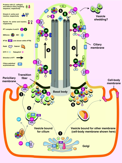
Cilia are specialized surface regions of eukaryotic cells that serve a variety of functions, ranging from motility to sensation and to regulation of cell growth and differentiation. The discovery that a number of human diseases, collectively known as ciliopathies, result from defective cilium function has expanded interest in these structures. Among the many properties of cilia, motility and intraflagellar transport have been most extensively studied. The latter is the process by which multiprotein complexes associate with microtubule motors to transport structural subunits along the axoneme to and from the ciliary tip. By contrast, the mechanisms by which membrane proteins and lipids are specifically targeted to the cilium are still largely unknown. In this Commentary, we review the current knowledge of protein and lipid targeting to ciliary membranes and outline important issues for future study. We also integrate this information into a proposed model of how the cell specifically targets proteins and lipids to the specialized membrane of this unique organelle.


Figures

Fig. 1.
Fig. 1.
Protein targeting to the ciliary membrane. The eukaryotic cilium is a distinct organelle that is separated from the cytoplasm by transition fibers that connect the basal body to the membrane and separate the ciliary membrane (green) from the periciliary (pale orange) (Reiter and Mostov, 2006) and cell-body membranes (dark orange) (Sloboda and Rosenbaum, 2007). Although cilia in certain cell types differ in the fine details of their structures, functions, mechanisms of assembly and regulation, some general principles have emerged in recent years; this figure attempts to integrate these general concepts. The ciliary membrane has a lipid composition that is distinct from that of the periciliary and cell-body membranes because it is highly enriched in sterols, glycolipids and sphingolipids (Tyler et al., 2009). This specialized composition is probably formed in the Golgi (Ejsing et al., 2009; Schuck and Simons, 2004). Various cell membranes have unique complements of membrane proteins, and one mechanism that might contribute to this specialized composition is the association of certain types of proteins with vesicles of specific lipid composition. The model shown is based on the current literature and recent findings from our own laboratory and outlines six stages: (1).Vesicles of different lipid composition and containing specific cargos form in the Golgi (Klemm et al., 2009). For example, those vesicles rich in sterols and sphinoglipids might load certain lipid raft-associated proteins such as the calflagins. One or more cilium-specific palmitoyl acyltransferases might reside in these vesicles to confer lipid-raft association to their substrates in order to sort them into these vesicles. These ciliary vesicles also load with other proteins that contain ciliary targeting sequences, whereas vesicles of other compositions might load other types of cargo (e.g. proteins bound for the cell-body membrane, shown here as one example). IFT20 also loads with the ciliary-bound vesicles and might serve as an adaptor to recruit other ciliary cargo. (2).Vesicles destined for the cilium interact with GTP-Rab8, which is produced by GDP-GTP exchange catalyzed by Rabin8 and the BBSome. This facilitates movement of the vesicles to the base of the cilium near the transition fiber (Hao and Scholey, 2009; Jin and Nachury, 2009), although the molecular mechanism for transport is not known. (3).Vesicles fuse with the periciliary membrane, and ciliary lipids and proteins enter the cilium. Some vesicles accumulate at the base of the cilium, giving rise to the ciliary necklace structure. The periciliary membrane and transition fiber can take different forms in different cell types. For example, the trypanosome flagellar pocket is a unique dynamic structure through which all endocytosis and exocytosis occurs, and which is much larger than the periciliary membrane of most cells. (4).IFT complexes, which consist of protein subcomplexes IFT-A and IFT-B and are possibly associated with the BBSome (Ou et al., 2005), move cargo along the length of the cilium. The BBSome might also interact directly with the ciliary membrane (Hao and Scholey, 2009). IFT is driven in the anterograde direction by kinesin 2 and in the retrograde direction by dynein 1b. A key component of IFT-B is DYF-11, which might promote a separate membrane association of the IFT complex through Rabaptin5 and GTP-Rab8 (Omori et al., 2008), although these interactions need further investigation because they were not confirmed in other studies (Follit et al., 2009). Ciliary membrane proteins might also be associated with the IFT complexes. (5).IFT complexes disassociate from their cargo at the ciliary tip, where anterograde motors become inactivated and retrograde motors become active. (6).Turnover products are recycled back to the base of the cilium. Note that not all components of IFT are included in this model, and specific elements are not drawn to scale.

Similar articles

Cited by

References

    1. Adhiambo C., Blisnick T., Toutirais G., Delannoy E., Bastin P. (2009). A novel function for the atypical small G protein Rab-like 5 in the assembly of the trypanosome flagellum. J. Cell Sci. 122, 834-841 - PubMed
    1. Baldari C. T., Rosenbaum J. (2009). Intraflagellar transport: it's not just for cilia anymore. Curr. Opin. Cell Biol. [Epub ahead of print] doi:10.1016/j.ceb.2009.10.010 - PMC - PubMed
    1. Bethani I., Lang T., Geumann U., Sieber J. J., Jahn R., Rizzoli S. O. (2007). The specificity of SNARE pairing in biological membranes is mediated by both proof-reading and spatial segregation. EMBO J. 26, 3981-3992 - PMC - PubMed
    1. Bhowmick R., Li M., Sun J., Baker S. A., Insinna C., Besharse J. C. (2009). Photoreceptor IFT complexes containing chaperones, guanylyl cyclase 1 and rhodopsin. Traffic 10, 648-663 - PMC - PubMed
    1. Blacque O. E., Leroux M. R. (2006). Bardet-Biedl syndrome: an emerging pathomechanism of intracellular transport. Cell. Mol. Life Sci. 63, 2145-2161 - PMC - PubMed

Publication types