

Phosphorylation of p53 serine 18 upregulates apoptosis to suppress Myc-induced tumorigenesis
- PMID: 20145032
- PMCID: PMC2829875
- DOI: 10.1158/1541-7786.MCR-09-0324
Phosphorylation of p53 serine 18 upregulates apoptosis to suppress Myc-induced tumorigenesis
Abstract
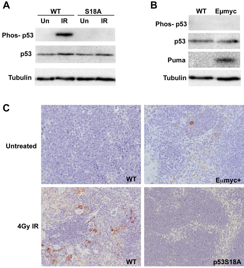
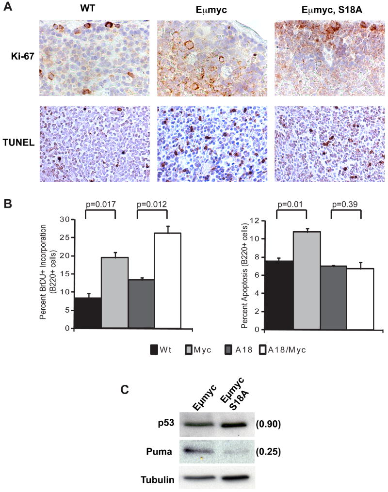
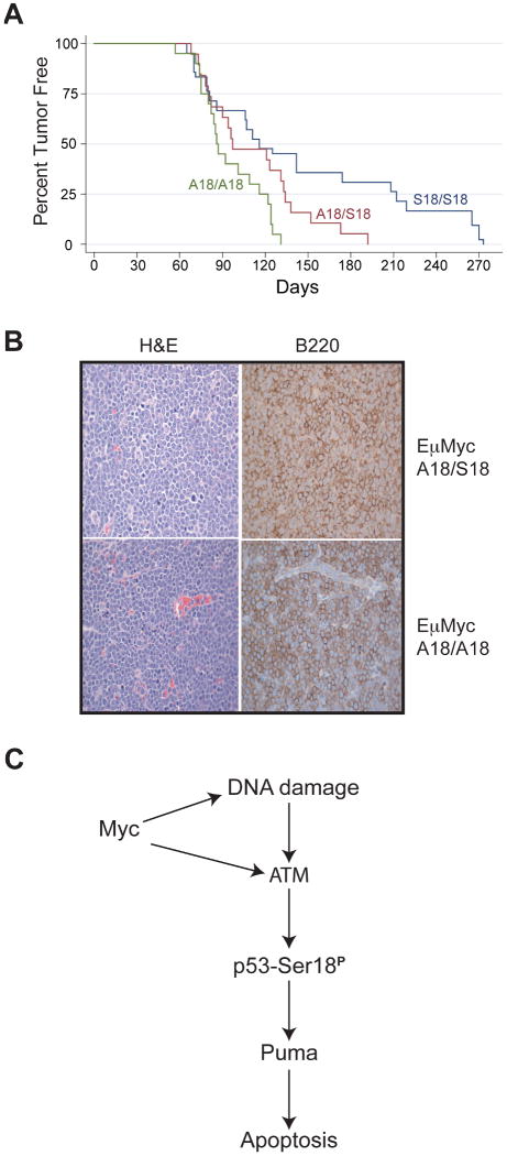
ATM and p53 are critical regulators of the cellular DNA damage response and function as potent tumor suppressors. In cells undergoing ionizing radiation, ATM is activated by double-strand DNA breaks and phosphorylates the NH(2) terminus of p53 at serine residue 18. We have previously generated mice bearing an amino acid substitution at this position (p53S18A) and documented a role for p53 phosphorylation in DNA damage-induced apoptosis. In this present study, we have crossed E mu myc transgenic mice with our p53S18A mice to explore a role for ATM-p53 signaling in response to oncogene-induced tumorigenesis. Similar to DNA damage induced by ionizing radiation, expression of c-Myc in pre-B cells induces p53 serine 18 phosphorylation and Puma expression to promote apoptosis. E mu myc transgenic mice develop B-cell lymphoma more rapidly when heterozygous or homozygous for p53S18A alleles. However, E mu myc-induced tumorigenesis in p53S18A mice is slower than that observed in E mu myc mice deficient for either p53 or ATM, indicating that both p53-induced apoptosis and p53-induced growth arrest contribute to the suppression of B-cell lymphoma formation in E mu myc mice. These findings further reveal that oncogene expression and DNA damage activate the same ATM-p53 signaling cascade in vivo to regulate apoptosis and tumorigenesis.
Figures
Similar articles
-
Oncogenes and the DNA damage response: Myc and E2F1 engage the ATM signaling pathway to activate p53 and induce apoptosis.Cell Cycle. 2006 Apr;5(8):801-3. doi: 10.4161/cc.5.8.2638. Epub 2006 Apr 17. Cell Cycle. 2006. PMID: 16582589
-
Targeting lysosomal degradation induces p53-dependent cell death and prevents cancer in mouse models of lymphomagenesis.J Clin Invest. 2008 Jan;118(1):79-88. doi: 10.1172/JCI33700. J Clin Invest. 2008. PMID: 18097482 Free PMC article.
-
ATM promotes apoptosis and suppresses tumorigenesis in response to Myc.Proc Natl Acad Sci U S A. 2006 Jan 31;103(5):1446-51. doi: 10.1073/pnas.0507367103. Epub 2006 Jan 23. Proc Natl Acad Sci U S A. 2006. PMID: 16432227 Free PMC article.
-
p53: guardian of the genome and policeman of the oncogenes.Cell Cycle. 2007 May 2;6(9):1006-10. doi: 10.4161/cc.6.9.4211. Epub 2007 May 28. Cell Cycle. 2007. PMID: 17457049 Review.
-
Exploiting synthetic lethal interactions for targeted cancer therapy.Cell Cycle. 2009 Oct 1;8(19):3112-9. doi: 10.4161/cc.8.19.9626. Cell Cycle. 2009. PMID: 19755856 Free PMC article. Review.
Cited by
-
Hexavalent chromium-induced apoptosis of granulosa cells involves selective sub-cellular translocation of Bcl-2 members, ERK1/2 and p53.Toxicol Appl Pharmacol. 2011 Mar 15;251(3):253-66. doi: 10.1016/j.taap.2011.01.011. Epub 2011 Jan 22. Toxicol Appl Pharmacol. 2011. PMID: 21262251 Free PMC article.
-
ATM phosphorylation of Mdm2 Ser394 regulates the amplitude and duration of the DNA damage response in mice.Cancer Cell. 2012 May 15;21(5):668-679. doi: 10.1016/j.ccr.2012.04.011. Cancer Cell. 2012. PMID: 22624716 Free PMC article.
-
Apoptosis is the essential target of selective pressure against p53, whereas loss of additional p53 functions facilitates carcinoma progression.Mol Cancer Res. 2011 Apr;9(4):430-9. doi: 10.1158/1541-7786.MCR-10-0277. Epub 2011 Mar 8. Mol Cancer Res. 2011. PMID: 21385880 Free PMC article.
-
DNA repair synthesis and ligation affect the processing of excised oligonucleotides generated by human nucleotide excision repair.J Biol Chem. 2014 Sep 19;289(38):26574-26583. doi: 10.1074/jbc.M114.597088. Epub 2014 Aug 8. J Biol Chem. 2014. PMID: 25107903 Free PMC article.
-
Regulation of the Mdm2-p53 signaling axis in the DNA damage response and tumorigenesis.Transl Cancer Res. 2016 Dec;5(6):707-724. doi: 10.21037/tcr.2016.11.75. Transl Cancer Res. 2016. PMID: 28690977 Free PMC article.
References
-
- Harris SL, Levine AJ. The p53 pathway: positive and negative feedback loops. Oncogene. 2005;24:2899–2908. - PubMed
-
- Vousden KH, Lu X. Live or let die: the cell's response to p53. Nat Rev Cancer. 2002;2:594–604. - PubMed
-
- Soussi T, Beroud C. Assessing TP53 status in human tumours to evaluate clinical outcome. Nat Rev Cancer. 2001;1:233–240. - PubMed
-
- Iwakuma T, Lozano G. MDM2, an introduction. Mol Cancer Res. 2003;1:993–1000. - PubMed
-
- Giaccia AJ, Kastan MB. The complexity of p53 modulation: emerging patterns from divergent signals. Genes Dev. 1998;12:2973–2983. - PubMed
Publication types
MeSH terms
Substances
Grants and funding
LinkOut - more resources
Full Text Sources
Molecular Biology Databases
Research Materials
Miscellaneous
