. 2010 Feb;8(2):232-45.
doi: 10.1158/1541-7786.MCR-09-0391. Epub 2010 Feb 9.

Cyclooxygenase-2 is a novel transcriptional target of the nuclear EGFR-STAT3 and EGFRvIII-STAT3 signaling axes

Affiliations

Cyclooxygenase-2 is a novel transcriptional target of the nuclear EGFR-STAT3 and EGFRvIII-STAT3 signaling axes

Hui-Wen Lo et al. Mol Cancer Res. 2010 Feb.

Abstract

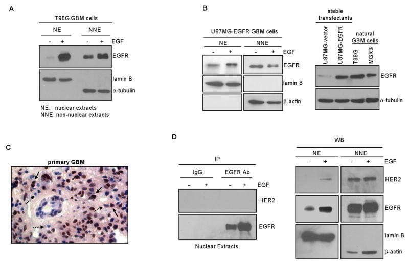
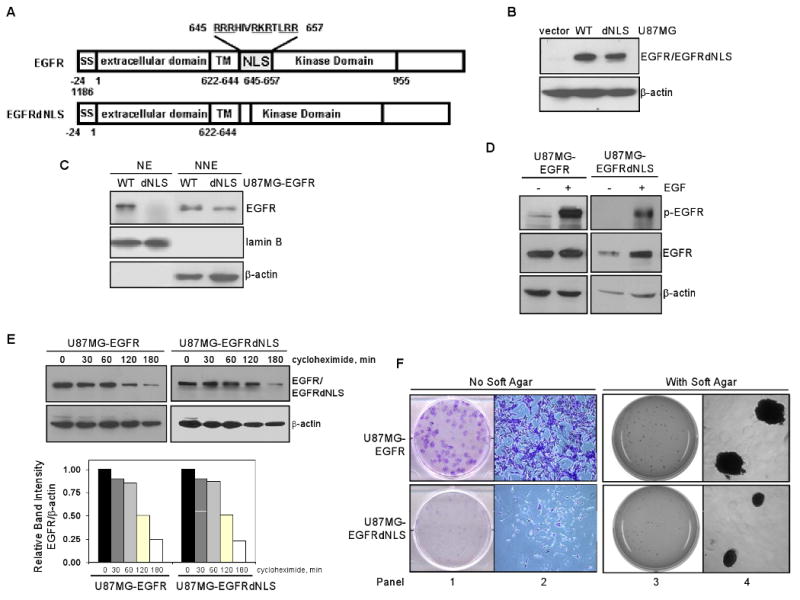
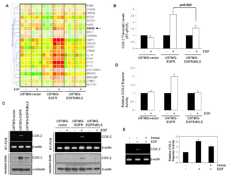
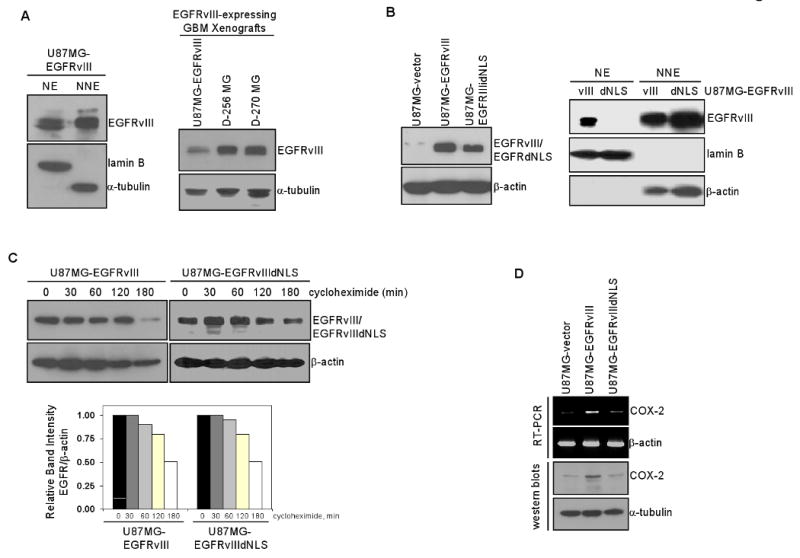
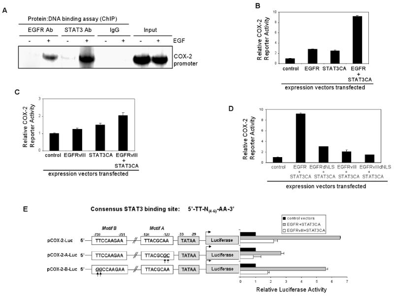
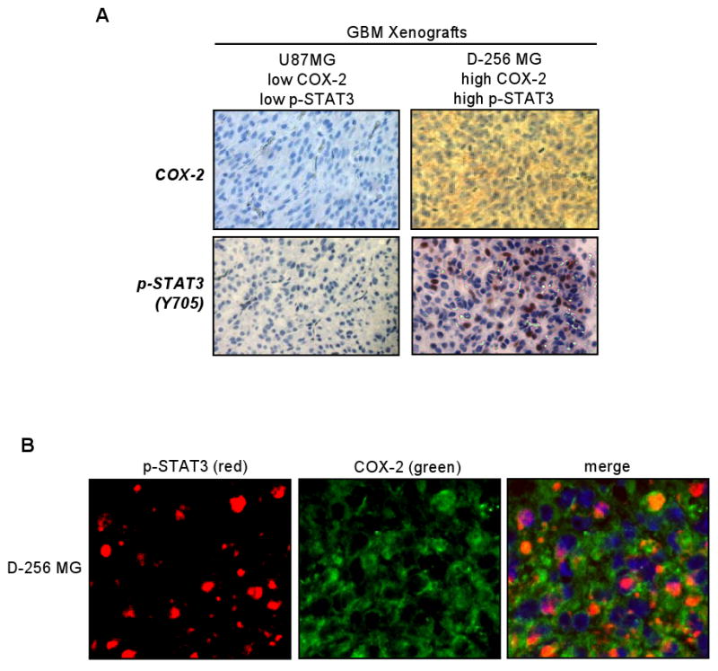
Emerging evidence indicates a novel mode of epidermal growth factor receptor (EGFR) signaling, notably, one involves EGFR nuclear translocalization and subsequent gene activation. To date, however, the significance of the nuclear EGFR pathway in glioblastoma (GBM) is unknown. Here, we report that EGFR and its constitutively activated variant EGFRvIII undergo nuclear translocalization in GBM cells, in which the former event requires EGF stimulation and the latter is constitutive. To gain insights into the effect of nuclear EGFR on gene expression in GBM, we created isogenic GBM cell lines, namely, U87MG-vector, U87MG-EGFR, and U87MG-EGFRdNLS that, respectively, express the control vector, EGFR, and nuclear entry-defective EGFR with a deletion of the nuclear localization signal (NLS). Microarray analysis shows that 19 genes, including cyclooxygenase-2 (COX-2), to be activated in U87MG-EGFR cells but not in U87MG-EGFRdNLS and U87MG-vector cells. Subsequent validation studies indicate that COX-2 gene is expressed at higher levels in cells with EGFR and EGFRvIII than those with EGFRdNLS and EGFRvIIIdNLS. Nuclear EGFR and its transcriptional cofactor signal transducer and activator of transcription 3 (STAT3) associate with the COX-2 promoter. Increased expression of EGFR/EGFRvIII and activated STAT3 leads to the synergistic activation of the COX-2 promoter. Promoter mutational analysis identified a proximal STAT3-binding site that is required for EGFR/EGFRvIII-STAT3-mediated COX-2 gene activation. In GBM tumors, an association exists between levels of COX-2, EGFR/EGFRvIII, and activated STAT3. Together, these findings indicate the existence of the nuclear EGFR/EGFRvIII signaling pathway in GBM and its functional interaction with STAT3 to activate COX-2 gene expression, thus linking EGFR-STAT3 and EGFRvIII-STAT3 signaling axes to proinflammatory COX-2 mediated pathway.


Figures

Figure 1
Figure 1. EGFR undergoes nuclear translocalization in human GBM cell lines and primary specimens
A. EGF induces EGFR nuclear transport in GBM cells with endogenous EGFR. Serum-starved human GBM T98G cells that express endogenous EGFR were stimulated with EGF (100 ng/ml) for 0 and 15 min, harvested and subjected to nuclear fractionation to obtain nuclear extracts (NE) and non-nuclear extracts (NNE). Both extracts were analyzed for EGFR, lamin B and α-tubulin expression via western blotting. Effectiveness of fractionation is indicated by the lack of cytoplasmic marker α-tubulin in NE and the absence of nuclear protein lamin B in NNE. B. EGFR undergoes EGF-activated nuclear transport in GBM cells engineered to stably express EGFR. Left panel: U87MG-EGFR stable transfectant cells were similarly treated and analyzed, as described in panel A. Right panel: Western blotting shows U87MG-EGFR cells to express EGFR at the levels comparable to natural T98G and MGR3 GBM cells who express endogenous EGFR. C. Nuclear EGFR is detected in a primary GBM specimen. We analyzed 12 primary GBM tumors for EGFR expression using IHC and found six tumors to express EGFR/EGFRvIII with one of them expressing significant levels of nuclear EGFR. EGFR-positive brown nuclei are pointed by solid arrows whereas EGFR-negative blue counterparts are marked by dashed arrows. D. Nuclear EGFR is not in complex with nuclear HER2 in T98G cells. Using T98G GBM cells know to expression both EGFR and HER2, we treated the cells with EGF for 0 and 15 min, fractionated the cells into the nuclear and on-nuclear fractions, immunoprecipitated nuclear EGFR using an EGFR antibody and subjected the immunoprecipitates to western blotting to detect HER2. As shown in the left panel, HER2 was not co-immunoprecipitated with nuclear EGFR despite nuclear EGFR was effectively immunoprecipitated by the EGFR antibody. IgG did not pull down EGFR or HER2, indicating specificity. As shown by the western blotting in the right panel, HER2 underwent a modest level of nuclear translocalization after EGF stimulation with the majority of the HER2 protein in the non-nuclear fraction.
Figure 2
Figure 2. Generation of GBM cells stably expressing mutant EGFR that is defective in nuclear entry
A. Structures of EGFR and EGFRdNLS, an EGFR mutant defective in nuclear entry. Top panel: EGFR contains a number of functional domains, namely, signal peptide (SS; aa -24 to -1), extracellular domain (1-621), transmembrane domain (TM; aa 622-644), NLS (aa 645-657) and kinase domain (658-955). Bottom panel: EGFRdNLS mutant contains a deletion of eight amino acids (aa 645-652) within the NLS region and a Ser insertion as aa residue 645. It has been shown that amino acid substitutions of the basic residues with this NLS region abolish the ability of EGFR to enter the nucleus (15, 38). B. EGFR expression in isogenic U87MG-vector, U87MG-EGFR and U87MG-EGFRdNLS cells. Western blotting shows that U87MG-vector cells do not express Myc-tagged EGFR protein and in contrast, U87MG-EGFR and U87MG-EGFRdNLS express increased and comparable levels of EGFR/EGFRdNLS. C. EGFRdNLS is absent in the cell nucleus. Efficiency of nuclear fractionation is indicated by the lack of cytoplasmic protein β-actin in NE and the absence of nuclear marker lamin B in NNE. D. EGFRdNLS undergoes EGF-induced autophosphorylation similar to EGFR. Isogenic U87MG-EGFR and U87MG-EGFRdNLS cells were serum starved, stimulated with EGFR for 0 and 15 min and subjected to western blotting to determine levels of p-EGFR (Y1068; indicator of receptor activation), EGFR and β-actin. E. EGFRdNLS has similar stability relative to EGFR. Isogenic U87MG-EGFR and U87MG-EGFRdNLS cells were treated with protein synthesis inhibitor cycloheximide (10 ug/ml) for 0-180 min and subjected to western blotting to determine levels of EGFR and β-actin. Intensity of the band signals were quantified using ImageJ software and protein half-life was subsequently computed. F. Nuclear EGFR is important for the clonogenic growth of EGFR-expressing GBM cells. Colony formation assays were performed in the absence (left two panels) and presence (right two panels) of soft agar. U87MG-EGFRdNLS cells formed significantly less colonies than U87MG-EGFR cells. As shown by the 5X images in the second panel, the colony from U87MG-EGFR cells contained more cells and was with intensive staining whereas the one from U87MG-EGFRdNLS cells contained significantly fewer cells. As shown by the high-resolution images (fourth panel), U87MG-EGFR cells formed larger colonies compared to U87MG-EGFRdNLS cells in the presence of soft agar, suggesting that nuclear EGFR is important for the anchorage-independent growth of U87MG cells. U87MG-vector cells did not form colonies in either assay.
Figure 3
Figure 3. Gene expression analysis identified the human COX-2 gene as a candidate nuclear EGFR target gene
A. Microarray shows 19 genes to be significantly induced by EGF in U87MG-EGFR cells, but not in U87MG-vector or U87MG-EGFRdNLS cells. Cells were treated with and without EGF and the levels of expression of over 47,000 human gene transcripts were determined. Data represent results of three independent experiments. ANOVA was conducted to derive p-values. Clustering analysis included a total of 22 transcripts, with 19 known genes, that were significantly activated by EGF in U87MG-EGFR cells, but not in U87MG-vector or U87MG-EGFRdNLS cells (p<0.05). An arrow points to COX-2. B. COX-2 transcripts are significantly up-regulated by EGF in U87MG-EGFR cells, but not in U87MG-EGFRdNLS or EGFR-vector cells. Microarray results was validated using RT-qPCR and the results show that the COX-2 transcription is significantly increased by EGF in U87MG-EGFR cells, but not in U87MG-EGFRdNLS or EGFR-vector cells. C. U87MG-EGFR cells express higher levels of COX-2 transcripts and protein than U87MG-EGFRdNLS or EGFR-vector cells. Three U87MG stable transfectants were subjected to RT-PCR (top panels) and western blotting (bottom panels) to determine levels of COX-2 expression. In the left panels, cells were growth under regular growth conditions with 10% FCS. In the right panels, cells were serum-starved for 24 hrs and treated with EGF (100 ng/ml) for 0 and 4 hrs. D. The human COX-2 promoter is significantly activated by EGF in U87MG-EGFR cells but not in U87MG-EGFRdNLS orU87MG-vector cells. A firefly luciferase reporter construct under the control of a 1-kb COX-2 promoter was transfected into the three U87MG isogenic cell lines. Twenty-four hrs later, the transfected cells were serum-starved for 24 hrs and stimulated with 100 ng/ml EGF for 0 and 4 hrs. A renilla luciferase vector, pRL-TK was co-transfected to control for transfection efficiency. Luciferase activity was determined as previously described (15). Relative reporter activity was determined by normalizing the firefly luciferase activity against that of the renilla luciferase. E: EGFR kinase inhibitor Iressa reduced EGF-induced COX-2 expression and promoter activation. U87MG-EGFR cells were serum starved for 24 hrs in the presence of Iressa (25 uM) or 1% DMSO for 24 hrs, treated with EGF for 0 and 1 hr, and analyzed for COX-2 expression via RT-PCR (left panel). Furthermore, aliquots of cells were transfected with pCOX-2-Luc and pRL-TK. Twenty-four hrs after transfections, the cells were similarly treated as described for the RT-PCR experiments and subjected to luciferase assay.
Figure 4
Figure 4. EGFRvIII undergoes nuclear translocalization and activates the human COX-2 gene in GBM cells
A. EGFRvIII is constitutively present in the nuclei of U87MG-EGFRvIII stable transfectants cells. Left panel: EGFRvIII is present in the nucleus of U87MG-EGFRvIII cells. Right panel: U87MG-EGFRvIII xenografts express EGFRvIII at the levels comparable to GBM xenografts with endogenous EGFRvIII (D-256 MG and D-270 MG). B. Characterization of isogenic U87MG-EGFRvIII and U87MG-EGFRvIIIdNLS cells. Left panel: Western blotting shows U87MG-EGFRvIII and U87MG-EGFRvIIIdNLS cells to express equivalent levels of the receptors. Right panel: Nuclear fractionation and western blotting shows EGFRvIIIdNLS to fail to enter the cell nucleus. C. EGFRvIII and EGFRvIIIdNLS proteins have similar half-life. Isogenic U87MG-EGFRvIII and U87MG-EGFRvIIIdNLS cells were treated with 10 ug/ml cycloheximide for 0-180 min and subjected to western blotting for EGFRvIII and β-actin. Band signals were quantified using ImageJ software and protein half-life was subsequently computed. The results indicate that NLS deletion does not affect EGFRvIII protein stability. D. Levels of COX-2 transcripts and protein are significantly higher in U87MG-EGFRvIII cells than U87MG-EGFRvIIIdNLS and U87MG-vector cells. Top panel: RT-PCR. Bottom panel: Western blotting.
Figure 5
Figure 5. STAT3 significantly enhances nuclear EGFR-mediated activation of the human COX-2 gene
A. Nuclear EGFR and nuclear STAT3 associate with the COX-2 gene promoter in an EGF-dependent fashion. In intracellular protein-DNA binding ChIP assay, we used an anti-EGFR antibody (Ab) and an anti-STAT3 Ab in immunoprecipitation whereas normal IgGs were used as negative controls. Input chromatins were used as loading controls for PCR. B. COX-2 promoter activity is enhanced by EGFR alone and STAT3CA alone but is significantly elevated by the combination of both. STAT3CA: constitutively activated STAT3 with two Cys substitutions that enable STAT3 molecules to dimerize spontaneously without phosphorylation. U87MG cells were transfected with pCOX-2-Luc and EGFR plasmid alone, STAT3CA plasmid alone or in combination for 48 hrs and then luciferase activity determined. pRL-TK vector was co-transfected as transfection efficiency control. Relative COX-2 reporter activity was determined by normalizing the firefly luciferase activity against that of the renilla luciferase. Three independent experiments were performed to derive means and standard deviations. C. The COX-2 promoter is activated by EGFRvIII. The experiment was conducted as described in panel B. Although the COX-2 promoter is activated by EGFRvIII, unlike the potent activation by EGFR-STAT3CA combination, STAT3CA only modestly enhances EGFRvIII-mediated activation of COX-2 promoter. D. Nuclear EGFR is important for EGFR-STAT3CA mediated activation of the COX-2 promoter. This was conducted as described in panel B. EGFRdNLS-STAT3CA co-transfection yields significantly lower activity than the EGFR-STAT3CA combination. In contrast, EGFRvIIIdNLS-STAT3CA co-transfection similarly activates the COX-2 promoter compared to EGFRvIII-STAT3CA combination. E. The EGFR-STAT3 signaling axis activates the COX-2 promoter via the STAT3-binding site proximal to the TATAA Box. Consensus STAT3-binding site is listed on the top. Web-based search engine, TFSearch, identifies two putative STAT3-binding sites in the human COX-2 promoter, namely, the proximal motif (motif A; nt -134 to -127) and the distant motif (motif B; nt -759 to 751). pCOX-2-A-Luc construct carries the mutant COX-2 promoter with two nucleotide substitutions with the proximal motif. pCOX-2-B-Luc construct contains the mutant COX-2 promoter with two nucleotide substitutions with the distant motif. The results show that mutations within the proximal motif, but not motif B, significantly decreased the ability of EGFR-STAT3CA and EGFRvIII-STAT3CA to activate the COX-2 promoter.
Figure 6
Figure 6. Increased COX-2 expression is detected in GBM xenografts with EGFR/EGFRvIII and activated STAT3
A. COX-2 is highly expressed in GBM xenografts with highly activated STAT3. Two GBM xenografts were analyzed for levels of COX-2 and p-STAT3 (Y705) via IHC. The D-256 MG GBM xenograft but not the U87MG xenograft expressed high levels of COX-2 and p-STAT3. The p-STAT3-positive nuclei are indicated by the brown signals whereas the negatively stained nuclei are in blue. B. The majority of p-STAT3-expressing GBM cells in the D-256 MG xenograft express COX-2. The D-256 MG GBM xenograft was analyzed for co-expression of p-STAT3 and COX-2 using double fluorescence staining-coupled IHC. Red fluorescence: p-STAT3. Green fluorescence: COX-2. Nuclei: blue. p-STAT3-positive nuclei: pink signals as merged products of red and blue fluorescence. The majority of p-STAT3-positive nuclei expressed high levels of COX-2. COX-2 is also expressed in cells without significant p-STAT3 expression, suggesting that p-STAT3-independent mechanisms may account for the transcriptional activation of the COX-2 gene in these cells.

Similar articles

Cited by

References

    1. Nakamura JL. The epidermal growth factor receptor in malignant gliomas: pathogenesis and therapeutic implications. Expert Opinion on Therapeutic Targets. 2007;11:463. - PubMed
    1. Lo HW, Hung MC. Nuclear EGFR signalling network in cancers: linking EGFR pathway to cell cycle progression, nitric oxide pathway and patient survival. Br J Cancer. 2006;94:184–8. - PMC - PubMed
    1. Yang Z, Bagheri-Yarmand R, Wang RA, et al. The epidermal growth factor receptor tyrosine kinase inhibitor ZD1839 (Iressa) suppresses c-Src and Pak1 pathways and invasiveness of human cancer cells. Clin Cancer Res. 2004;10:658–67. - PubMed
    1. Sugawa N, Ekstrand AJ, James CD, Collins VP. Identical splicing of aberrant epidermal growth factor receptor transcripts from amplified rearranged genes in human glioblastomas. Proc Natl Acad Sci U S A. 1990;87:8602–6. - PMC - PubMed
    1. Nishikawa R, Ji XD, Harmon RC, et al. A mutant epidermal growth factor receptor common in human glioma confers enhanced tumorigenicity. Proc Natl Acad Sci U S A. 1994;91:7727–31. - PMC - PubMed

Publication types

MeSH terms