Comparative Study
. 2010 Feb;133(Pt 2):375-88.
doi: 10.1093/brain/awp298. Epub 2010 Feb 9.

Pharmacological prion protein silencing accelerates central nervous system autoimmune disease via T cell receptor signalling

Affiliations
Comparative Study

Pharmacological prion protein silencing accelerates central nervous system autoimmune disease via T cell receptor signalling

Wei Hu et al. Brain. 2010 Feb.

Abstract

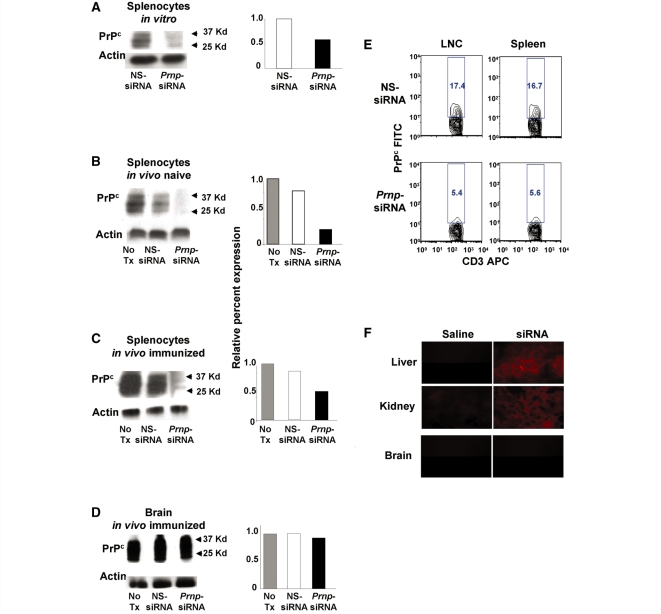
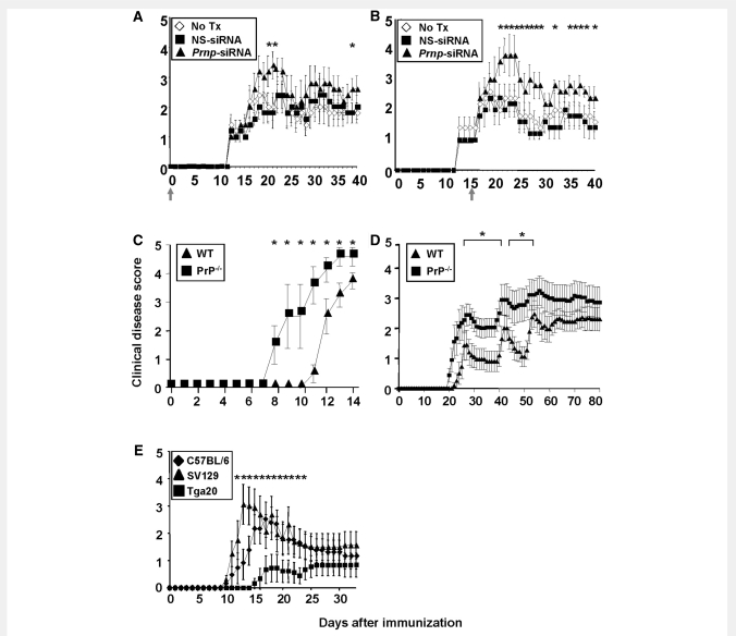
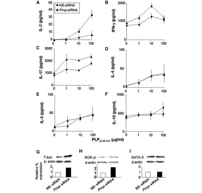
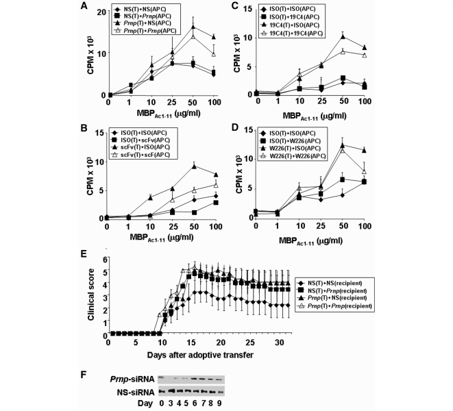
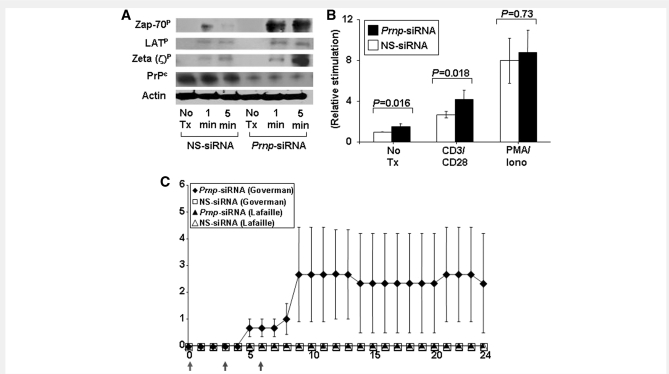
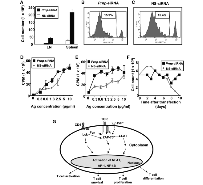
The primary biological function of the endogenous cellular prion protein has remained unclear. We investigated its biological function in the generation of cellular immune responses using cellular prion protein gene-specific small interfering ribonucleic acid in vivo and in vitro. Our results were confirmed by blocking cellular prion protein with monovalent antibodies and by using cellular prion protein-deficient and -transgenic mice. In vivo prion protein gene-small interfering ribonucleic acid treatment effects were of limited duration, restricted to secondary lymphoid organs and resulted in a 70% reduction of cellular prion protein expression in leukocytes. Disruption of cellular prion protein signalling augmented antigen-specific activation and proliferation, and enhanced T cell receptor signalling, resulting in zeta-chain-associated protein-70 phosphorylation and nuclear factor of activated T cells/activator protein 1 transcriptional activity. In vivo prion protein gene-small interfering ribonucleic acid treatment promoted T cell differentiation towards pro-inflammatory phenotypes and increased survival of antigen-specific T cells. Cellular prion protein silencing with small interfering ribonucleic acid also resulted in the worsening of actively induced and adoptively transferred experimental autoimmune encephalomyelitis. Finally, treatment of myelin basic protein(1-11) T cell receptor transgenic mice with prion protein gene-small interfering ribonucleic acid resulted in spontaneous experimental autoimmune encephalomyelitis. Thus, central nervous system autoimmune disease was modulated at all stages of disease: the generation of the T cell effector response, the elicitation of T effector function and the perpetuation of cellular immune responses. Our findings indicate that cellular prion protein regulates T cell receptor-mediated T cell activation, differentiation and survival. Defects in autoimmunity are restricted to the immune system and not the central nervous system. Our data identify cellular prion protein as a regulator of cellular immunological homoeostasis and suggest cellular prion protein as a novel potential target for therapeutic immunomodulation.


Figures

Figure 1
Figure 1
Decreased PrPc expression levels on peripheral leucocytes but not in the CNS in mice treated with Prnp-siRNA. (A) Transfection with Prnp-siRNA decreased PrPc expression in murine splenocytes, compared with nonsense (NS)-siRNA treatment. On Day 3 after in vivo treatment of (B) unimmunized SJL/J mice and (C) SJL/J mice immunized with PLPp139−151 with 50 μg of Prnp-siRNA, PrPc expression was decreased in splenocytes, but not in (D) brain homogenate of immunized mice. (A–D) Densitometry was performed, and relative PrPc expression was determined by normalizing the PrPc to β-actin levels. (E) Using flow cytometry three days after immunization and siRNA treatment, lymph node cells (LNC) and splenocytes were gated on CD3 and PrPc. The top two panels show the percentage of PrPc+ cells within the CD3+ cell population from nonsense-siRNA-treated murine cells. The lower two panels show the percentage of PrPc+ cells within the CD3+ cell population from Prnp-siRNA-treated murine cells. PrPc expression was decreased by ∼70% following Prnp-siRNA treatment. (F) After injecting Cy3-labelled siRNA or DY547-labelled siRNA intravenously, fluorescence was detectable 72 h later in liver and kidney tissue but not in the brain of naïve mice or mice in which EAE had been induced by active immunization (data not shown).
Figure 2
Figure 2
Prnp-siRNA treated and PrP-deficient (PrP−/−) mice develop more severe EAE whereas mice overexpressing PrPC are protected. (A and B) In vivo silencing of PrPc worsens the clinical course of actively induced EAE in SJL/J mice. (A) In an EAE ‘prevention’ experiment, SJL/L mice were injected intravenously with a single dose of Prnp-siRNA or nonsense (NS)-siRNA at the time of immunization with PLPp139−151, as indicated by a grey arrow. Prnp-siRNA treatment resulted in clinically more severe disease. (B) In an EAE ‘treatment’ experiment, Prnp-siRNA or nonsense-siRNA was injected at the time of clinical onset of EAE (grey arrow). Mice treated with Prnp-siRNA had a significantly worse initial clinical exacerbation. (C) Male and (D) female PrP−/− mice had earlier disease onset and significantly higher disease scores than male wild-type (WT) mice. (E) Tga20 mice had a delayed disease onset, and developed only mild EAE compared to C57BL/6 and Sv129 wild-type mice.
Figure 3
Figure 3
Silencing PrPc in antigen specific splenocytes increases the expression of Th1 cytokines by upregulating T box expressed in T cells (T-bet). Splenocytes from SJL/J mice immunized with PLPp139–151 and treated concomitantly intravenously with 50 µg of Prnp-siRNA or nonsense (NS)-siRNA, were brought into single cell suspension 3 days after immunization and were pulsed with PLPp139–151. Interleukin (IL)-2, interferon (IFN)-γ, IL-17, IL-4, IL-5 and IL-10 cytokine expression was measured by ELISA. Silencing of PrPc significantly increased the expression of (A) IL-2, (B) IFN-γ and (C) IL-17. In contrast, silencing of PrPc had no effect on the expression of (D) IL-4, (E) IL-5 and (F) IL-10. (G) Protein expression of T box expressed in T cells and (H) retinoic acid receptor related orphan receptor (ROR)γt in splenocytes of mice treated with Prnp-siRNA was increased compared with nonsense-siRNA-treated cells. (I) In contrast, the expression of GATA binding protein (GATA)-3 was not altered. The data are shown as mean clinical score ± standard deviation.
Figure 4
Figure 4
Silencing PrPc in antigen-specific T cells increases T cell proliferation and activation markers. (A) Lymph node cells (LNC) and (B) splenocytes from SJL/J mice immunized with PLPp139–151 and treated with Prnp-siRNA or nonsense (NS)-RNA at the time of immunization were brought into single cell suspension on day 10 after immunization. Both (A) Lymph node cells and (B) splenocytes from Prnp-siRNA-treated mice showed increased proliferation. (C) Prnp-siRNA treatment also significantly increased the mean fluorescence intensity (MFI) of the activation marker CD25 in D10 T cells activated by anti-CD3, with or without anti-CD28 monoclonal antibody. (D) There was less of an effect of Prnp-siRNA treatment on the expression of CD69, an early T cell activation marker. Proliferation was measured as counts per minute (CPM).
Figure 5
Figure 5
Antigen-specific T cells are the main target of Prnp-siRNA. (A) Irradiated splenocytes from B10PL mice that served as antigen presenting cells (APC), or MBP1–11 TCR transgenic CD4+ T cells were treated with Prnp-siRNA and/or nonsense-siRNA, and incubated with antigen. Silencing of PrPc in T cells results in increased proliferation, whereas silencing of PrPc in antigen presenting cells had no apparent effect on T cell proliferation. Targeting PrPc in T cells with a (B) single chain variable fragment (scFv), or two monoclonal mouse anti-mouse PrP IgG1 monoclonal antibodies (C) 19C4, (D) W226 also resulted in the augmented proliferation of antigen-specific T cells. (E) Mice treated with Prnp-siRNA as recipients of Prnp-siRNA-treated adoptively transferred T cells, as recipients of direct Prnp-siRNA treatment, or both, developed significantly worse EAE than controls. (F) PrPc expression after Prnp-siRNA treatment is suppressed for 5 days.
Figure 6
Figure 6
Silencing PrPc on T cells increases ZAP-70 phosphorylation and NFAT/AP-1 reporter activity. (A) Phosphorylation of ZAP-70 and the zeta chain of the TCR/CD3 complex was increased in activated CD4+ T cells treated with Prnp-siRNA. (B) Transcription of an NFAT/AP-1 luciferase reporter was also significantly increased in activated and unstimulated T cells treated with Prnp-siRNA. In contrast, NFAT/AP-1 reporter activity was not altered in PMA/ionomycin-stimulated T cells. (C) MBP peptide AC1–11 TCR transgenic mice on a B10.PL background (I-Au) generated by Joan Goverman and coworkers developed EAE after in vivo intravenous treatment with Prnp-siRNA on Days 0, 3 and 6 of the observation period (indicated by grey arrows), but not after treatment with nonsense-siRNA. In contrast, MBP1–11 TCR transgenic mice generated by Lafaille and coworkers did not develop disease.
Figure 7
Figure 7
Silencing of PrPc improves T cell survival. Prnp-siRNA or nonsense-siRNA-transfected purified OVA323–339 TCR transgenic CD45.2+ CD4+ T cells were transferred intravenously into C57BL/6 CD45.1+ wild-type mice that were immunized with OVA323–339 in complete freund's adjuvant 24 h after cell transfer. (A) A significantly higher number of Prnp-siRNA-transfected CD45.2+ donor OVA323–339 TCR transgenic CD4+ T cells were recovered from lymph nodes and spleens than nonsense-siRNA-transfected cells. At Day 5 post transfer, there was no difference with regard to in vivo antigen-specific proliferation between OVA323–339 TCR transgenic CD4+ T cells transfected with (B) Prnp-siRNA, or with (C) nonsense-siRNA. When (D) lymph nodes and (E) splenocytes of recipient mice were resected on Day 3 after immunization, and recall assays were performed with OVA323–339, a significant increase in cell proliferation in both compartments was observed. (F) The number of purified antigen-activated CD4+ MBP1–11 TCR transgenic T cells transfected with nonsense-siRNA started to decline in vitro on Day 3, and none of the cells were viable on Day 10. Cell numbers remained relatively stable in the CD4+ MBP1–11 TCR transgenic T cells treated with Prnp-siRNA. (G) Schematic of proposed mechanisms of action of PrPc in antigen-specific T cells.

Similar articles

Cited by

References

    1. Antoine N, Cesbron JY, Coumans B, Jolois O, Zorzi W, Heinen E. Differential expression of cellular prion protein on human blood and tonsil lymphocytes. Haematologica. 2000;85:475–80. - PubMed
    1. Bueler H, Fischer M, Lang Y, Bluethmann H, Lipp HP, DeArmond SJ, et al. Normal development and behaviour of mice lacking the neuronal cell-surface PrP protein. Nature. 1992;356:577–82. - PubMed
    1. Burthem J, Urban B, Pain A, Roberts DJ. The normal cellular prion protein is strongly expressed by myeloid dendritic cells. Blood. 2001;98:3733–8. - PubMed
    1. Cashman NR, Loertscher R, Nalbantoglu J, Shaw I, Kascsak RJ, Bolton DC, et al. Cellular isoform of the scrapie agent protein participates in lymphocyte activation. Cell. 1990;61:185–92. - PubMed
    1. Chan AC, Iwashima M, Turck CW, Weiss A. ZAP-70: a 70 kd protein-tyrosine kinase that associates with the TCR zeta chain. Cell. 1992;71:649–62. - PubMed

Publication types