. 2010 Apr;78(4):1659-69.
doi: 10.1128/IAI.01010-09. Epub 2010 Feb 9.

UpaH is a newly identified autotransporter protein that contributes to biofilm formation and bladder colonization by uropathogenic Escherichia coli CFT073

Affiliations

UpaH is a newly identified autotransporter protein that contributes to biofilm formation and bladder colonization by uropathogenic Escherichia coli CFT073

Luke P Allsopp et al. Infect Immun. 2010 Apr.

Abstract

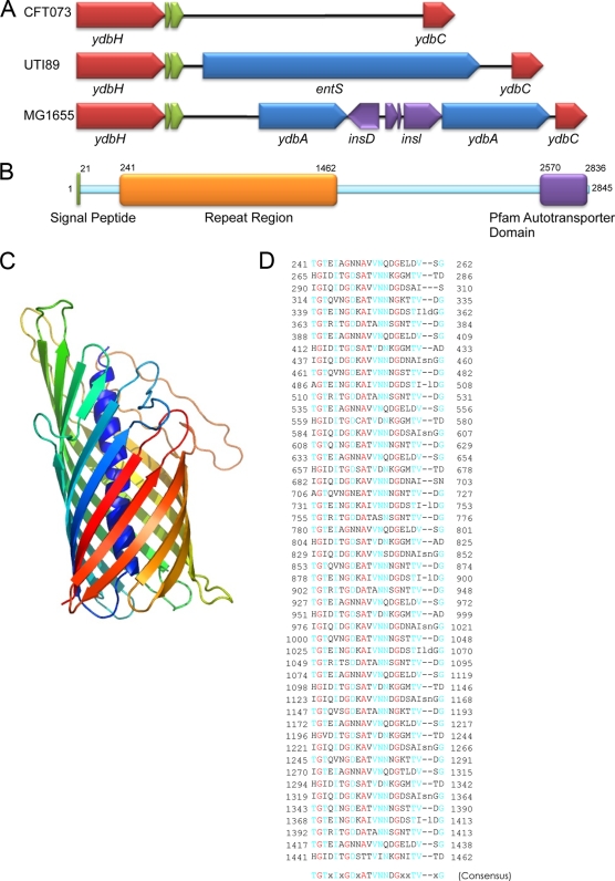
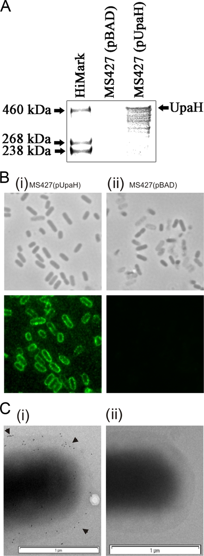
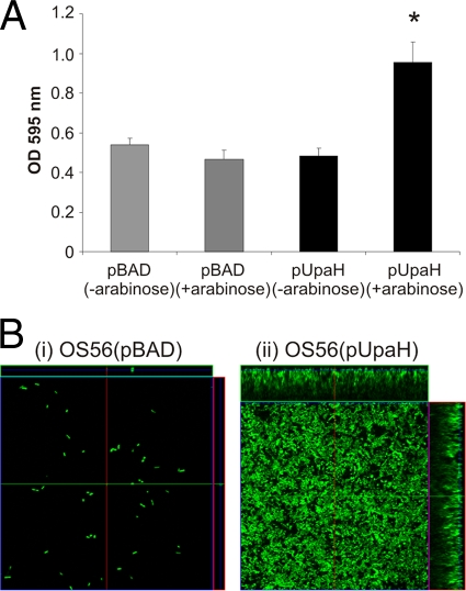
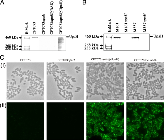
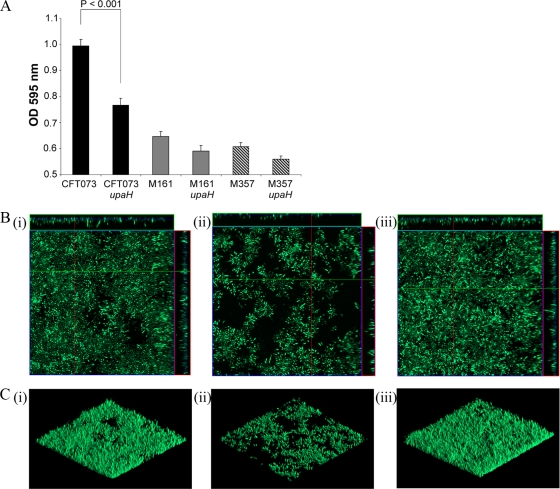
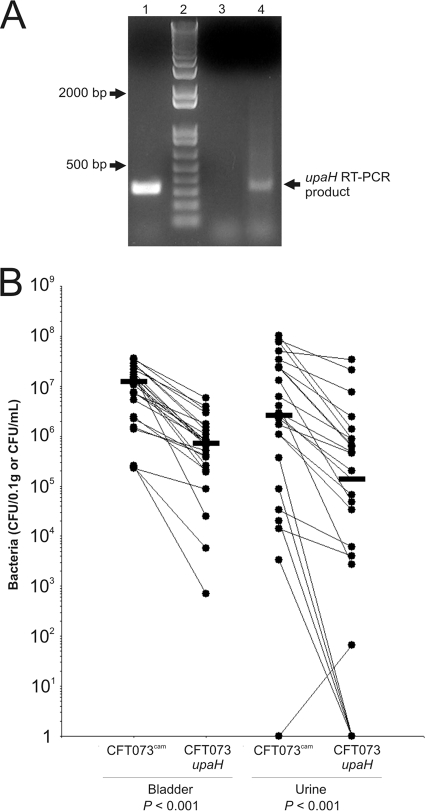
Escherichia coli is the primary cause of urinary tract infection (UTI) in the developed world. The major factors associated with virulence of uropathogenic E. coli (UPEC) are fimbrial adhesins, which mediate specific attachment to host receptors and trigger innate host responses. Another group of adhesins is represented by the autotransporter (AT) subgroup of proteins. In this study, we identified a new AT-encoding gene, termed upaH, present in a 6.5-kb unannotated intergenic region in the genome of the prototypic UPEC strain CFT073. Cloning and sequencing of the upaH gene from CFT073 revealed an intact 8.535-kb coding region, contrary to the published genome sequence. The upaH gene was widely distributed among a large collection of UPEC isolates as well as the E. coli Reference (ECOR) strain collection. Bioinformatic analyses suggest beta-helix as the predominant structure in the large N-terminal passenger (alpha) domain and a 12-strand beta-barrel for the C-terminal beta-domain of UpaH. We demonstrated that UpaH is expressed at the cell surface of CFT073 and promotes biofilm formation. In the mouse UTI model, deletion of the upaH gene in CFT073 and in two other UPEC strains did not significantly affect colonization of the bladder in single-challenge experiments. However, in competitive colonization experiments, CFT073 significantly outcompeted its upaH isogenic mutant strain in urine and the bladder.


Figures

FIG. 1.
FIG. 1.
(A) Physical map indicating the genomic location of upaH in E. coli CFT073, UTI89, and MG1655. (B) Schematic illustration of the domain organization of UpaH from E. coli CFT073. Indicated are the signal peptide, the repeat region within the passenger domain, and the transmembrane β-domain. Numbers indicate amino acid positions of region boundaries. (C) Predicted structure of the β-domain of UpaH. Shown is a ribbon diagram of the structure of the β-domain (residues 2528 to 2845) obtained by homology modeling based on the crystal structure of the NalP β-domain (43), colored blue (N terminus) through green and yellow to red (C terminus). (D) Amino acid sequence of the repeats present in UpaH. Amino acids in red indicate ≤98% conservation; amino acids in blue indicate ≤55% conservation.
FIG. 2.
FIG. 2.
(A) Western blot analysis of UpaH performed using whole-cell lysates prepared from E. coli MS427(pBAD) and MS427(pUpaH) grown in the presence of 0.1% arabinose. (B) Phase-contrast and immunofluorescence microscopy employing UpaH-specific antiserum against cells of E. coli strains MS427(pUpaH) (i) or MS427(pBAD) (ii) grown in the presence of arabinose. Overnight cultures were fixed and incubated with anti-UpaH serum followed by incubation with goat anti-rabbit IgG coupled to Alexa Fluor 488. (C) Immunogold electron microscopy of E. coli MS427(pUpaH) (i) and MS427(pBAD) (ii) grown in the presence of arabinose and labeled with anti-UpaH serum followed by protein A-gold (10 nm) conjugate. Gold particles were observed on the surface of E. coli MS427(pUpaH) but not MS427(pBAD).
FIG. 3.
FIG. 3.
UpaH mediates biofilm formation. (A) Biofilm formation in polystyrene microtiter plates by E. coli MS427(pBAD) and MS427(pUpaH) following static growth in the absence (−) or presence (+) of 0.1% arabinose. Biofilm growth was quantified by solubilization of crystal violet-stained cells with ethanol-acetone and determination of the absorbance at 595 nm. Results represent averages of a minimum of 8 replicates ± SEM. Significant biofilm growth was observed for E. coli MS427(pUpaH) following induction of UpaH expression with arabinose (*, P < 0.001 as determined by analysis of variance [NOVA]). (B) Dynamic-flow-chamber assay examining biofilm formation of E. coli OS56(pBAD) and OS56(pUpaH) grown in the presence of 0.1% arabinose. Biofilm development was monitored by confocal scanning laser microscopy after 24 h. The images are representative horizontal sections collected within each biofilm and vertical sections (to the right of and below each larger panel, representing the yz plane and the xz plane, respectively) at the positions indicated by the white lines. E. coli MS427(pUpaH) produced a significant biofilm in this assay.
FIG. 4.
FIG. 4.
(A) Western blot analysis of UpaH performed using whole-cell lysates prepared from E. coli CFT073, CFT073upaH, CFT073upaH(pBAD), and CFT073upaH(pUpaH). A band corresponding to UpaH was detected in E. coli CFT073 and CFT073upaH(pUpaH) but not in E. coli CFT073upaH and CFT073upaH(pBAD). (B) Western blot analysis of UpaH performed using whole-cell lysates prepared from E. coli M161, M161upaH, M357, and M357upaH. A band corresponding to UpaH was detected in E. coli M161 and M357 but not in E. coli M161upaH or M357upaH. (C) Phase-contrast (i) or immunofluorescence (ii) microscopy employing UpaH-specific antiserum against cells of E. coli CFT073, CFT073upaH, CFT073upaH(pUpaH), or CFT073PcLupaH. Overnight cultures were fixed and incubated with anti-UpaH serum, followed by incubation with goat anti-rabbit IgG coupled to Alexa Fluor 488. A positive reaction indicating surface localization of UpaH was detected only for CFT073upaH(pUpaH) and CFT073PcLupaH.
FIG. 5.
FIG. 5.
(A) Biofilm formation in polystyrene microtiter plates by three sets of isogenic strains with respect to upaH: E. coli CFT073 and CFT073upaH; E. coli M161 and M161upaH; and E. coli M357 and M357upaH. Results represent averages of OD595 readings of at least 4 independent experiments ± SEM. There was no difference in the growth of wild-type and upaH mutant strains. (B) Dynamic-flow-chamber assay examining biofilm formation of E. coli CFT073 (i), E. coli CFT073upaH (ii), and E. coli CFT073PcLupaH (iii). Biofilm development was monitored by confocal scanning laser microscopy after 40 h. The images are representative horizontal sections collected within each biofilm and vertical sections (to the right of and below each larger panel, representing the yz plane and the xz plane, respectively) at the positions indicated by the white lines. (C) Three-dimensional image demonstrating the structure of the biofilm formed by E. coli CFT073 (i), E. coli CFT073upaH (ii), and E. coli CFT073PcLupaH (iii).
FIG. 6.
FIG. 6.
(A) RT-PCR analysis of upaH transcription by CFT073 during colonization of the mouse urinary tract. Urine from six mice infected with CFT073 was collected and pooled at 18 h postinoculation and added directly into a 2× volume of RNAprotect (Qiagen). CFT073 cells were concentrated by centrifugation, and total RNA was extracted. Lane 1, upaH-specific PCR product (328 bp) obtained from genomic DNA (positive control); lane 2, 1-Kb Plus DNA ladder (Invitrogen); lane 3, PCR employing RNA prior to cDNA synthesis as a template (negative control); lane 4, upaH-specific PCR product (328 bp) obtained using the cDNA as the template. (B) Competitive mixed-infection experiment. C57BL/6 mice (n = 24) were challenged with a 1:1 mixture of E. coli CFT073cam and CFT073upaH. Total CFU were enumerated on selective medium to differentiate between CFT073cam (chloramphenicol resistant) and CFT073upaH (kanamycin resistant). Each symbol represents the total number of CFU for an individual mouse per 0.1 g of bladder tissue or the total number of CFU per ml urine. Lines connect values for the same mouse. Horizontal bars represent the median values. No significant colonization of the kidneys was observed.

Similar articles

Cited by

References

    1. Altschul, S. F., T. L. Madden, A. A. Schaffer, J. Zhang, Z. Zhang, W. Miller, and D. J. Lipman. 1997. Gapped BLAST and PSI-BLAST: a new generation of protein database search programs. Nucleic Acids Res. 25:3389-3402. - PMC - PubMed
    1. Anderson, G. G., J. J. Palermo, J. D. Schilling, R. Roth, J. Heuser, and S. J. Hultgren. 2003. Intracellular bacterial biofilm-like pods in urinary tract infections. Science 301:105-107. - PubMed
    1. Barnard, T. J., N. Dautin, P. Lukacik, H. D. Bernstein, and S. K. Buchanan. 2007. Autotransporter structure reveals intra-barrel cleavage followed by conformational changes. Nat. Struct. Mol. Biol. 14:1214-1220. - PMC - PubMed
    1. Benz, I., and M. A. Schmidt. 1992. AIDA-I, the adhesin involved in diffuse adherence of the diarrhoeagenic Escherichia coli strain 2787 (O126:H27), is synthesized via a precursor molecule. Mol. Microbiol. 6:1539-1546. - PubMed
    1. Bertani, G. 1951. Studies on lysogenesis. I. The mode of phage liberation by lysogenic Escherichia coli. J. Bacteriol. 62:293-300. - PMC - PubMed

Publication types

MeSH terms

Associated data

LinkOut - more resources