. 2010 Mar 16;107(11):5076-81.
doi: 10.1073/pnas.0908790107. Epub 2010 Feb 9.

Abnormal development of the cerebral cortex and cerebellum in the setting of lamin B2 deficiency

Affiliations

Abnormal development of the cerebral cortex and cerebellum in the setting of lamin B2 deficiency

Catherine Coffinier et al. Proc Natl Acad Sci U S A. .

Abstract

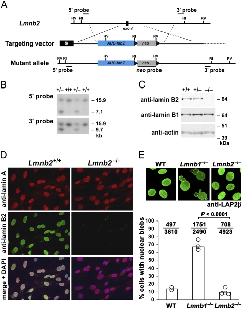
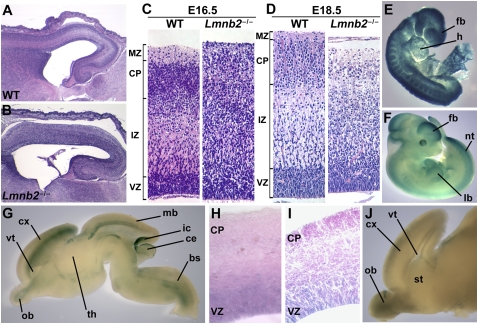
Nuclear lamins are components of the nuclear lamina, a structural scaffolding for the cell nucleus. Defects in lamins A and C cause an array of human diseases, including muscular dystrophy, lipodystrophy, and progeria, but no diseases have been linked to the loss of lamins B1 or B2. To explore the functional relevance of lamin B2, we generated lamin B2-deficient mice and found that they have severe brain abnormalities resembling lissencephaly, with abnormal layering of neurons in the cerebral cortex and cerebellum. This neuronal layering abnormality is due to defective neuronal migration, a process that is dependent on the organized movement of the nucleus within the cell. These studies establish an essential function for lamin B2 in neuronal migration and brain development.


Conflict of interest statement

The authors declare no conflict of interest.

Figures

Fig. 1.
Fig. 1.
Inactivation of mouse Lmnb2. (A) Gene-targeting strategy to replace the coding sequences of Lmnb2 exon 1 with a lacZ cassette, beginning at the translation initiation site (ATG). The positions of relevant EcoRI and EcoRV sites and the 5′, 3′, and neo probes for Southern blot analysis are shown. (B) Southern blots identifying gene-targeting events in mouse embryonic stem cells. Genomic DNA was digested with EcoRV. (C) Western blots of mouse embryonic fibroblast extracts with antibodies against lamin B1 and lamin B2. Actin was used as a loading control. (D) Immunofluorescence microscopy of Lmnb2+/+ and Lmnb2−/− embryonic fibroblasts with antibodies against lamin A and lamin B2. (Bottom) Merged images with DAPI staining for nuclear DNA. (E) Lmnb2−/− fibroblasts did not have a higher frequency of cells with nuclear blebs than WT cells. (Upper) Confocal images of Lmnb2+/+, Lmnb1−/−, and Lmnb2−/− fibroblasts stained with a monoclonal antibody against LAP2β, a protein of the inner nuclear membrane. (Lower) graph showing the percentage of cells with nuclear blebs. Each open circle represents an independent cell line (two WT, three Lmnb1−/−, and four Lmnb2−/− cell lines). The ratio above each bar indicates the number of cells with nuclear blebs over the total number of cells evaluated.
Fig. 2.
Fig. 2.
Abnormal patterning of the cerebral cortex in the setting of Lmnb2 deficiency. (AD) Lmnb2 deficiency alters neuronal layering in the cerebral cortex. H&E staining of paraffin-embedded sections of cerebral cortex from E16.5 WT (A) and Lmnb2−/− (B) embryos. (C) Higher-magnification views of sections from the same E16.5 WT and Lmnb2−/− embryos. Cortical layers are indicated on the Left, from Top to Bottom: MZ, marginal zone; CP, cortical plate; IZ, intermediate zone; VZ, ventricular zone. Neuronal progenitors proliferate in the VZ; postmitotic cells leave the VZ and migrate along glial fibers to the CP. (D) Sections from E18.5 embryos showing similar defects. (E and F) Whole-mount β-galactosidase staining of Lmnb2+/− embryos at E8.5 (E) and E11.5 (F). Staining was ubiquitous at E8.5; at E11.5, staining was prominent in the forebrain (fb), midbrain, hindbrain, limb buds (lb), tailbud, somites, neural tube (nt), and retina. heart (h). (G) Whole-mount staining of an E16.5 Lmnb2+/− brain cut sagittally; β-galactosidase expression is found in the cortex (cx), olfactory bulb (ob), midbrain (mb), brainstem (bs), inferior colliculus (ic) and superficial layer of the cerebellum (ce); ventricle (vt); hypothalamus (th). (H) 40-μm section of the cortex of an E16.5 Lmnb2+/− embryo and (I) 20-μm section of the cortex of a newborn Lmnb2+/− pup, after β-galactosidase staining, revealing Lmnb2 expression in the VZ. (J) β-Galactosidase staining of the brain of a newborn Lmnb2+/− mouse, revealing Lmnb2 expression in the ob, cx, and vt.
Fig. 3.
Fig. 3.
Lmnb2 deficiency causes defective neuronal migration in the brain. (A) Birthdating experiment demonstrating defective neuronal migration in the cortex of Lmnb2−/− embryos. Pregnant females were injected with BrdU at E13.5; brains were collected at E18.5, sectioned, and stained with DAPI and a rat monoclonal antibody against BrdU. Neurons born at E13.5, the time of the BrdU injection, stain very intensely for BrdU and were present within the lower levels of the cortical plate in WT mice. Neurons born later (therefore containing less BrdU) migrate to more superficial layers of the cortex. In Lmnb2−/− embryos, intense BrdU staining is noted in the most superficial layers of the cortical plate, consistent with an inability of the neurons born later to migrate past older BrdU-positive neurons. Arrow indicates the orientation of neuronal migration. (B) Sections from the same experiment stained with a sheep polyclonal antibody against BrdU (red) and a rat monoclonal against Ctip2, a marker for cortical layers V and VI (green). In WT embryos, neurons with the strongest BrdU staining were found in deep layers of the cortical plate together with Ctip2 staining. In contrast, in the Lmnb2−/− embryos, BrdU-positive and Ctip2-positive cells were both found in the more superficial part of the cortex. (C) Immunostaining of cortical sections of E19.5 WT and Lmnb2−/− embryos with antibodies against Ctip2 (green) and NeuN (red). In WT brains, a large number of mature neurons positive for NeuN were detected above layers V and VI (positive for Ctip2), whereas in Lmnb2−/− brains, many NeuN-positive cells were found below Ctip2-positive cells. (D) Immunostaining of cortical sections of E19.5 WT and Lmnb2−/− embryos with antibodies specific for Ctip2 (green) and FoxP1, a marker for layers III–V (red). In Lmnb2−/− embryos, FoxP1-positive neurons accumulated in lower levels of the cortex. (E) Immunostaining of cortical sections of E19.5 WT and Lmnb2−/− embryos with antibodies against Cux1, a marker for layers II–IV (green) and FoxP1 (red). In WT brains, Cux1-positive neurons were mainly located above the cells that stain for FoxP1, whereas in Lmnb2−/− brains, many Cux1-positive cells were located below FoxP1-positive cells. (F) Immunostaining of cortical sections of E19.5 WT and Lmnb2−/− embryos with antibodies specific for FoxP2, a marker for layer VI (green), and for Ctip2 (red). In WT embryos, Ctip2 staining was detected in the upper part of the FoxP2 territory, corresponding to layer V, whereas in Lmnb2−/− embryos the two cell populations were intermixed.
Fig. 4.
Fig. 4.
Abnormal cerebellar morphology in Lmnb2−/− mice. (A) Cross-sections of cerebellum in newborn WT and Lmnb2−/− mice. The cerebellum of Lmnb2−/− embryos was smooth and smaller in size; asterisks show fissures in the WT cerebellum (absent in the Lmnb2−/− cerebellum). (B) Cerebellar sections of E19.5 WT and Lmnb2−/− embryos stained with H&E (Top) and with an antibody against calbindin (Bottom). Calbindin stains the layer of Purkinje cells (arrow in WT section; layer is also visible in the H&E-stained section). Note the reduced thickness of the external granule layer in Lmnb2−/− cerebellum (arrowhead). Cp, choroid plexus. (C) Cross-section of the cerebellum from an E16.5 Lmnb2+/− embryo and a newborn Lmnb2+/− mouse (P0) after whole-mount staining for β-galactosidase. Lmnb2 expression is noted in the superficial layer of the cerebellum, the external granule layer (arrowhead) and in the vicinity of the Purkinje cell layer (arrow). (D) Immunostaining of a cerebellar section of an E17.5 Lmnb2+/− embryo with antibodies against lamin B1 (green) and lamin B2 (red). Nuclear DNA was stained with DAPI. Lamins B1 and B2 are detected in all cells, with a stronger signal in the Purkinje layer (arrow).

Comment in

Similar articles

Cited by

References

    1. Stewart CL, Roux KJ, Burke B. Blurring the boundary: The nuclear envelope extends its reach. Science. 2007;318:1408–1412. - PubMed
    1. Zheng Y, Tsai MY. The mitotic spindle matrix: A fibro-membranous lamin connection. Cell Cycle. 2006;5:2345–2347. - PubMed
    1. Dechat T, et al. Nuclear lamins: Major factors in the structural organization and function of the nucleus and chromatin. Genes Dev. 2008;22:832–853. - PMC - PubMed
    1. Hutchison CJ. Lamins: Building blocks or regulators of gene expression? Nat Rev Mol Cell Biol. 2002;3:848–858. - PubMed
    1. Worman HJ, Fong LG, Muchir A, Young SG. Laminopathies and the long strange trip from basic cell biology to therapy. J Clin Invest. 2009;119:1825–1836. - PMC - PubMed

Publication types

LinkOut - more resources