

NOTCH signaling is required for formation and self-renewal of tumor-initiating cells and for repression of secretory cell differentiation in colon cancer
- PMID: 20145124
- PMCID: PMC4010106
- DOI: 10.1158/0008-5472.CAN-09-2557
NOTCH signaling is required for formation and self-renewal of tumor-initiating cells and for repression of secretory cell differentiation in colon cancer
Abstract
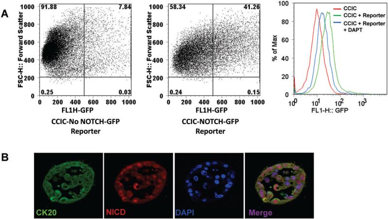
NOTCH signaling is critical for specifying the intestinal epithelial cell lineage and for initiating colorectal adenomas and colorectal cancers (CRC). Based on evidence that NOTCH is important for the maintenance and self-renewal of cancer-initiating cells in other malignancies, we studied the role of NOTCH signaling in colon cancer-initiating cells (CCIC). Tumors formed by CCICs maintain many properties of the primary CRCs from which they were derived, such as glandular organization, cell polarity, gap junctions, and expression of characteristic CRC molecular markers. Furthermore, CCICs have the property of self-renewal. In this study, we show that NOTCH signaling is 10- to 30-fold higher in CCIC compared with widely used colon cancer cell lines. Using small-molecule inhibition and short hairpin RNA knockdown, we show that NOTCH prevents CCIC apoptosis through repression of cell cycle kinase inhibitor p27 and transcription factor ATOH1. NOTCH is also critical to intrinsic maintenance of CCIC self-renewal and the repression of secretory cell lineage differentiation genes such as MUC2. Our findings describe a novel human cell system to study NOTCH signaling in CRC tumor initiation and suggest that inhibition of NOTCH signaling may improve CRC chemoprevention and chemotherapy.
Figures


Similar articles
-
NOTCH Signaling Regulates Asymmetric Cell Fate of Fast- and Slow-Cycling Colon Cancer-Initiating Cells.Cancer Res. 2016 Jun 1;76(11):3411-21. doi: 10.1158/0008-5472.CAN-15-3198. Epub 2016 Apr 11. Cancer Res. 2016. PMID: 27197180 Free PMC article.
-
The class I HDAC inhibitor MGCD0103 induces cell cycle arrest and apoptosis in colon cancer initiating cells by upregulating Dickkopf-1 and non-canonical Wnt signaling.Oncotarget. 2010 Nov;1(7):596-605. doi: 10.18632/oncotarget.194. Oncotarget. 2010. PMID: 21317455 Free PMC article.
-
Prostaglandin E2 Promotes Colorectal Cancer Stem Cell Expansion and Metastasis in Mice.Gastroenterology. 2015 Dec;149(7):1884-1895.e4. doi: 10.1053/j.gastro.2015.07.064. Epub 2015 Aug 7. Gastroenterology. 2015. PMID: 26261008 Free PMC article.
-
Colon cancer stem cells: promise of targeted therapy.Gastroenterology. 2010 Jun;138(6):2151-62. doi: 10.1053/j.gastro.2009.12.063. Gastroenterology. 2010. PMID: 20420952 Review.
-
Role of Notch signaling in colon homeostasis and carcinogenesis.Cancer Sci. 2011 Nov;102(11):1938-42. doi: 10.1111/j.1349-7006.2011.02049.x. Epub 2011 Sep 6. Cancer Sci. 2011. PMID: 21801279 Free PMC article. Review.
Cited by
-
Understanding the Principles of Pattern Formation Driven by Notch Signaling by Integrating Experiments and Theoretical Models.Front Physiol. 2020 Jul 31;11:929. doi: 10.3389/fphys.2020.00929. eCollection 2020. Front Physiol. 2020. PMID: 32848867 Free PMC article. Review.
-
Notch Signaling in Osteogenesis, Osteoclastogenesis, and Angiogenesis.Am J Pathol. 2019 Aug;189(8):1495-1500. doi: 10.1016/j.ajpath.2019.05.005. Am J Pathol. 2019. PMID: 31345466 Free PMC article. Review.
-
LGR5 positivity defines stem-like cells in colorectal cancer.Carcinogenesis. 2014 Apr;35(4):849-58. doi: 10.1093/carcin/bgt377. Epub 2013 Nov 26. Carcinogenesis. 2014. PMID: 24282287 Free PMC article.
-
Molecular identification and targeting of colorectal cancer stem cells.Oncotarget. 2010 Oct;1(6):387-395. doi: 10.18632/oncotarget.173. Oncotarget. 2010. PMID: 21311095 Free PMC article. Review.
-
Nuclear factor of activated T-cells 5 increases intestinal goblet cell differentiation through an mTOR/Notch signaling pathway.Mol Biol Cell. 2014 Sep 15;25(18):2882-90. doi: 10.1091/mbc.E14-05-0998. Epub 2014 Jul 23. Mol Biol Cell. 2014. PMID: 25057011 Free PMC article.
References
-
- Jemal A. Cancer Statistics CA: A Cancer Journal for Clinicians. 2007;57:43–66. - PubMed
-
- Korinek V, Barker N, Moerer P, et al. Depletion of epithelial stem-cell compartments in the small intestine of mice lacking Tcf-4. Nature Genetics. 1998;19:379–83. - PubMed
-
- Korinek V, Barker N, Morin PJ, et al. Constitutive Transcriptional Activation by a beta -Catenin-Tcf Complex in APC−/− Colon Carcinoma. Science. 1997;275:1784–7. - PubMed
-
- Andreu P, Colnot S, Godard C, et al. Crypt-restricted proliferation and commitment to the Paneth cell lineage following Apc loss in the mouse intestine. Development. 2005;132:1443–51. - PubMed
Publication types
MeSH terms
Substances
Grants and funding
LinkOut - more resources
Full Text Sources
Other Literature Sources
Miscellaneous
