. 2010 Feb 15;70(4):1679-88.
doi: 10.1158/0008-5472.CAN-09-2740. Epub 2010 Feb 9.

Heterogeneous nuclear ribonucleoprotein H blocks MST2-mediated apoptosis in cancer cells by regulating A-Raf transcription

Affiliations

Heterogeneous nuclear ribonucleoprotein H blocks MST2-mediated apoptosis in cancer cells by regulating A-Raf transcription

Jens Rauch et al. Cancer Res. .

Abstract

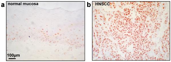
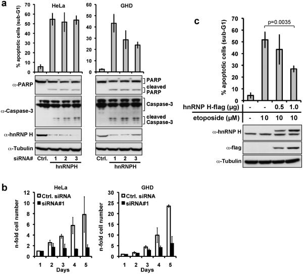
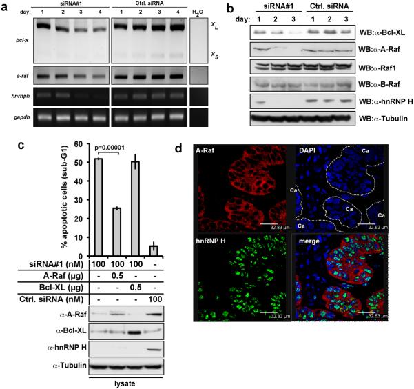
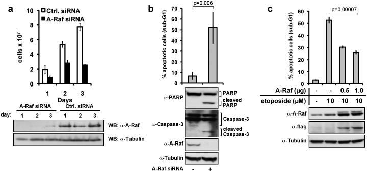
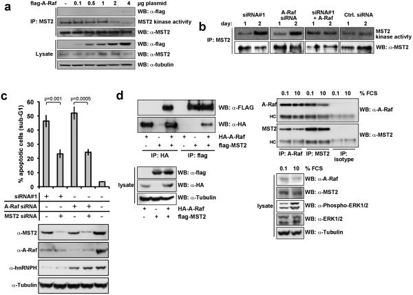
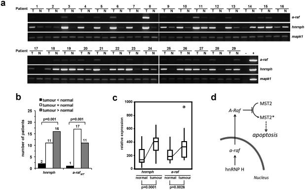
A-Raf belongs to the family of oncogenic Raf kinases that are involved in mitogenic signaling by activating the mitogen-activated protein (MAP)/extracellular signal-regulated kinase (ERK) kinase (MEK)-ERK pathway. Low kinase activity of A-Raf toward MEK suggested that A-Raf might have alternative functions. Here, we show that A-Raf prevents cancer cell apoptosis contingent on the expression of the heterogeneous nuclear ribonucleoprotein H (hnRNP H) splice factor, which is required for the correct transcription and expression of a-raf. Apoptosis was prevented by A-Raf through sequestration and inactivation of the proapoptotic MST2 kinase. Small interfering RNA-mediated knockdown of hnRNP H or A-Raf resulted in MST2-dependent apoptosis. In contrast, enforced expression of either hnRNP H or A-Raf partially counteracted apoptosis induced by etoposide. In vivo expression studies of colon specimens corroborated the overexpression of hnRNP H in malignant tissues and its correlation with A-Raf levels. Our findings define a novel mechanism that is usurped in tumor cells to escape naturally imposed apoptotic signals.


Figures

Figure 1
Figure 1. hnRNP H is overexpressed in HNSCC
HnRNP H expression was detected in normal mucosa (a) and carcinoma tissue (b) (HNSCC). Immunohistochemistry was performed on cryosections of human tissue specimens.
Figure 2
Figure 2. Knock-down of hnRNP H induces apoptosis in carcinoma cell lines
(a) HeLa cells and GHD cells were transfected with with three different hnRNP H specific siRNA oligonucleotides (siRNA#1-3) or scrambled control siRNA (Ctrl. siRNA) at 100nM final concentration. Three days after transfection, apoptosis was measured by flow cytometry assessing fragmented DNA content (subG1) (upper panel). Additionally, cells were lysed, and expression of hnRNP H, Caspase-3, and PARP was assessed by immunoblotting (lower panels). Shown are mean values with SD from three independent experiments. (b) Numbers of HeLa and GHD-1 cells were determined in a time kinetic following siRNA#1 (black bars), or control siRNA (open bars) transfection. Results represent the mean with standard deviation (SD) of three independent experiments. (c) HnRNP H overexpression partly counteracts etoposide-induced apoptosis. HeLa cells were transfected with indicated amounts of a hnRNP H expression plasmid, and treated with 10μM etoposide 24 hours later. 72 hours post transfection the proportion of apoptotic cells was assessed by measuring DNA fragmentation by flow cytometry. Shown are mean values with SD from three independent experiments (upper panel). Additionally, cells were lysed, and hnRNP H- and flag- expression was assessed by immunoblotting (lower panel).
Figure 3
Figure 3. hnRNP H controls bcl-x and a-raf mRNA levels and splicing, but only A-Raf antagonises apoptosis induced by hnRNP H depletion
(a) bcl-xl/s, a-raf, and hnRNP H mRNA expression in HeLa cells was analyzed by semi-quantitative RT-PCR four days after transfection with control (Ctrl. siRNA) or hnRNP H-specific siRNA (siRNA#1). gapdh mRNA levels were determined as loading controls. Shown is a representative result from three independent experiments. (b) Bcl-xl, A-Raf, Raf-1, B-Raf and hnRNP H protein expression was analyzed by immunoblotting three days post-transfection with control (Ctrl. siRNA) or hnRNP H-specific siRNA (siRNA#1). As loading control tubulin protein levels were determined. Shown are representative results from three independent experiments. (c) HeLa cells were co-transfected with A-Raf or Bcl-xl expression plasmids and siRNAs as indicated. Apoptosis was determined assessing DNA fragmentation by flow cytometry three days post-transfection. The data represent mean percentage of apoptosis with SD of three independent experiments (upper panel). hnRNP H- , A-Raf- and Bcl-XL-expression was assessed by immunoblotting (lower panel). (d) HnRNP H and A-Raf co-localise in primary carcinomas. HnRNP H and A-Raf were visualized using double staining with the respective antibodies.
Figure 4
Figure 4. A-Raf mediates carcinoma cell survival
(a) HeLa cells were transfected with 100nM A-Raf siRNA oligonucleotides or control siRNA. Cell numbers were assessed at the indicated time points after transfection with A-Raf or control siRNAs. Shown are mean values with SD from two independent experiments. In parallel, the expression of A-Raf was monitored in whole cell lysates for three days post transfection by immunoblotting. Shown are representative results from three independent experiments (lower panels). (b) Apoptosis was assessed by DNA fragmentation three days after transfection with A-Raf or control siRNAs. Shown are mean values with SD from three independent experiments. Additionally, cells were lysed, and expression of A-Raf, Caspase-3, and PARP was assessed by immunoblotting (lower panels). (c) A-Raf overexpression counteracts etoposide-induced apoptosis. HeLa cells were transfected with increasing amounts of an A-Raf expression plasmid (flag-tagged A-Raf), and treated with 10μM etoposide 24 hours later. 72 hrs post transfection the proportion of apoptotic cells was assessed upon measuring DNA fragmentation by flow cytometry (upper panel). Shown are mean values with SD from three independent experiments. In parallel, the expression of A-Raf was monitored in whole cell lysates by immunoblotting (lower panel).
Figure 5
Figure 5. HnRNP H and A-Raf suppress apoptosis via inhibition of MST2 kinase activation
(a) Increasing amounts of Flag-A-Raf expression plasmids were transiently transfected into HEK293 cells, and MST2 immunoprecipitates were examined for kinase activity using an in-gel kinase assay. An equal aliquot was used for immunoblotting to assure that equal amounts of MST2 had been immunoprecipitated. Lysates were immunoblotted for expression of the transfected Flag-A-Raf and tubulin as loading control. Shown are the representative results from three independent experiments. (b) HeLa cells were transiently transfected with hnRNP H (siRNA#1), A-Raf, and control siRNAs at final concentrations of 100nM. Where indicated, A-Raf expression plasmid (1.0μg) was co-transfected. The kinase activity of MST2 immunoprecipitates was assessed by in-gel kinase assays at day one and two after transfection (upper panel). The amount of MST2 immunoprecipitated from whole cell lysates was visualized by immunoblotting (lower panel). (c) HeLa cells were transfected with hnRNP H siRNA, A-Raf siRNA or MST2 siRNA as indicated. Total amounts of siRNA were adjusted using control siRNA. Three days post transfection apoptosis was determined by assessing DNA fragmentation using flow cytometry. Shown are mean values with SD from three independent experiments (upper panel). In parallel, the expression of hnRNP H, A-Raf, and MST2 was monitored in whole cell lysates by immunoblotting (lower panel). (d) A-Raf and MST2 interact in cultured cells. (left panel) Flag-tagged MST2 and HA-tagged A-Raf were transiently transfected in HEK293 cells. After 24hrs MST2 and A-Raf were immunoprecipitated (IP) with flag-tag or HA-tag specific antibodies, and analysed by immunoblotting (WB) (upper panel). Lysates were immunoblotted for expression of the transfected HA-A-Raf, flag-tagged MST2 and tubulin as loading control (lower panel) (right panel )Endogenous A-Raf and MST2 were immunoprecipitated from lysates of HeLa cells, which had been serum starved (0.1% FCS, 16 hours) or treated full medium (10% FCS) after serum-starvation using specifc antibodies for A-Raf or MST2, and analysed by immunoblotting. As an isotype control an antibody specific for Enolase was used (upper panel). Lysates were immunoblotted for expression of A-Raf, MST2, phosphorylated ERK1/2, ERK1/2, and tubulin as loading control (lower panel). HC – heavy chain immunoglobulin. Shown are the representative results from three independent experiments.
Figure 6
Figure 6. Expression of hnRNP H and a-raf in colon carcinomas and normal adjacent tissues
(a) The expression of hnRNP H and a-raf mRNA was assessed in colon tumours (T) and normal adjacent tissues (N) by semi-quantitative RT-PCR as described in materials and methods. Mapk1 mRNA levels were determined as a loading control. (b) Significantly elevated number of patients with high expression of hnRNP H and a-raf in tumours. The expression level of hnRNP H and a-raf in tumour tissue was correlated to each corresponding normal tissue after standardization of all values using mapk1. Patients were divided into three groups according to their relative expression levels of hnRNP H and a-raf mRNA in tumour tissue. Black bars (tumour ​<​normal) indicate tissues where expression in the tumour is lower than in normal tissues, white bars (tumour =normal) indicate no significant difference between normal and tumour tissue, and grey bars tumour ​>​normal) indicate tissues where expression in the tumour is significantly higher than in normal tissues. Shown are the numbers of patients in each group. The distribution of relative expression was calculated using the Chi-square test. (c) Relative expression of hnRNP H and a-raf mRNA according to disease state of the tissue. Significantly elevated expressions of hnRNP H and a-raf were observed in tumour tissues. Shown are boxplots where boxes indicate the median (line) and IQR, whiskers show the range, and asterisks indicate outliers. (d) Model for HnRNP H-mediated A-Raf transcription and regulation of MST2-Dependent Apoptosis. hnRNP H is necessary for proper splicing of mature A-Raf. A-Raf binds to MST2 and thus, is a potent inhibitor of MST2-dependent apoptosis.

Similar articles

Cited by

References

    1. Honore B, Rasmussen HH, Vorum H, et al. Heterogeneous nuclear ribonucleoproteins H, H′, and F are members of a ubiquitously expressed subfamily of related but distinct proteins encoded by genes mapping to different chromosomes. J Biol Chem. 1995;270:28780–9. - PubMed
    1. Chou MY, Rooke N, Turck CW, Black DL. hnRNP H is a component of a splicing enhancer complex that activates a c-src alternative exon in neuronal cells. Mol Cell Biol. 1999;19:69–77. - PMC - PubMed
    1. Garneau D, Revil T, Fisette JF, Chabot B. Heterogeneous nuclear ribonucleoprotein F/H proteins modulate the alternative splicing of the apoptotic mediator Bcl-x. J Biol Chem. 2005;280:22641–50. - PubMed
    1. Dominguez C, Allain FH. NMR structure of the three quasi RNA recognition motifs (qRRMs) of human hnRNP F and interaction studies with Bcl-x G-tract RNA: a novel mode of RNA recognition. Nucleic Acids Res. 2006;34:3634–45. - PMC - PubMed
    1. Wang E, Dimova N, Cambi F. PLP/DM20 ratio is regulated by hnRNPH and F and a novel G-rich enhancer in oligodendrocytes. Nucleic Acids Res. 2007;35:4164–78. - PMC - PubMed

Publication types

MeSH terms

Substances