. 2010 Apr 9;285(15):11596-606.
doi: 10.1074/jbc.M109.076984. Epub 2010 Feb 9.

Trypanosoma cruzi induces the reactive oxygen species-PARP-1-RelA pathway for up-regulation of cytokine expression in cardiomyocytes

Affiliations

Trypanosoma cruzi induces the reactive oxygen species-PARP-1-RelA pathway for up-regulation of cytokine expression in cardiomyocytes

Xueqing Ba et al. J Biol Chem. .

Abstract

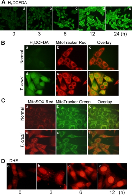
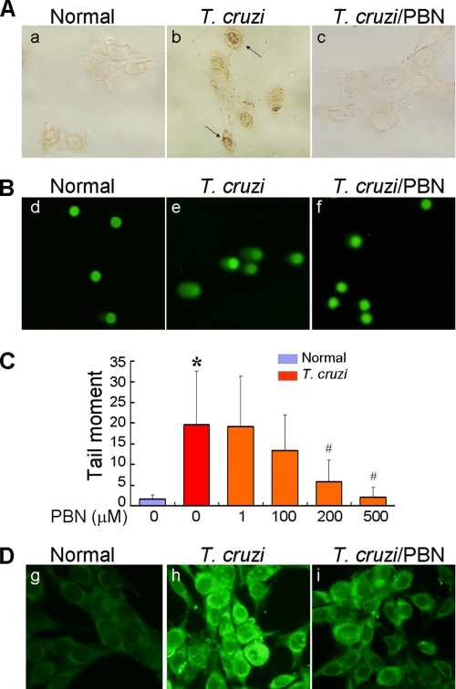
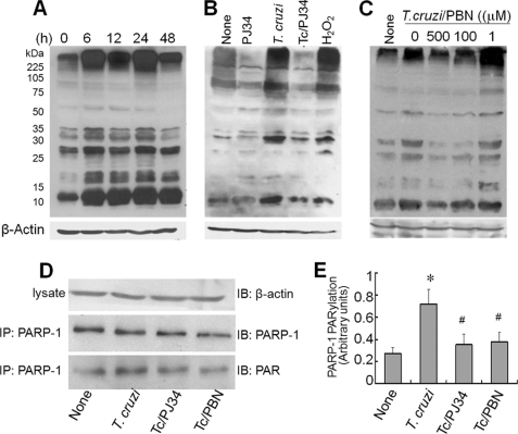
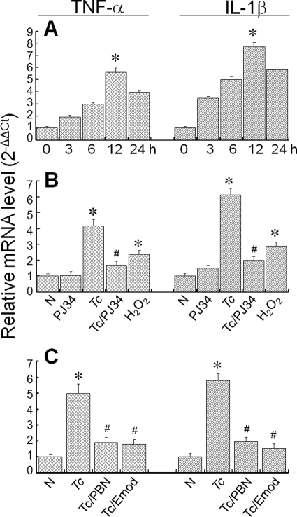
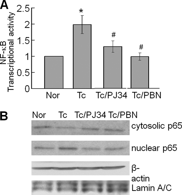
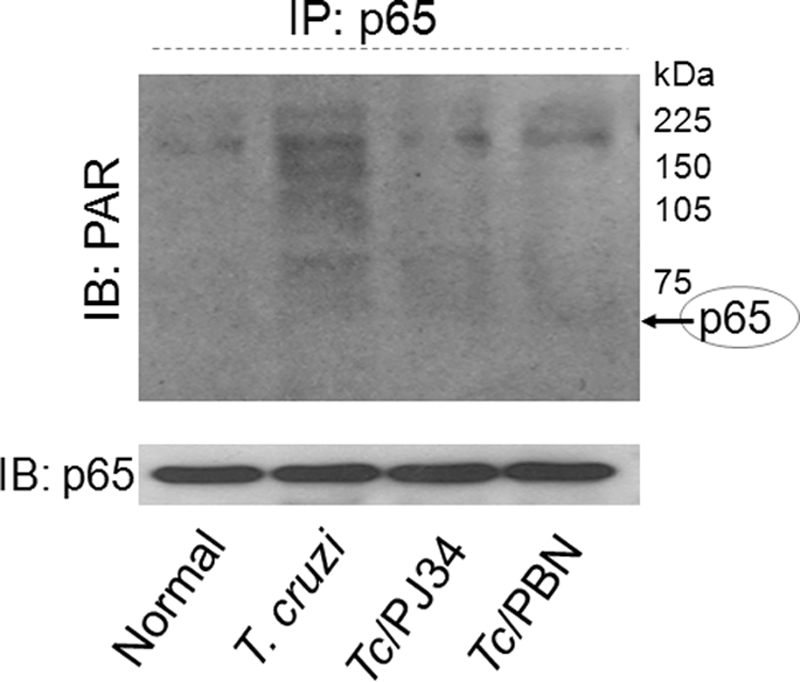
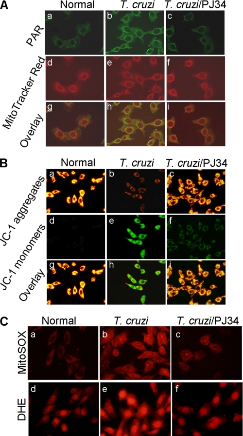
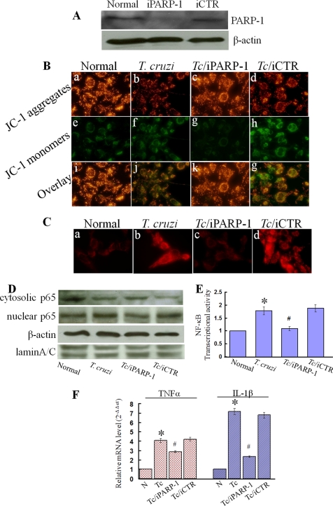
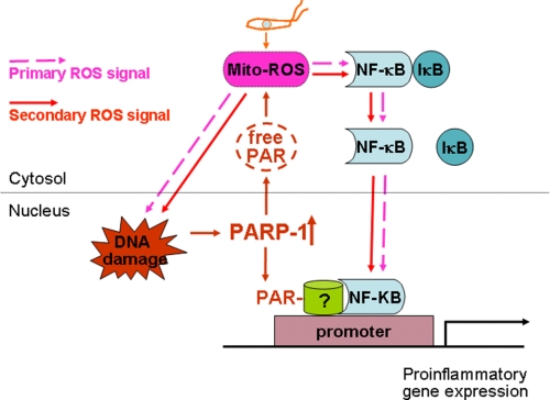
In this study, we demonstrate that human cardiomyocytes (AC16) produce reactive oxygen species (ROS) and inflammatory cytokines in response to Trypanosoma cruzi. ROS were primarily produced by mitochondria, some of which diffused to cytosol of infected cardiomyocytes. These ROS resulted in an increase in 8-hydroxyguanine lesions and DNA fragmentation that signaled PARP-1 activation evidenced by poly(ADP-ribose) (PAR) modification of PARP-1 and other proteins in infected cardiomyocytes. Phenyl-alpha-tert-butylnitrone blocked the mitochondrial ROS (mtROS) formation, DNA damage, and PARP-1 activation in infected cardiomyocytes. Further inhibition studies demonstrated that ROS and PARP-1 signaled TNF-alpha and IL-1beta expression in infected cardiomyocytes. ROS directly signaled the nuclear translocation of RelA (p65), NF-kappaB activation, and cytokine gene expression. PARP-1 exhibited no direct interaction with p65 and did not signal its translocation to nuclei in infected cardiomyocytes. Instead, PARP-1 contributed to PAR modification of p65-interacting nuclear proteins and assembly of the NF-kappaB transcription complex. PJ34 (PARP-1 inhibitor) also prevented mitochondrial poly(ADP-ribosyl)ation (PARylation) and ROS formation. We conclude that T. cruzi-mediated mtROS provide primary stimulus for PARP-1-NF-kappaB activation and cytokine gene expression in infected cardiomyocytes. PAR modification of mitochondrial membranes then results in a feedback cycle of mtROS formation and DNA damage/PARP-1 activation. ROS, either through direct modulation of cytosolic NF-kappaB, or via PARP-1-dependent PAR modification of p65-interacting nuclear proteins, contributes to cytokine gene expression. Our results demonstrate a link between ROS and inflammatory responses in cardiomyocytes infected by T. cruzi and provide a clue to the pathomechanism of sustained inflammation in Chagas disease.


Figures

FIGURE 1.
FIGURE 1.
T. cruzi-induced ROS production in AC16 cardiomyocytes. A, AC16 cardiomyocytes were infected with T. cruzi (cell:parasite ratio, 1:3). Shown are representative fluorescent micrographs of AC16 cells infected for 0–24 h and stained with H2DCFDA (detects intracellular ROS, green). B, normal (panels a–c) and infected (panels d–f) cardiomyocytes at 12 h pi were stained with H2DCFDA (panels a and d) and MitoTracker Red (red, localizes to mitochondria, panels b and e). Overlay images (panels c and f, yellow) indicated mitochondria are the major source of intracellular ROS in infected cardiomyocytes. C, overlay images (panels c and f, yellow) of cardiomyocytes stained with MitoSOX Red (red, detects mtROS, panels a and d) and MitoTracker Green (green, localizes to mitochondria, panels b and e) validates mitochondrial production of ROS in infected cardiomyocytes at 12 h pi (panels d–f). D, dihydroethidium (DHE) is oxidized by cytosolic ROS to fluorescent ethidium bromide that intercalates DNA yielding a bright red nuclear fluorescence. A gradual increase in DHE fluorescence during 0–12 h pi indicates that mtROS are diffused to cytosol in infected cardiomyocytes.
FIGURE 2.
FIGURE 2.
ROS elicit DNA oxidative damage and DNA fragmentation in T. cruzi-infected cardiomyocytes. AC16 cells (wt and infected) were treated with 500 μm PBN for 12 h. A, cardiomyocytes were fixed and permeabilized, and immunostaining was performed with anti-8-oxoG antibody (brown nuclei). B, a Comet assay was performed to detect DNA fragmentation. Shown are representative micrographs of SYBR-green-stained nuclear DNA and Comet tails in normal, infected, and PBN-treated/infected cardiomyocytes. C, quantitation of tail moment, indicative of DNA fragmentation, was performed as described under “Experimental Procedures.” Data (mean ± S.D.) are representative of three independent experiments (p < 0.01; *, normal versus T. cruzi-infected; #, infected/PBN-treated versus infected/untreated). D, PBN scavenges ROS in infected cardiomyocytes. AC16 cardiomyocytes (normal and infected) were incubated for 12 h (± PBN), labeled with 10 μm H2DCFDA (green), and ROS-specific signal detected by fluorescence microscopy.
FIGURE 3.
FIGURE 3.
ROS stimulates PARP-1 activation in cardiomyocytes infected by T. cruzi. A, PAR modification. AC16 cells were infected with T. cruzi for 0–48 h. Cell lysates (40 μg of protein) were resolved by SDS-PAGE, and protein PAR modification was detected using anti-PAR antibody. B and C, enhanced PAR modification of cellular proteins is dependent upon PARP-1 and ROS activation. AC16 cells (wt and infected) were incubated with 10 μm PJ34 (PARP-1 inhibitor), 50 μm H2O2, or 0–500 μm PBN for 12 h. Cell lysates (40 μg of protein) were resolved by SDS-PAGE, and Western blotting was performed with anti-PAR antibody. D and E, PARP-1 is PAR-modified in a ROS-dependent manner. AC16 cells were infected with T. cruzi for 12 h (±10 μm PJ34 or 500 μm PBN). Cell lysates were subjected to immunoprecipitation (IP) with anti-PARP antibody, and immunoblotting (IB) was performed with anti-PARP-1 and anti-PAR-1 antibodies. Densitometric analysis of PAR-modified PARP-1, normalized to β-actin signal, is shown in E. Data (mean ± S.D.) are representative of three independent experiments (p < 0.01; *, normal versus T. cruzi-infected; #, infected/PBN-treated versus infected/untreated). All membranes were re-probed with β-actin antibody to validate equal loading of samples.
FIGURE 4.
FIGURE 4.
ROS and PARP-1 induce cytokine gene expression in infected cardiomyocytes. A, the mRNAs for TNF-α and IL-1β are increased in infected cells. Total RNA was isolated from cardiomyocytes at 0–24 h pi, and real-time RT-PCR performed as described under “Experimental Procedures.” After normalization to β-actin mRNA, relative increase in TNF-α and IL-1β mRNA levels in infected cardiomyocytes was plotted in comparison to normal controls. Data are presented as mean ± S.D. (p < 0.01; *, normal versus T. cruzi-infected). B and C, ROS, PARP-1, and NF-κB inhibition attenuated the enhanced cytokine mRNA level in infected cardiomyocytes. Cardiomyocytes (normal and infected) were incubated for 12 h in the presence or absence of 10 μm PJ34, 50 μm H2O2, 500 μm PBN, or 50 μg/ml emodin (inhibits NF-κB activation), and a real-time RT-PCR was performed to measure the TNF-α and IL-1β mRNA levels. Data (mean ± S.D.) are representative of triplicate experiments (p < 0.01; *, normal versus T. cruzi-infected; #, infected/PBN-treated versus infected/untreated).
FIGURE 5.
FIGURE 5.
A, ROS and PARP-1 signal NF-κB activation in T. cruzi-infected cardiomyocytes. AC16 cells were transiently transfected with pNF-κB-luc (with 5× NF-κB binding site). Cells were infected with T. cruzi and incubated in the presence or absence of 10 μm PJ34 or 500 μm PBN for 12 h. The relative NF-κB transcriptional activity was measured by firefly luciferase activity normalized to Renilla luciferase activity. The transcriptional activity of NF-κB in normal cells was considered as baseline and valued 1 or 100%. Data (mean ± S.D.) are representative of three independent experiments (p < 0.01; *, normal versus T. cruzi-infected; #, infected/PBN-treated versus infected/untreated). B, PARP-1 and ROS inhibitors decreased the nuclear translocation of RelA (p65) in infected cardiomyocytes. AC16 cells (wt and infected, ± PJ34 or PBN) were incubated for 12 h, and cytosolic and nuclear fractions were prepared as described under “Experimental Procedures.” The p65 expression level was detected by immunoblotting with specific antibody. Membranes were re-probed with antibody to β-actin and Lamin A/C to normalize the cytosolic and nuclear fractions, respectively.
FIGURE 6.
FIGURE 6.
PAR modification of p65-associated proteins was enhanced in an ROS-dependent manner. AC16 cells (normal and infected) were incubated in the presence of 10 μm PJ34 or 500 μm PBN for 12 h. Immunoprecipitation was performed with RelA (p65)-specific antibody. Immune complexes were resolved on 10% SDS-PAGE, and immunoblotting was performed using antibodies specific for PAR polymer and RelA (p65) subunit of the NF-κB complex.
FIGURE 7.
FIGURE 7.
PARP-1 activation contributes to a loss of mitochondrial membrane potential and enhanced ROS production in infected cardiomyocytes. AC16 cells (normal and infected) were incubated in presence or absence of 10 μm PJ34 for 12 h. A, shown are the representative fluorescence micrographs of normal (panels a, d, and g), T. cruzi-infected (panels b, e, and h), and infected/PJ34-treated (panels c, f, and i) cardiomyocytes, stained with anti-PAR antibody (panels a–c, green) and MitoTracker Red (localizes to mitochondria, panels d–f). Overlay images (panels g–i, yellow) demonstrate mitochondrial localization of PAR in infected cardiomyocytes. B, shown are the representative fluorescence micrographs of JC-1-stained wt and infected cardiomyocytes (±PJ34). Note the accumulation of green monomers in infected cardiomyocytes (panel e) was inhibited by PJ34 (panel f). Overlay images (panels g–i) show that a majority of mitochondria fluoresced red in normal (panel g) and PJ34-treated/infected cardiomyocytes (panel i), whereas mitochondria in infected cardiomyocytes fluoresced green (panels h). C, shown are fluorescent micrographs of wt and infected cardiomyocytes (±PJ34) stained with MitoSOX Red (detects mitochondrial O2˙̄, panels a–c), and DHE (detects intracellular/cytosolic ROS, panels d–f, red) probes. Note that mitochondrial and cytosolic ROS were decreased by PJ34 treatment of infected cardiomyocytes.
FIGURE 8.
FIGURE 8.
Genetic interference of PARP-1 rescued mitochondrial membrane potential, reduced mitochondrial ROS, and decreased NF-κB-mediated transcription of inflammatory genes in infected cardiomyocytes. A, AC16 cells were transfected with small RNAi of PARP-1 (iPARP-1) or negative control (iCTR) for 48 h, and immunoblotting was performed with anti-PARP-1 and anti-β-actin (loading control) antibodies. B–F, cells were transfected with PARP-1 or control RNAi (as above) and infected with T. cruzi for 12-h (B) JC-1 staining. Note the accumulation of green monomers and the loss of red aggregates in infected cardiomyocytes (panels b, f, and j) was inhibited by PARP-1 RNAi (panels c, g, and k) but not control RNAi (panels d, h, and g). C, MitoSOX Red staining. Note the mitochondrial ROS in infected cardiomyocytes (panel b) was inhibited by PARP-1 RNAi (panel c) but not control RNAi (panel d). D and E, the cytosolic and nuclear fractions of wt and infected cardiomyocytes (±PARP-1 or control RNAi) were prepared as described under “Experimental Procedures.” The p65 expression level was detected by immunoblotting with specific antibody. Membranes were re-probed with antibody to β-actin and Lamin A/C. Densitometric analysis of cytosolic and nuclear p65, normalized to β-actin and Lamin A/C signal, respectively, is shown in panel E. F, a real-time RT-PCR was performed to measure the TNF-α and IL-1β mRNA levels in wt and infected cardiomyocytes (±PARP-1 or control RNAi). Data (mean ± S.D.) are representative of triplicate experiments (p < 0.01; *, normal versus T. cruzi-infected; #, infected/PARP-1-interfered versus infected/untreated).
FIGURE 9.
FIGURE 9.
Potential mechanism of ROS-PARP-1-RelA signaling of cytokine gene expression in T. cruzi-infected cardiomyocytes. ROS induced by T. cruzi infection elicit nuclear DNA damage and PARP-1 activation that then sustains an autocatalytic cycle of mitochondrial and intracellular ROS production, PARP-1 activation, and PAR formation. ROS (produced in response to T. cruzi infection) and by PARP-1/PAR-dependent mitochondrial alterations provide primary and secondary stimuli, respectively, for nuclear translocation of NF-κB, NF-κB activation, and cytokine gene expression. PARP-1 activation and enhanced PAR modification of p65-associated proteins may also signal optimal transcriptional activation of NF-κB-dependent pro-inflammatory gene expression.

Similar articles

Cited by

References

    1. World Health Organization (2006) Report of the Scientific Working Group on Chagas Disease, UNDP/World Bank/WHO
    1. Tanowitz H. B., Machado F. S., Jelicks L. A., Shirani J., de Carvalho A. C., Spray D. C., Factor S. M., Kirchhoff L. V., Weiss L. M. (2009) Prog. Cardiovasc. Dis. 51, 524–539 - PMC - PubMed
    1. Talvani A., Rocha M. O., Barcelos L. S., Gomes Y. M., Ribeiro A. L., Teixeira M. M. (2004) Clin. Infect. Dis. 38, 943–950 - PubMed
    1. Machado F. S., Souto J. T., Rossi M. A., Esper L., Tanowitz H. B., Aliberti J., Silva J. S. (2008) Microbes Infect. 10, 1558–1566 - PMC - PubMed
    1. Rossi M. A., Ramos S. G., Bestetti R. B. (2003) Front. Biosci. 8, 94–109 - PubMed

Publication types