. 2010 Apr 9;285(15):11489-97.
doi: 10.1074/jbc.M109.074088. Epub 2010 Feb 9.

Mechanistic differences between two conserved classes of small heat shock proteins found in the plant cytosol

Affiliations

Mechanistic differences between two conserved classes of small heat shock proteins found in the plant cytosol

Eman Basha et al. J Biol Chem. .

Abstract

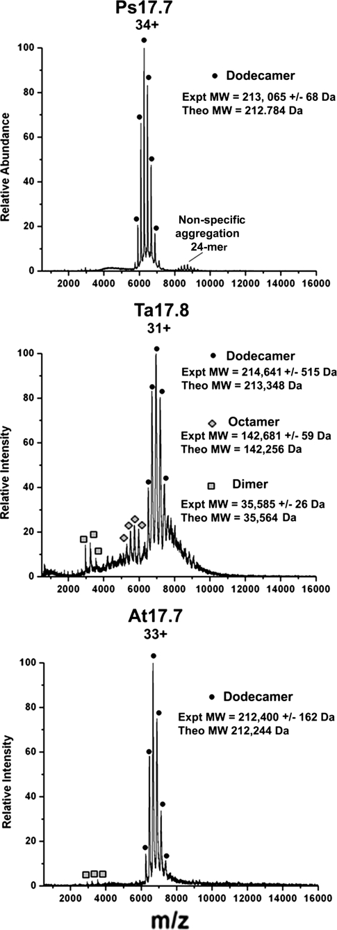
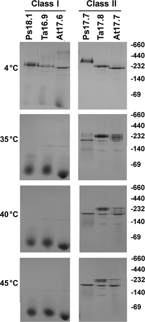
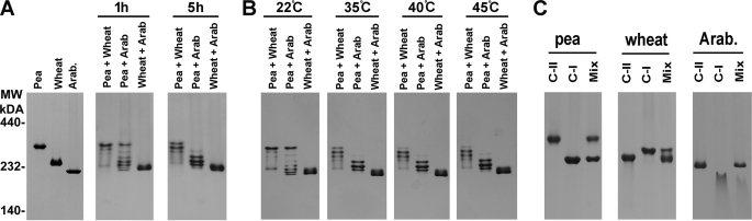
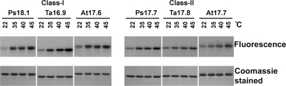
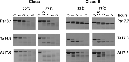
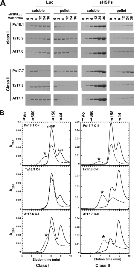
The small heat shock proteins (sHSPs) and alpha-crystallins are highly effective, ATP-independent chaperones that can bind denaturing client proteins to prevent their irreversible aggregation. One model of sHSP function suggests that the oligomeric sHSPs are activated to the client-binding form by dissociation at elevated temperatures to dimers or other sub-oligomeric species. Here we examine this model in a comparison of the oligomeric structure and chaperone activity of two conserved classes of cytosolic sHSPs in plants, the class I (CI) and class II (CII) proteins. Like the CI sHSPs, recombinant CII sHSPs from three divergent plant species, pea, wheat, and Arabidopsis, are dodecamers as determined by nano-electrospray mass spectrometry. While at 35 to 45 degrees C, all three CI sHSPs reversibly dissociate to dimers, the CII sHSPs retain oligomeric structure at high temperature. The CII dodecamers are, however, dynamic and rapidly exchange subunits, but unlike CI sHSPs, the exchange unit appears larger than a dimer. Differences in dodecameric structure are also reflected in the fact that the CII proteins do not hetero-oligomerize with CI sHSPs. Binding of the hydrophobic probe bis-ANS and limited proteolysis demonstrate CII proteins undergo significant, reversible structural changes at high temperature. All three recombinant CII proteins more efficiently protect firefly luciferase from insolubilization during heating than do the CI proteins. The CI and CII proteins behave strictly additively in client protection. In total, the results demonstrate that different sHSPs can achieve effective protection of client proteins by varied mechanisms.


Figures

FIGURE 1.
FIGURE 1.
Class II sHSPs are dodecameric. Nano-electrospray ionization mass spectra of the class II sHSPs showing the charge species envelope. Calculated experimental and theoretical masses are listed. Deviation of the experimental mass from the calculated mass is due to retention of buffer and water solvent molecules under the gentle nano-electrospray ionization conditions used, which are required to retain oligomeric structure.
FIGURE 2.
FIGURE 2.
Class II sHSP oligomers do not dissociate to stable dimers. The indicated sHSPs (15 μl of 24 μm) were separated on 4–20% acrylamide blue-native gels maintained at the indicated temperatures. Gels were stained with Coomassie Blue. Positions of protein molecular weight markers in kDa are shown on the right.
FIGURE 3.
FIGURE 3.
Class II sHSPs form unique hetero-oligomers. Non-denaturing PAGE was used to examine oligomeric species of purified sHSPs alone and after mixing for different times or at different temperatures. sHSPs (24 μm) were mixed together to a final concentration of 12 μm each in 25 mm Tris pH 7.5 and 15 μl were loaded on the gel. Gels were stained with Coomassie Blue. A, left panel, each class II sHSP analyzed alone. Middle and right panels, mixed proteins were incubated at room temperature for 1 or 5 h as indicated, before electrophoresis. B, mixed proteins were incubated at the indicated temperatures for 15 min before electrophoresis. C, indicated CI and CII sHSPs were separated alone or after mixing at room temperature. Separation of all samples was on 4–15% PAGE.
FIGURE 4.
FIGURE 4.
CI and CII sHSPs bind bis-ANS at elevated temperatures. 100 μl of 12 μm sHSPs were incubated for 60 min at the indicated temperature. The fluorescent probe bis-ANS was added at a 10-fold molar excess for the last 10 min of treatment. The samples were subjected to UV cross-linking by irradiation for 10 min at 256 nm. Following cross-linking 20 μl of each sample was run on a 10–17% gradient SDS-PAGE gel. Bis-ANS binding was visualized by fluorescence under UV illumination, and the gel was stained with Coomassie Blue to show equivalent amount of protein.
FIGURE 5.
FIGURE 5.
CI and CII proteins exhibit altered conformation at 37 °C as revealed by partial proteolysis. 24 μm protein was incubated with trypsin at a 1:400 w/w ratio (trypsin:sHSP) in low salt buffer at either room temperature (22 °C) or 37 °C for the indicated times (hr). Treatments were stopped by boiling in SDS sample buffer. Samples (20 μl) were loaded on 10–17% acrylamide gradient SDS gels and stained with Coomassie Blue. Note that shorter times were used for the 37 °C digestions to visualize comparable fragmentation.
FIGURE 6.
FIGURE 6.
CII sHSP effectively protect Luc from heat-induced aggregation and form stable sHSP-Luc complexes. A, 1 μm Luc was incubated for 8 min at 42 °C with sHSPs at the molar ratios as indicated. Samples were separated into soluble and pellet fractions and equal fraction volumes were analyzed by SDS-PAGE. Gels were stained with Coomassie Blue. Panels on the left show the amount of soluble or pelleted Luc, whereas panels on the right show soluble or pelleted sHSP. Protection of Luc is evident from the amount found in the soluble fraction compared with the control in the absence of sHSP. With the exception of PsHsp17.1, all the sHSP remains soluble with heating in the presence of substrate. B, sHSP-Luc complexes separated by size exclusion chromatography. 1 μm Luc was incubated with 12 μm sHSPs for 8 min at 22 °C (solid line) or 42 °C (dashed line), except PsHsp17.7 C-II, which was used at 6 μm. Insoluble material was removed by centrifugation, and the supernatant was loaded onto TSKgel G5000 PWXL column at room temperature and eluted in low salt buffer at a flow rate of 1 ml/min. Asterisks mark positions of the sHSP-Luc complexes. Note that in heated samples there is no longer a peak at the position of native Luc, because Luc is either associated with the sHSP, or insoluble and removed by centrifugation prior to loading on the column. Arrows indicate elution of MW standards, left to right: Vo (void volume), 660, 158, and 44 kDa.
FIGURE 7.
FIGURE 7.
Protection of client proteins by CI and CII proteins is additive. SDS-PAGE and Coomassie Blue staining of the soluble and pellet fractions obtained after heating of Luc or MDH with sHSPs. A, 1 μm Luc was incubated for 8 min at 42 °C with the indicated ratio of sHSP, using a combination of Arabidopsis CI and CII sHSPs mixed at ratios of 4:0, 3:1, 2:2, 1:3, 0:4. B, aggregation-protection of MDH. 3 μm MDH was incubated with sHSPs at the indicated ratios for 1 h at 45 °C. After heating, the mixtures were separated into soluble and pellet fractions and equal fraction volumes were analyzed. Only Luc and MDH are shown. sHSPs in the samples were fully soluble under all conditions (not shown).

Similar articles

Cited by

References

    1. van Montfort R., Slingsby C., Vierling E. (2001) Adv. Protein Chem. 59, 105–156 - PubMed
    1. Lee G. J., Pokala N., Vierling E. (1995) J. Biol. Chem. 270, 10432–10438 - PubMed
    1. Lee G. J., Roseman A. M., Saibil H. R., Vierling E. (1997) EMBO J. 16, 659–671 - PMC - PubMed
    1. Haslbeck M., Walke S., Stromer T., Ehrnsperger M., White H. E., Chen S., Saibil H. R., Buchner J. (1999) EMBO J. 18, 6744–6751 - PMC - PubMed
    1. van Montfort R. L., Basha E., Friedrich K. L., Slingsby C., Vierling E. (2001) Nat. Struct. Biol. 8, 1025–1030 - PubMed

Publication types

MeSH terms

Substances

LinkOut - more resources