. 2010 Apr;95(4):542-50.
doi: 10.3324/haematol.2009.010736. Epub 2010 Feb 9.

Hematopoietic stem cells in co-culture with mesenchymal stromal cells--modeling the niche compartments in vitro

Affiliations

Hematopoietic stem cells in co-culture with mesenchymal stromal cells--modeling the niche compartments in vitro

Duohui Jing et al. Haematologica. 2010 Apr.

Abstract

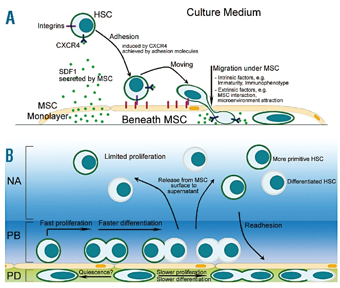
Background: Hematopoietic stem cells located in the bone marrow interact with a specific microenvironment referred to as the stem cell niche. Data derived from ex vivo co-culture systems using mesenchymal stromal cells as a feeder cell layer suggest that cell-to-cell contact has a significant impact on the expansion, migratory potential and 'stemness' of hematopoietic stem cells. Here we investigated in detail the spatial relationship between hematopoietic stem cells and mesenchymal stromal cells during ex vivo expansion.

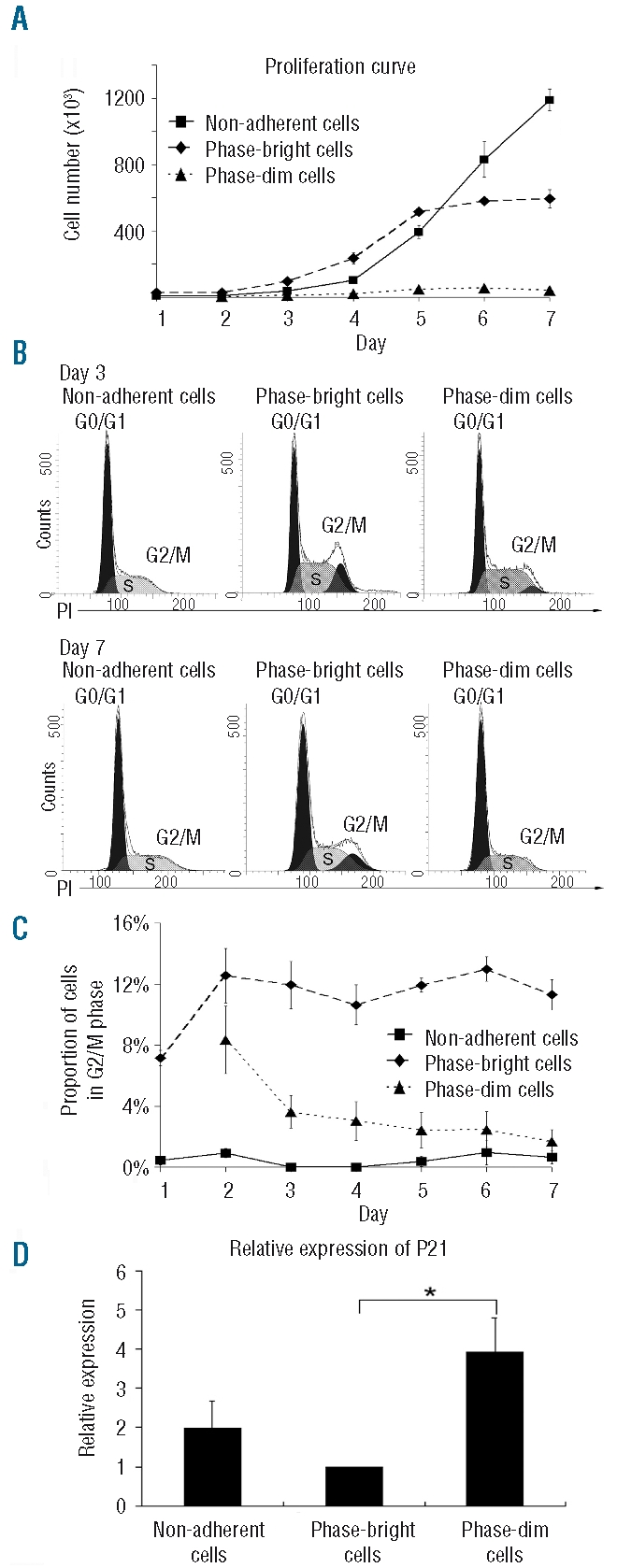
Design and methods: In the co-culture system, we defined three distinct localizations of hematopoietic stem cells relative to the mesenchymal stromal cell layer: (i) those in supernatant (non-adherent cells); (ii) those adhering to the surface of mesenchymal stromal cells (phase-bright cells) and (iii) those beneath the mesenchymal stromal cells (phase-dim cells). Cell cycle, proliferation, cell division and immunophenotype of these three cell fractions were evaluated from day 1 to 7.

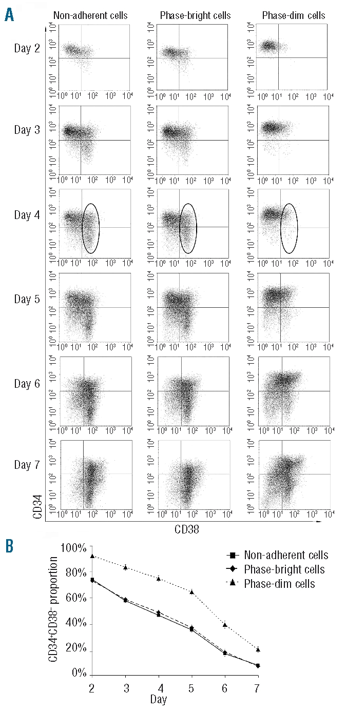
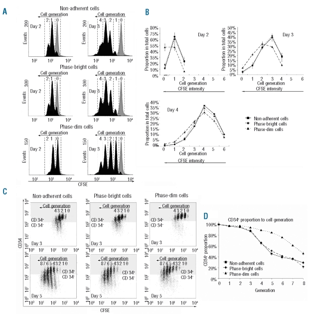
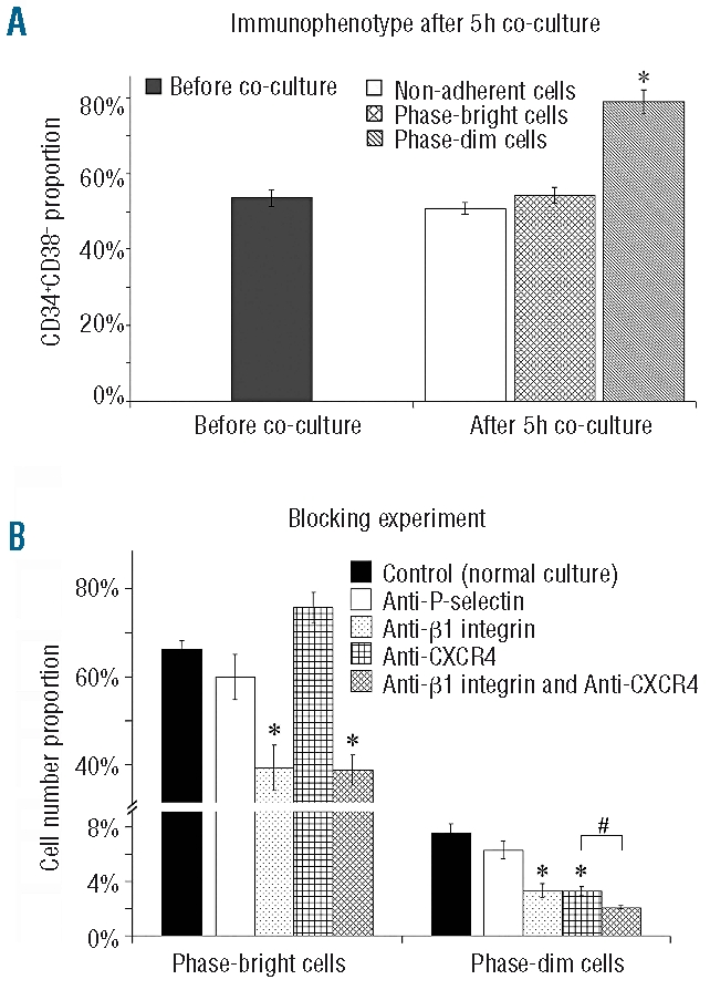
Results: Phase-bright cells contained the highest proportion of cycling progenitors during co-culture. In contrast, phase-dim cells divided much more slowly and retained a more immature phenotype compared to the other cell fractions. The phase-dim compartment was soon enriched for CD34(+)/CD38(-) cells. Migration beneath the mesenchymal stromal cell layer could be hampered by inhibiting integrin beta1 or CXCR4.

Conclusions: Our data suggest that the mesenchymal stromal cell surface is the predominant site of proliferation of hematopoietic stem cells, whereas the compartment beneath the mesenchymal stromal cell layer seems to mimic the stem cell niche for more immature cells. The SDF-1/CXCR4 interaction and integrin-mediated cell adhesion play important roles in the distribution of hematopoietic stem cells in the co-culture system.


Figures

Figure 1.
Figure 1.
Distinct compartmentalization of HSC cultured on MSC. (A, B) Scanning electron microscopy analysis revealed that HSC are either above the MSC layer (black asterisks) or beneath (white arrow). HSC migrating underneath the MSC layer were also observed (outlined white asterisks). (C) Individual frames taken from a time-lapse video depict an individual HSC moving above (black arrow) and below (white arrow) the MSC layer for a period of 54 min. M indicates MSC. (D) Phase-contrast microscopy analysis shows that HSC located above or beneath the MSC layer appeared as phase-bright and - dim cells, respectively. (E) Confocal laser scanning microscopy after immunolabeling for CD45. (F) Phase-bright cells on the surface of the MSC layer. (G) MSC layer. (H) Phase-dim cells beneath the MSC layer. (I) Phase contrast microscopy of the cells beneath the MSC layer (phase-dim cells), after removal of the phase-bright cells. (J) A cross-section is shown using immunofluorescence imaging (actin, green; nucleus, blue; CD45, red). Scale bar: 20 μm.
Figure 2.
Figure 2.
Cell proliferation and cell cycle status. (A) Proliferation kinetics of the three cell fractions (N=4). (B) Representative propidium iodide (PI) staining of cells from the three distinct compartments at day 3 and 7. (C) Dynamics of the three cell fractions in G2/M phase during 1 week of co-culture (N=3). (D) Relative expression of p21 in the three compartments at day 5 (N=3, *P<0.05). The data were normalized to the p21 expression in phase-bright cells, which was arbitrarily set at one.
Figure 3.
Figure 3.
Immunophenotype of the cells in the three compartments. (A) Representative FACS analysis of the three cell fractions from day 2 to 7. The CD38+ cell fraction at day 4 is shown within the circle. (B) Proportion of CD34+CD38 cells in the three cell fractions during 1 week of co-culture (N=7).
Figure 4.
Figure 4.
Tracking cell division in the three distinct compartments using CFSE staining. (A) Representative FACS analysis of CFSE staining at days 2 and 3. Cell generations were identified according to the control cells which were treated with mitomycin at day 0 (gray peak). (B) Distributions of cell generations of the three cell fractions at days 2, 3 and 4 (N=3). The pattern of CD34 expression in the different cell generations was studied. (C) Representative FACS analysis of CD34 and CFSE double staining at days 3 and 5. The dots in the gray region represent cells positive for CD34, the dots in the white region represent cells negative for CD34. Cell generations were identified according to the control cells which were treated with mitomycin at day 0. (D) CD34+ expression over eight cell generations within the three cell fractions (N = 8).
Figure 5.
Figure 5.
(A) Enrichment of CD34+CD38cells in the phase-dim fraction. Cells containing around 50% CD34+CD38 cells were plated on the MSC layer. After 5 h the proportion of CD34+CD38 cells among the phase-dim fraction was enriched up to 80% (N=4, *P<0.01). (B) Blocking experiments for P-selectin, integrin β1 and CXCR4. By blocking integrin β1 or CXCR4 or both, percentages of phase-bright or phase-dim cells were down-regulated (N = 4, *P<0.01 (relative to control); #P<0.05 (relative to individual blocking for integrin β1 or CXCR4).
Figure 6.
Figure 6.
Graphic of the in vitro HSC/MSC co-culture system. (A) Migration towards and retention of HSC to MSC is mediated by SDF1/CXCR4 and adhesion molecules such as integrins. (B) The MSC layer serves in vitro as a boundary between two distinct compartments, i.e. the MSC surface mediating cell proliferation and a niche-like compartment beneath the layer. Here beneath the layer, phase-dim (PD) cells show a delayed cell-cycle activity and a more immature phenotype. In contrast, phase-bright (PB) HSC on the MSC surface revealed significantly more proliferation activity. We assume that cells from the MSC surface are released into the supernatant, the third microenvironment (non-adherent cells, NA) in the co-culture system. The graphic illustrates the dynamic interplay of HSC in the three compartments.

Similar articles

Cited by

References

    1. Osawa M, Hanada K, Hamada H, Nakauchi H. Long-term lymphohematopoietic reconstitution by a single CD34-low/negative hematopoietic stem cell. Science. 1996;273(5272):242–5. - PubMed
    1. Akashi K, Traver D, Miyamoto T, Weissman IL. A clonogenic common myeloid progenitor that gives rise to all myeloid lineages. Nature. 2000;404(6774):193–7. - PubMed
    1. Metcalf D. On hematopoietic stem cell fate. Immunity. 2007;26(6):669–73. - PubMed
    1. Gordon MY. Stem cells for regenerative medicine – biological attributes and clinical application. Exp Hematol. 2008;36(6):726–32. - PubMed
    1. Sorrentino BP. Clinical strategies for expansion of haematopoietic stem cells. Nat Rev Immunol. 2004;4(11):878–88. - PubMed

Publication types