Review
. 2010 Jun;95(6):1004-15.
doi: 10.3324/haematol.2009.017079. Epub 2010 Feb 9.

PIM serine/threonine kinases in the pathogenesis and therapy of hematologic malignancies and solid cancers

Affiliations
Review

PIM serine/threonine kinases in the pathogenesis and therapy of hematologic malignancies and solid cancers

Laurent Brault et al. Haematologica. 2010 Jun.

Abstract

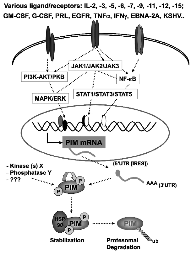
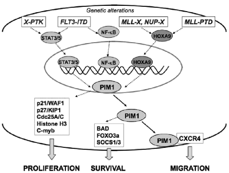
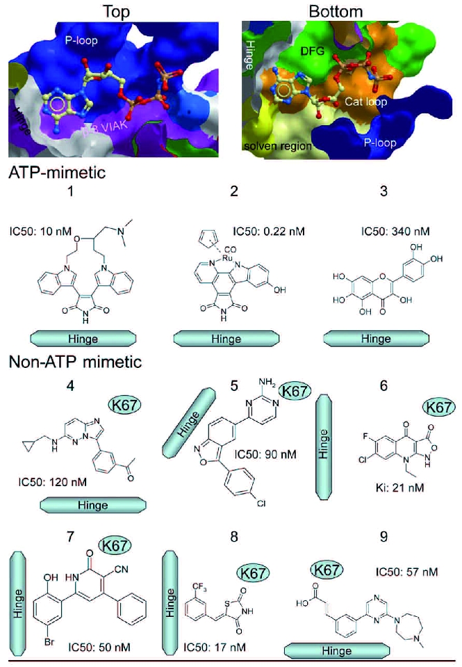
The identification as cooperating targets of Proviral Integrations of Moloney virus in murine lymphomas suggested early on that PIM serine/threonine kinases play an important role in cancer biology. Whereas elevated levels of PIM1 and PIM2 were mostly found in hematologic malignancies and prostate cancer, increased PIM3 expression was observed in different solid tumors. PIM kinases are constitutively active and their activity supports in vitro and in vivo tumor cell growth and survival through modification of an increasing number of common as well as isoform-specific substrates including several cell cycle regulators and apoptosis mediators. PIM1 but not PIM2 seems also to mediate homing and migration of normal and malignant hematopoietic cells by regulating chemokine receptor surface expression. Knockdown experiments by RNA interference or dominant-negative acting mutants suggested that PIM kinases are important for maintenance of a transformed phenotype and therefore potential therapeutic targets. Determination of the protein structure facilitated identification of an increasing number of potent small molecule PIM kinase inhibitors with in vitro and in vivo anticancer activity. Ongoing efforts aim to identify isoform-specific PIM inhibitors that would not only help to dissect the kinase function but hopefully also provide targeted therapeutics. Here, we summarize the current knowledge about the role of PIM serine/threonine kinases for the pathogenesis and therapy of hematologic malignancies and solid cancers, and we highlight structural principles and recent progress on small molecule PIM kinase inhibitors that are on their way into first clinical trials.


Figures

Figure 1.
Figure 1.
Regulation of PIM1 expression. Binding of several ligands leads to activation of a complex network of signaling pathways that results in upregulation of PIM1 mRNA. Binding of PIM1 to heat shock protein 90 (HSP90) protects from proteosomal degradation. Most experimental data has been generated using PIM1; very little is known about regulation of PIM2 and PIM3. There is increasing evidence for modification of PIM kinases through as yet unkown protein kinases and/or phosphatases.
Figure 2.
Figure 2.
Human PIM kinases amino acid sequences alignment. ClustalW2 alignment with protein sequence from Swiss-Prot (accession numbers: P11309 (PIM1), Q9P1W9 (PIM2) and Q86V86 (PIM3)). The kinase domain is shown in gray; the ATP binding domain in orange. S (Serine, = red), T (Threonine, =blue), Y (Tyrosine, =green) are potential phosphorylation sites. Reported phosphorylation sites are shown in yellow. Reported mutation in a case of colon cancer is indicated in black (PIM1Y144H); reported point mutations in DBA cases are shown in pink (PIM1C17Y and PIM1P311T); residues that have been reported to be modified by somatic hypermutation in human B-cell malignancies are marked in blue
Figure 3.
Figure 3.
Potential downstream substrates of over-expressed PIM1 in hematologic malignancies. Functionally collaborating genetic alterations such as mutated protein tyrosine kinases and fusion genes involving transcriptional regulators such as mixed lineage leukemia (MLL) lead to constitutive activation of major signaling mediators, like STAT5 or HOXA9, shown to be transcriptional activators of PIM1. Elevated PIM1 levels support cellular proliferation through modification of cell cycle regulators, survival through modification of regulators of apoptosis, as well as homing and migration through modification of the CXCR4 chemokine receptor.
Figure 4.
Figure 4.
Structural aspects of PIM1. (A) Structural overview of the PIM1 crystal structure in complex with the non-hydrolysable ATP analogue AMPPNP. Regulatory elements (the phosphate binding (P)-loop, the magnesium binding motif DFG, the hinge region, the catalytic loop and the activation segment (A)-loop and the β-hairpin insert) are labelled and highlighted using different colours. Some conserved secondary structure elements are labelled. (B) Details of the ATP binding site (boxed in A) showing the main interactions formed by the co-factor with PIM1. The unusual presence of a proline residue (P123) prevents formation of a second hydrogen bond with ATP. (C) Details of the interaction of the phosphate moieties of AMPP-NP and Mg2+ with the D186FG motif, the conserved lysine (K67) and glutamate (E89) and the catalytic aspartate (D167). The enlarged region corresponds to the boxed area in B.
Figure 5.
Figure 5.
Classes of identified small molecule PIM kinase inhibitors. Structural elements contributing to the PIM1 ATP binding site are shown in the upper 2 panels. The binding surface of the upper lobe (top) and lower lobe (bottom) are shown. Contributions to the binding surface are colour coded: hinge (gray), P-loop (blue), beta 3 and VIAK motif (magenta), solvent region (yellow) and catalytic loop (orange). Examples of the main chemical classes of inhibitors are shown in the lower panel (see text for details). Inhibitors may interact with the kinase hinge region in an ATP mimetic (forming hydrogen bonds with the hinge backbone) or non-ATP mimetic way. The binding mode of each inhibitor is indicated by showing its orientation towards the kinase hinge region and the active site lysine (K67) which offers an alternative anchor point for non-ATP mimetic inhibitors. No experimental structures are available for the compounds 6, 7 and 8 and the shown binding mode has been determined by comparison with known PIM inhibitor complexes, so far unpublished structural data and docking.

Similar articles

Cited by

References

    1. Cuypers HT, Selten G, Quint W, Zijlstra M, Maandag ER, Boelens W, et al. Murine leukemia virus-induced T-cell lymphoma-genesis: integration of proviruses in a distinct chromosomal region. Cell. 1984;37(1):141–50. - PubMed
    1. Selten G, Cuypers HT, Boelens W, Robanus-Maandag E, Verbeek J, Domen J, et al. The primary structure of the putative oncogene pim-1 shows extensive homology with protein kinases. Cell. 1986;46(4):603–11. - PubMed
    1. van Lohuizen M, Verbeek S, Krimpenfort P, Domen J, Saris C, Radaszkiewicz T, et al. Predisposition to lymphomagenesis in pim-1 transgenic mice: cooperation with c-myc and N-myc in murine leukemia virus-induced tumors. Cell. 1989;56(4):673–82. - PubMed
    1. Berns A, Breuer M, Verbeek S, van Lohuizen M. Transgenic mice as a means to study synergism between oncogenes. Int J Cancer Suppl. 1989;4:22–5. - PubMed
    1. Saris CJ, Domen J, Berns A. The pim-1 oncogene encodes two related protein-serine/threonine kinases by alternative initiation at AUG and CUG. Embo J. 1991;10(3):655–64. - PMC - PubMed

Publication types

MeSH terms

Substances