

Establishment and characterization of a new hypoxia-resistant cancer cell line, OCUM-12/Hypo, derived from a scirrhous gastric carcinoma
- PMID: 20145613
- PMCID: PMC2833244
- DOI: 10.1038/sj.bjc.6605543
Establishment and characterization of a new hypoxia-resistant cancer cell line, OCUM-12/Hypo, derived from a scirrhous gastric carcinoma
Abstract
Background: Many kinds of solid tumour have heterogeneously a hypoxic environment. Tumour hypoxia reported to be associated with more aggressive tumour phenotypes such as high metastatic ability and resistance to various anti-cancer therapies which may lead to a poorer prognosis. However, the mechanisms by which hypoxia affects the aggressive phenotypes remain unclear.
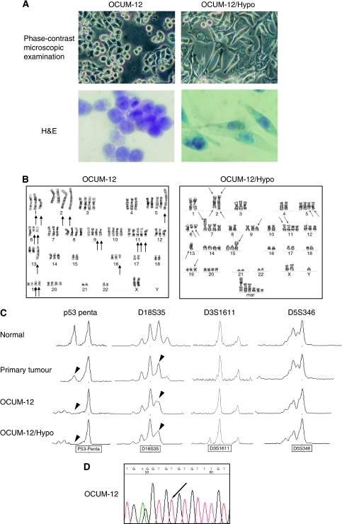
Methods: We established a scirrhous gastric carcinoma cell line (OCUM-12) from ascites associated with scirrhous gastric carcinoma, and a hypoxia-resistant cancer cell line (OCUM-12/Hypo) was cloned from OCUM-12 cells by continuous exposure to 1% oxygen.
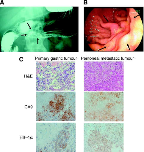
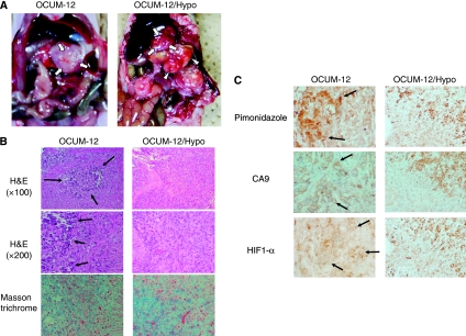
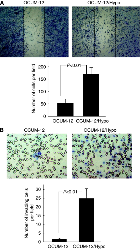
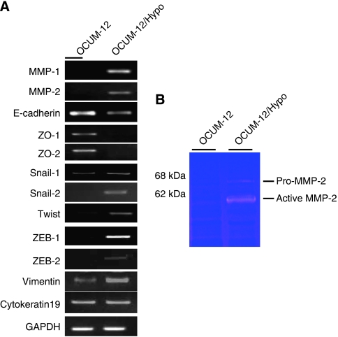
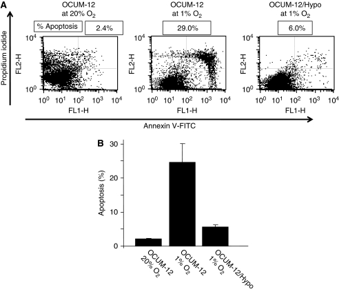
Results: Histologic findings from orthotopic tumours derived from parent OCUM-12 cells and daughter OCUM-12/Hypo cells revealed poorly differentiated adenocarcinoma with extensive fibrosis that resembled human scirrhous gastric cancer. Necrotic lesions were frequently detected in the OCUM-12 tumours but were rarely found in the OCUM-12/Hypo tumours, although both types had multiple hypoxic loci. Apoptosis rate of OCUM-12 cells was increased to 24.7% at 1% O(2), whereas that of OCUM-12/Hypo was 5.6%. The OCUM-12/Hypo orthotopic models developed multiple metastases to the peritoneum and lymph nodes, but the OCUM-12 models did not. OCUM-12/Hypo cells showed epithelial-to-mesenchymal transition and high migratory and invasive activities in comparison with OCUM-12 cells. The mRNA expression levels of both E-cadherin and zonula occludens ZO-1 and ZO-2 decreased in OCUM-12/Hypo cells, and that of vimentin, Snail-1, Slug/Snail-2, Twist, ZEB-1, ZEB-2, matrix metalloproteinase-1 (MMP-1), and MMP-2 were increased in OCUM-12/Hypo cells.
Conclusion: OCUM-12 and OCUM-12/Hypo may be useful for the elucidation of disease progression associated with scirrhous gastric cancer in the setting of chronic hypoxia.
Figures
Similar articles
-
Peritoneal metastatic model for human scirrhous gastric carcinoma in nude mice.Clin Exp Metastasis. 1996 Jan;14(1):43-54. doi: 10.1007/BF00157685. Clin Exp Metastasis. 1996. PMID: 8521616
-
HIF-1α is a crucial factor in the development of peritoneal dissemination via natural metastatic routes in scirrhous gastric cancer.Int J Oncol. 2013 Nov;43(5):1431-40. doi: 10.3892/ijo.2013.2068. Epub 2013 Aug 21. Int J Oncol. 2013. PMID: 23970191
-
Establishment of a new scirrhous gastric cancer cell line with loss of heterozygosity at E-cadherin locus.Int J Oncol. 2001 Nov;19(5):1029-33. doi: 10.3892/ijo.19.5.1029. Int J Oncol. 2001. PMID: 11605005
-
[Pathogenesis and progression of scirrhous carcinoma].Gan To Kagaku Ryoho. 1994 Oct;21(14):2364-70. Gan To Kagaku Ryoho. 1994. PMID: 7944478 Review. Japanese.
-
[Dysfunction of E-cadherin due to mutation of beta-catenin in a scirrhous gastric cancer cell line].Nihon Rinsho. 1995 Jul;53(7):1590-4. Nihon Rinsho. 1995. PMID: 7629993 Review. Japanese.
Cited by
-
Downregulation of malic enzyme 3 facilitates progression of gastric carcinoma via regulating intracellular oxidative stress and hypoxia-inducible factor-1α stabilization.Cell Mol Life Sci. 2024 Aug 30;81(1):375. doi: 10.1007/s00018-024-05388-9. Cell Mol Life Sci. 2024. PMID: 39212717 Free PMC article.
-
Snail1 mediates hypoxia-induced melanoma progression.Am J Pathol. 2011 Dec;179(6):3020-31. doi: 10.1016/j.ajpath.2011.08.038. Epub 2011 Oct 11. Am J Pathol. 2011. PMID: 21996677 Free PMC article.
-
The HIF-1α as a Potent Inducer of the Hallmarks in Gastric Cancer.Cancers (Basel). 2022 May 30;14(11):2711. doi: 10.3390/cancers14112711. Cancers (Basel). 2022. PMID: 35681691 Free PMC article. Review.
-
ZO-2 Is a Master Regulator of Gene Expression, Cell Proliferation, Cytoarchitecture, and Cell Size.Int J Mol Sci. 2019 Aug 24;20(17):4128. doi: 10.3390/ijms20174128. Int J Mol Sci. 2019. PMID: 31450555 Free PMC article. Review.
-
CD9-positive exosomes from cancer-associated fibroblasts stimulate the migration ability of scirrhous-type gastric cancer cells.Br J Cancer. 2018 Mar 20;118(6):867-877. doi: 10.1038/bjc.2017.487. Epub 2018 Feb 13. Br J Cancer. 2018. PMID: 29438363 Free PMC article.
References
-
- Albini A, Iwamoto Y, Kleinman HK, Martin GR, Aaronson SA, Kozlowski JM, McEwan RN (1987) A rapid in vitro assay for quantitating the invasive potential of tumor cells. Cancer Res 47: 3239–3245 - PubMed
-
- Benchimol S, Fuks A, Jothy S, Beauchemin N, Shirota K, Stanners CP (1989) Carcinoembryonic antigen, a human tumor marker, functions as an intercellular adhesion molecule. Cell 57: 327–334 - PubMed
-
- Brown JM (1979) Evidence for acutely hypoxic cells in mouse tumours, and a possible mechanism of reoxygenation. Br J Radiol 52: 650–656 - PubMed
Publication types
MeSH terms
Substances
LinkOut - more resources
Full Text Sources
Medical
Research Materials
Miscellaneous
