

Oxygen-sensitive outcomes and gene expression in acute ischemic stroke
- PMID: 20145654
- PMCID: PMC2913550
- DOI: 10.1038/jcbfm.2010.7
Oxygen-sensitive outcomes and gene expression in acute ischemic stroke
Abstract
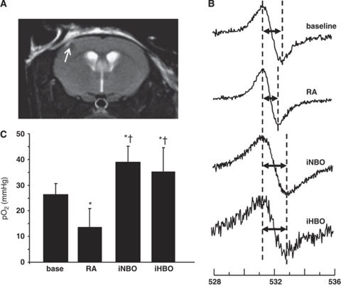
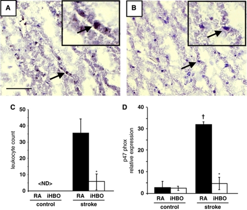
Acute ischemic stroke (AIS) results in focal deprivation of blood-borne factors, one of them being oxygen. The purpose of this study was two-fold: (1) to identify therapeutic conditions for supplemental oxygen in AIS and (2) to use transcriptome-wide screening toward uncovering oxygen-sensitive mechanisms. Transient MCAO in rodents was used to delineate the therapeutic potential of normobaric (NBO, 100% O(2), 1ATA) and hyperbaric oxygen (HBO, 100% O(2), 2ATA) during ischemia (iNBO, iHBO) and after reperfusion (rNBO, rHBO). Stroke lesion was quantified using 4.7 T MRI at 48 h. Supplemental oxygen during AIS significantly attenuated percent stroke hemisphere lesion volume as compared with that in room air (RA) controls, whereas identical treatment immediately after reperfusion exacerbated lesion volume (RA=22.4+/-1.8, iNBO=9.9+/-3.6, iHBO=6.6+/-4.8, rNBO=29.8+/-3.6, rHBO=35.4+/-7.6). iNBO and iHBO corrected penumbra tissue pO(2) during AIS as measured by EPR oxymetry. Unbiased query of oxygen-sensitive transcriptome in stroke-affected tissue identified 5,769 differentially expressed genes. Candidate genes were verified by real-time PCR using neurons laser-captured from the stroke-affected somatosensory cortex. Directed microarray analysis showed that supplemental oxygen limited leukocyte accumulation to the infarct site by attenuation of stroke-inducible proinflammatory chemokine response. The findings provide key information relevant to understanding oxygen-dependent molecular mechanisms in the AIS-affected brain.
Figures


Similar articles
-
Effect of large dose hyperbaric oxygenation therapy on prognosis and oxidative stress of acute permanent cerebral ischemic stroke in rats.Neurol Res. 2008 May;30(4):389-93. doi: 10.1179/174313208X300413. Neurol Res. 2008. PMID: 18544257
-
Neuroprotection by hyperbaric oxygenation after experimental focal cerebral ischemia monitored by MRI.Stroke. 2004 May;35(5):1175-9. doi: 10.1161/01.STR.0000125868.86298.8e. Epub 2004 Apr 1. Stroke. 2004. PMID: 15060313
-
Reduced cerebral monocarboxylate transporters and lactate levels by ethanol and normobaric oxygen therapy in severe transient and permanent ischemic stroke.Brain Res. 2015 Apr 7;1603:65-75. doi: 10.1016/j.brainres.2015.01.040. Epub 2015 Jan 30. Brain Res. 2015. PMID: 25641040
-
Oxygen therapy in acute ischemic stroke - experimental efficacy and molecular mechanisms.Curr Mol Med. 2009 Mar;9(2):227-41. doi: 10.2174/156652409787581619. Curr Mol Med. 2009. PMID: 19275631 Review.
-
The effects of hyperbaric oxygen therapy on the brain with middle cerebral artery occlusion.J Cell Physiol. 2021 Mar;236(3):1677-1694. doi: 10.1002/jcp.29955. Epub 2020 Jul 21. J Cell Physiol. 2021. PMID: 32692455 Review.
Cited by
-
Ischemic stroke-induced polyaxonal innervation at the neuromuscular junction is attenuated by robot-assisted mechanical therapy.Exp Neurol. 2021 Sep;343:113767. doi: 10.1016/j.expneurol.2021.113767. Epub 2021 May 25. Exp Neurol. 2021. PMID: 34044000 Free PMC article.
-
Method parameters' impact on mortality and variability in rat stroke experiments: a meta-analysis.BMC Neurosci. 2013 Apr 1;14:41. doi: 10.1186/1471-2202-14-41. BMC Neurosci. 2013. PMID: 23548160 Free PMC article.
-
A Review of Low-Frequency EPR Technology for the Measurement of Brain pO2 and Oxidative Stress.Appl Magn Reson. 2021 Oct;52(10):1379-1394. doi: 10.1007/s00723-021-01384-5. Epub 2021 Jul 16. Appl Magn Reson. 2021. PMID: 35340811 Free PMC article.
-
Oxygen-inducible glutamate oxaloacetate transaminase as protective switch transforming neurotoxic glutamate to metabolic fuel during acute ischemic stroke.Antioxid Redox Signal. 2011 May 15;14(10):1777-85. doi: 10.1089/ars.2011.3930. Epub 2011 Mar 28. Antioxid Redox Signal. 2011. PMID: 21361730 Free PMC article.
-
Glutamate oxaloacetate transaminase enables anaplerotic refilling of TCA cycle intermediates in stroke-affected brain.FASEB J. 2017 Apr;31(4):1709-1718. doi: 10.1096/fj.201601033R. Epub 2017 Jan 17. FASEB J. 2017. PMID: 28096234 Free PMC article.
References
-
- Anderson DC, Bottini AG, Jagiella WM, Westphal B, Ford S, Rockswold GL, Loewenson RB. A pilot study of hyperbaric oxygen in the treatment of human stroke. Stroke. 1991;22:1137–1142. - PubMed
-
- Badr AE, Yin W, Mychaskiw G, Zhang JH. Dual effect of HBO on cerebral infarction in MCAO rats. Am J Physiol Regul Integr Comp Physiol. 2001;280:R766–R770. - PubMed
-
- Bederson JB, Pitts LH, Tsuji M, Nishimura MC, Davis RL, Bartkowski H. Rat middle cerebral artery occlusion: evaluation of the model and development of a neurologic examination. Stroke. 1986;17:472–476. - PubMed
-
- Benedek A, Moricz K, Juranyi Z, Gigler G, Levay G, Harsing LG, Jr, Matyus P, Szenasi G, Albert M. Use of TTC staining for the evaluation of tissue injury in the early phases of reperfusion after focal cerebral ischemia in rats. Brain Res. 2006;1116:159–165. - PubMed
Publication types
MeSH terms
Substances
Grants and funding
LinkOut - more resources
Full Text Sources
Other Literature Sources
Medical
