

Case report: Osteonecrosis of the femoral head after hip arthroscopy
- PMID: 20146035
- PMCID: PMC2947685
- DOI: 10.1007/s11999-010-1256-1
Case report: Osteonecrosis of the femoral head after hip arthroscopy
Abstract
Background: Hip arthroscopy is a common orthopaedic procedure used as a diagnostic and therapeutic tool with a multitude of surgical indications. The complication rate is reportedly between 1.3% and 23.3%. Major complications are related to traction, fluid extravasation, and iatrogenic chondral injury. Although osteonecrosis is a concern with any surgical procedure about the hip, this complication has been primarily a theoretical concern with hip arthroscopy.
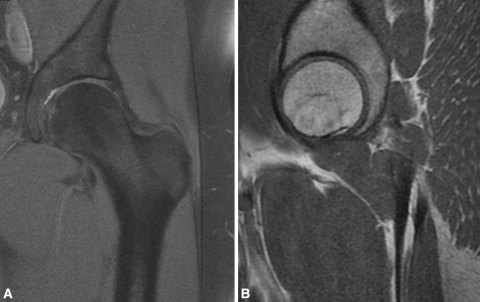
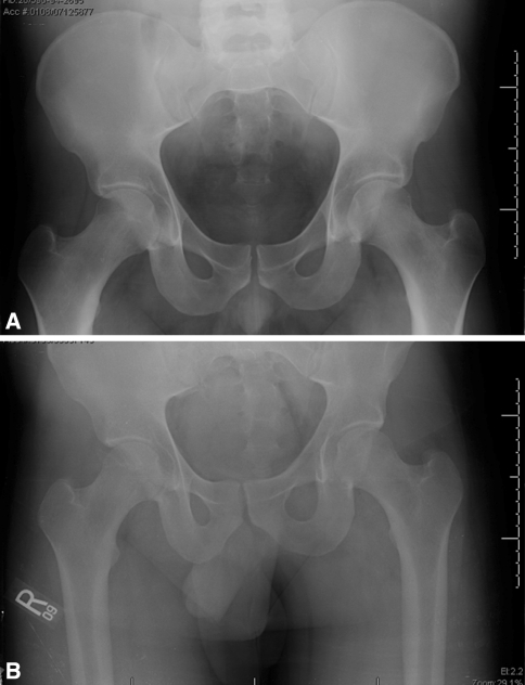
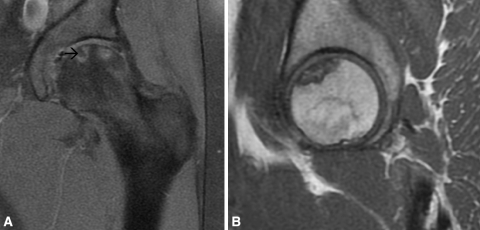
Case description: We report the case of a 24-year-old man who presented with a 2-year history of left hip pain. He underwent hip arthroscopy to include débridement of a torn labrum and removal of a prominent pincer lesion for femoroacetabular impingement. Traction was initiated by applying manual traction to the traction bar until 10 mm of joint distraction was obtained. Traction was removed at 90 minutes. At the 3-month followup, MRI showed osteonecrosis in the subcapital region of the left femoral head.
Literature review: It generally is agreed the magnitude and duration of traction during hip arthroscopy increase the risk of traction-related injuries. Only one previous case of femoral head osteonecrosis associated with hip arthroscopy has been reported, and this may have resulted from the initial traumatic event. Based on anatomic studies, the use of standard arthroscopic portals would not put at risk any dominant normal vascular structures supplying the femoral head. In contrast, the literature shows that femoral head osteonecrosis may develop secondary to a combination of increased intraarticular pressure and traction.
Purposes and clinical relevance: We suspect this case of femoral head osteonecrosis after hip arthroscopy was caused by traction used in the procedure.
Figures
Similar articles
-
Clinical outcomes following arthroscopic treatment of femoro-acetabular impingement using a minimal traction approach and an initial capsulotomy. Minimum two year follow-up.Int Orthop. 2018 Nov;42(11):2549-2554. doi: 10.1007/s00264-018-3904-0. Epub 2018 Mar 23. Int Orthop. 2018. PMID: 29572638
-
Avascular necrosis of the femoral head after hip arthroscopy.Hip Int. 2011 Sep-Oct;21(5):623-6. doi: 10.5301/HIP.2011.8693. Hip Int. 2011. PMID: 21960450
-
Acetabular labral tear complicating idiopathic osteonecrosis of the femoral head treated by labral repair with hip arthroscopy: a case report.J Med Case Rep. 2014 Nov 18;8:372. doi: 10.1186/1752-1947-8-372. J Med Case Rep. 2014. PMID: 25404056 Free PMC article.
-
[Hip arthroscopy. Minimal invasive diagnosis and therapy of the diseased or injured hip joint].Unfallchirurg. 2001 Jan;104(1):2-18. doi: 10.1007/s001130050682. Unfallchirurg. 2001. PMID: 11381758 Review. German.
-
Beyond the Scope Open Treatment of Femoroacetabular Impingement.Bull Hosp Jt Dis (2013). 2018 Mar;76(1):47-54. Bull Hosp Jt Dis (2013). 2018. PMID: 29537957 Review.
Cited by
-
Early experience with a comprehensive hip preservation service intended to improve clinical care, education, and academic productivity.Clin Orthop Relat Res. 2012 Dec;470(12):3446-52. doi: 10.1007/s11999-012-2549-3. Clin Orthop Relat Res. 2012. PMID: 22926493 Free PMC article.
-
The Use of Superselective Arteriography in the Evaluation of the Influence of Intracapsular Hip Joint Pressure on the Blood Flow of the Femoral Head.Med Princ Pract. 2016;25(2):123-9. doi: 10.1159/000442019. Epub 2015 Oct 30. Med Princ Pract. 2016. PMID: 26517358 Free PMC article.
-
Complications of arthroscopic surgery of the hip.Bone Joint Res. 2012 Jul 1;1(7):131-44. doi: 10.1302/2046-3758.17.2000108. Print 2012 Jul. Bone Joint Res. 2012. PMID: 23610683 Free PMC article.
-
Long-term Outcome of Multiple Small-diameter Drilling Decompression Combined with Hip Arthroscopy versus Drilling Alone for Early Avascular Necrosis of the Femoral Head.Chin Med J (Engl). 2017 Jun 20;130(12):1435-1440. doi: 10.4103/0366-6999.207470. Chin Med J (Engl). 2017. PMID: 28584206 Free PMC article.
-
Clinical outcomes following arthroscopic treatment of femoro-acetabular impingement using a minimal traction approach and an initial capsulotomy. Minimum two year follow-up.Int Orthop. 2018 Nov;42(11):2549-2554. doi: 10.1007/s00264-018-3904-0. Epub 2018 Mar 23. Int Orthop. 2018. PMID: 29572638
References
-
- Burman MS. Arthroscopy or the direct visualization of joints. J Bone Joint Surg Am. 1931;4:669–695.
-
- Byrd J. Complications associated with hip arthroscopy. In: Byrd J, editor. Operative Hip Arthroscopy. New York, NY: Thieme; 1998. pp. 171–176.
Publication types
MeSH terms
LinkOut - more resources
Full Text Sources
