. 2010 May;67(10):1723-33.
doi: 10.1007/s00018-010-0278-x. Epub 2010 Feb 10.

P2X7, NMDA and BDNF receptors converge on GSK3 phosphorylation and cooperate to promote survival in cerebellar granule neurons

Affiliations

P2X7, NMDA and BDNF receptors converge on GSK3 phosphorylation and cooperate to promote survival in cerebellar granule neurons

Felipe Ortega et al. Cell Mol Life Sci. 2010 May.

Abstract

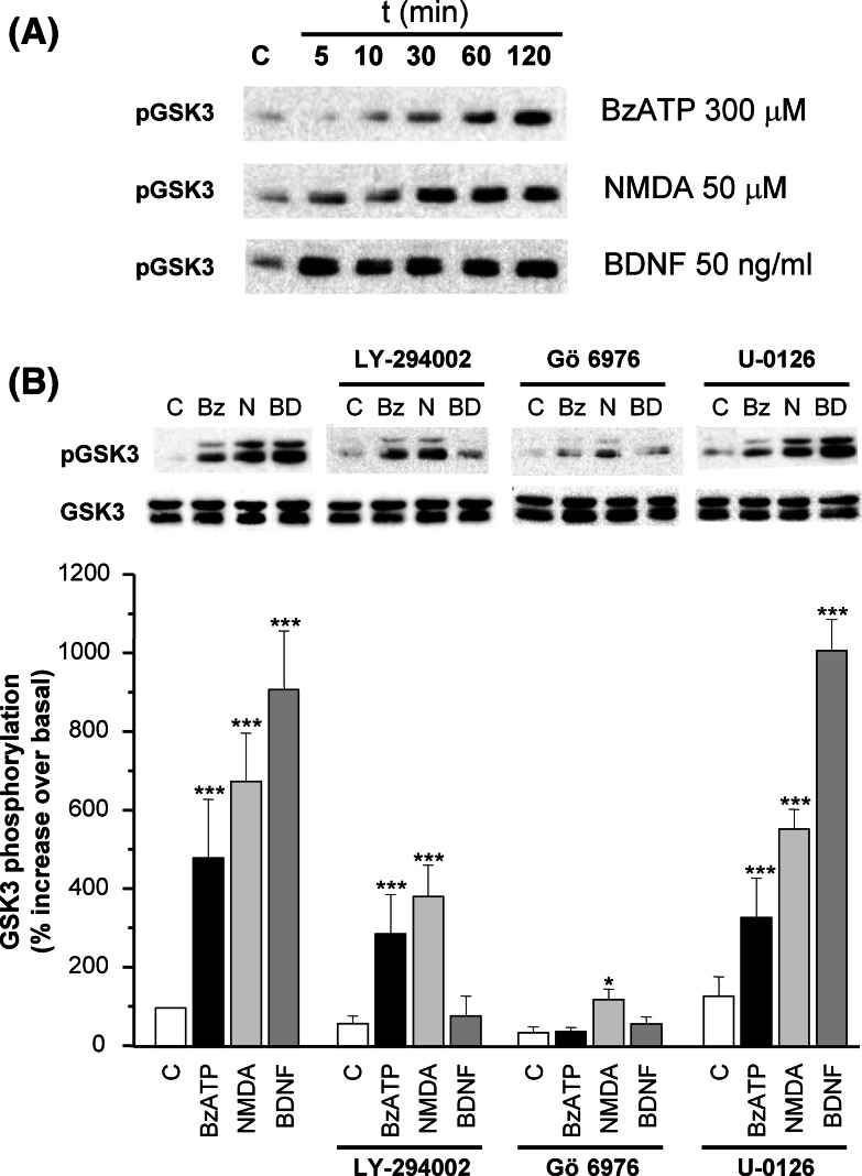
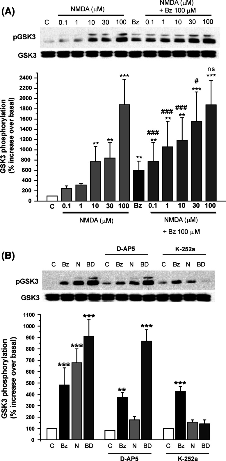
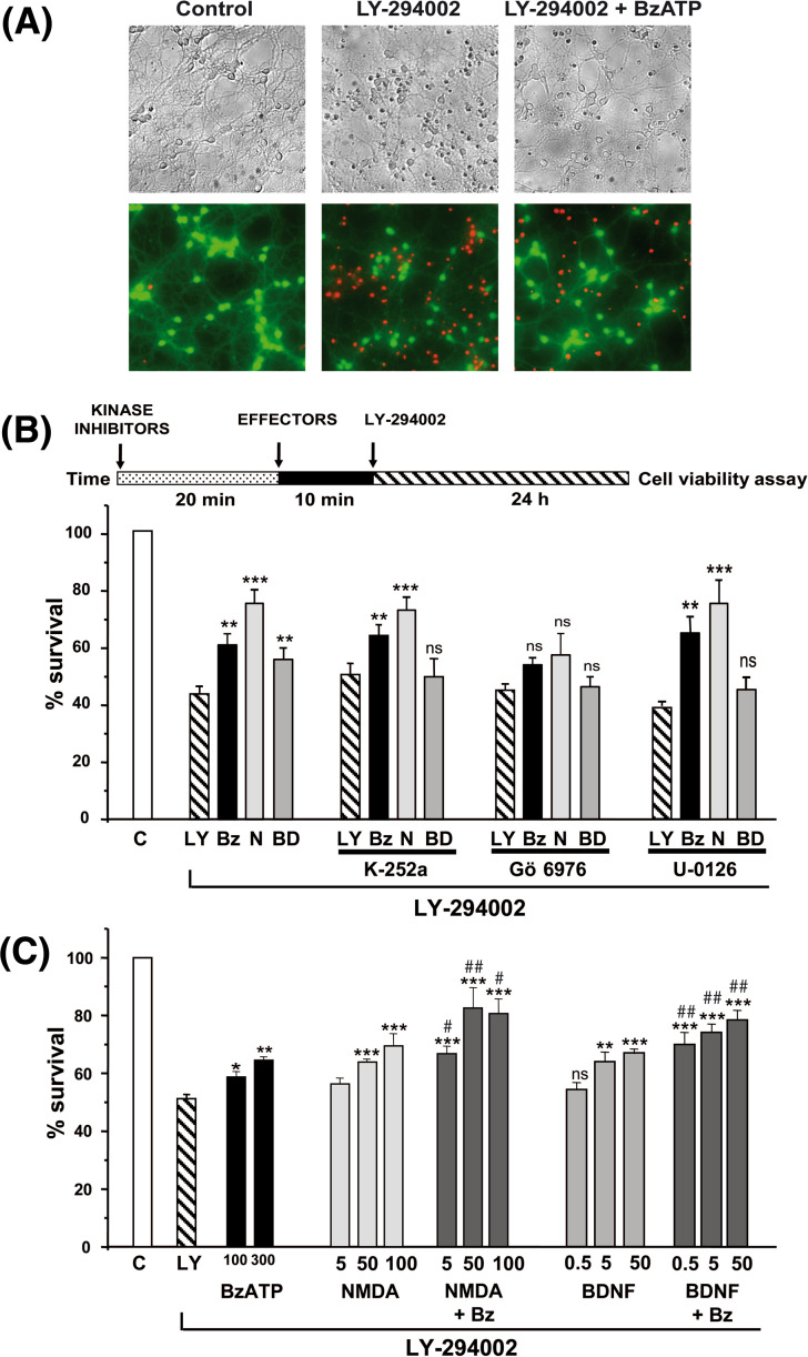
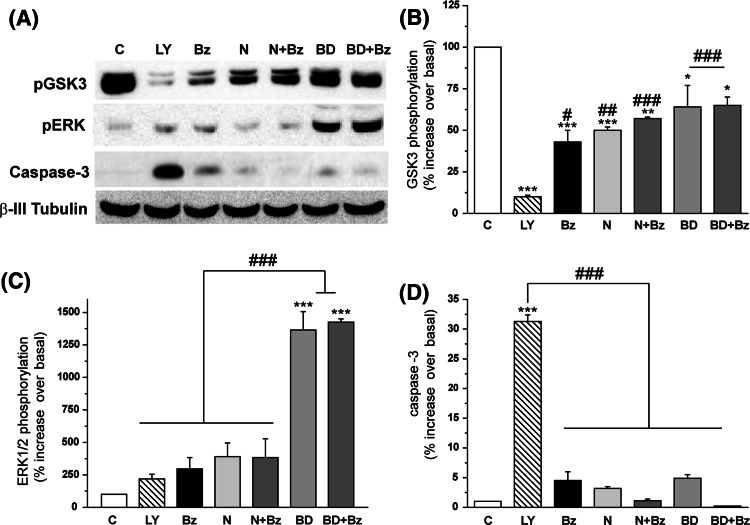
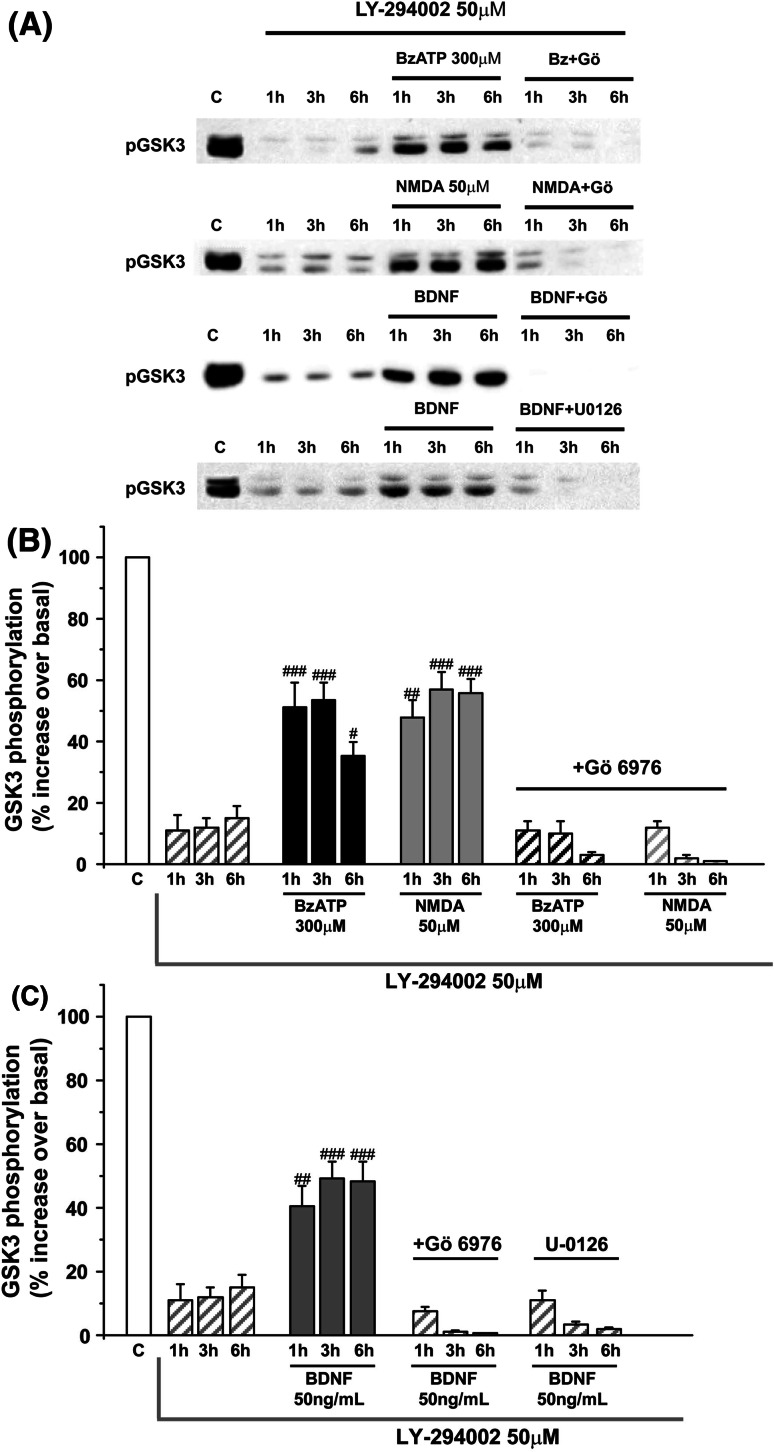
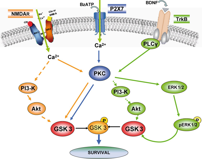
Glycogen synthase kinase-3 (GSK3) is a key player in the regulation of neuronal survival. Herein, we report evidence of an interaction between P2X7 receptors with NMDA and BDNF receptors at the level of GSK3 signalling and neuroprotection. The activation of these receptors in granule neurons led to a sustained pattern of GSK3 phosphorylation that was mainly PKC-dependent. BDNF was the most potent at inducing GSK3 phosphorylation, which was also dependent on PI3K. The P2X7 agonist, BzATP, exhibited additive effects with both NMDA and BDNF to rescue granule neurons from cell death induced by PI3K inhibition. This survival effect was mediated by the PKC-dependent GSK3 pathway. In addition, ERK1/2 proteins were also involved in BDNF protective effect. These results show the function of ATP in amplifying neuroprotective actions of glutamate and neurotrophins, and support the role of GSK3 as an important convergence point for these survival promoting factors in granule neurons.


Figures

Fig. 1
Fig. 1
GSK3 phosphorylation induced by BzATP, NMDA and BDNF in granule neurons. a Time-course of GSK3 phosphorylation. Granule cells were stimulated with 300 μM BzATP, 50 μM NMDA and 50 ng/ml BDNF, at different incubation times. b Effect of various signalling pathway inhibitors on GSK3 phosphorylation. Granule cells in culture were submitted to different treatments: 20 min incubation with signalling inhibitors, 50 μM LY-294002, 0.5 μM Gö 6976 and 10 μM U-0126. Then cells were stimulated for 10 min with 300 μM BzATP, 50 μM NMDA and 50 ng/ml BDNF in the continuous presence of these inhibitors. Then cells were harvested and phosphorylation of GSK3 was analysed by immunoblotting. Histograms represent the percentage increase with respect to non-stimulated cells (100% control value), and were obtained by normalization of densitometric values of phospho-proteins with respect to total forms of GSK3. The blots correspond to representative experiments and values are the mean ± SD of at least three experiments performed from different cultures. Data were analysed by Dunnet and Tukey tests, and were statistically significant at *P < 0.05 and ***P < 0.01 when the effects were compared to the respective control for each condition (open bars in the absence stimulation)
Fig. 2
Fig. 2
Convergence of BzATP and NMDA in GSK3 phosphorylation. a Effect of combined treatment of BzATP and NMDA on GSK3 phosphorylation. Granule neurons were stimulated for 10 min with different NMDA concentrations (from 0.1 to 100 μM) in the absence or presence of co-stimulation with 100 μM BzATP concentration. b Effect of NMDA antagonist and kinase inhibitor of TrkB receptor. Cells were treated for 5 min with 10 μM D-AP5 and 100 nM K-252a, and then 300 μM BzATP, 50 μM NMDA and 50 ng/ml BDNF were added for additional 10 min. Then cells were harvested and phosphorylation of GSK3 was analysed by immunoblotting, as described in “Material and methods”. Data were obtained by normalization of densitometric values of phospho-GSK3 with respect to total GSK3. The blots correspond to representative experiments and values are the mean ± SD of at least three experiments performed from different cultures. Data were analysed by Dunnet and Tukey tests, and were statistically significant at ***P < 0.001 and **P < 0.01 when the effects were compared to the controls (empty bars in the absence stimulation) (a, b), and at ### P < 0.001, and # P < 0.05, when the values at each NMDA concentration was compared in the absence and presence of BzATP a. Bz BzATP, N NMDA, BD BDNF
Fig. 3
Fig. 3
Effect of BzATP, NMDA and BDNF in the regulation of neuronal survival in viability assays. The different treatments were added directly to granule neurons maintained in complete culture medium. The PI3K inhibitor, LY-294002, was added at 50 μM concentration, and cell viability was analyzed 24 h later by the LIVE/DEAD viability/cytotoxicity kit a and the MTT assay (b, c). a, b Survival effects of BzATP, NMDA and BDNF. In a granule neurons were stimulated in the absence (control) or presence of 300 μM BzATP for 10 min, before the addition of LY-294002, except in the control. The micrographs show the same field in visible (upper panels) and calcein/ethidium bromide staining (lower panels). The apoptotic nuclei are stained red, and viable cells are stained green. b Schematic representation of the experimental design followed in survival studies. Granule neurons were preincubated in the absence and presence of the kinase inhibitors: 200 nM K-252a, 0.5 μM Gö 6976 and 10 μM U-0126 for 20 min previous to the addition of the agonists, 300 μM BzATP, 50 μM NMDA and 50 ng/ml BDNF. After 10 min, the PI3K inhibitor LY-294002 was added and cell viability tested 24 h later. c Additive survival effects of BzATP with NMDA and BDNF. The following stimulations were done before the addition of LY-294002: 100 and 300 μM BzATP, 5, 50 and 100 μM of NMDA, and 0.5, 5 and 50 ng/ml BDNF. The stimulations with NMDA and BDNF were carried out in the absence or presence of co-stimulation with 300 μM BzATP (dark grey bars). Data are the mean ± SD of at least three experiments performed in duplicate from different cultures. Data were analysed by Dunnet and Tukey tests and were statistically significant at ***P < 0.001, **P < 0.01 and *P < 0.05 when treatments with LY-294002 (dashed bars in b and c) were taken as reference, and at ##P < 0.001 and #P < 0.05 when each value was compared in the presence or absence of BzATP (dark grey bars). LY LY-294002, Bz BzATP, N NMDA, BD BDNF
Fig. 4
Fig. 4
Analysis of the effect of LY-294002 treatment on phosphorylation levels of GSK3 and ERK1/2, and caspase-3 activation. Granule neurons maintained in complete culture medium were treated in the presence or absence of 300 μM BzATP, 50 μM NMDA and 50 ng/ml BDNF for 10 min before the addition of 50 μM LY-294002. Cell extracts were obtained at 6 h incubation time with LY-294002 treatment and analysed by immunoblotting for GSK3 and ERK1/2 phosphorylation (ac), and for the presence of the 17 kDa active caspase-3 fragment (a, d). Histograms represent the percentage increase with respect to non-stimulated cells (100% control value), and were obtained by normalization of densitometric values of phospho-proteins and caspase-3, with respect to that obtained for β-tubulin, employed as a control charge. Values are the mean ± SD of at least three experiments performed from different cultures. The blots correspond to representative experiments a. Data were analysed by Dunnet and Tukey tests and were statistically significant at ***P < 0.001, **P < 0.01 and *P < 0.05 when compared to the control of untreated cells (empty bars), and at ### P < 0.001, ## P < 0.01 and # P < 0.05, when compared with LY-294002 treatment (dashed bars), as indicated in the figure. LY LY-294002, Bz BzATP, N NMDA, BD BDNF
Fig. 5
Fig. 5
Effect of PKC and MAPK inhibition on GSK3 phosphorylation levels under conditions of PI3K inhibition. Granule neurons maintained in complete culture medium were treated in the presence or absence of 300 μM BzATP, 50 μM NMDA and 50 ng/ml BDNF for 10 min before the addition of 50 μM LY-294002. When required, the transducing inhibitors, 0.5 μM Gö 6976 and 10 μM U-0126 were added for 20 min before the addition of the three stimuli. Cell extracts were obtained at different incubation times of 1, 3 and 6 h with LY-294002 treatment, and analysed by immunoblotting for GSK3 phosphorylation, as described before. In a the blots corresponding to representative experiments are shown. In b the phosphorylation values for BzATP and NMDA treatments are shown, and in c for BDNF. Values are the mean ± SD of at least three experiments performed from different cultures. Data were analysed by Dunnet and Tukey tests, and were statistically significant at ### P < 0.001, ## P < 0.01 and # P < 0.05, when the values at each LY-294002 incubation time were compared with respect to the treatments with Gö 6976 and U-0126. LY LY-294002, Bz BzATP, N NMDA, BD BDNF
Fig. 6
Fig. 6
Schematic diagram of P2X7, NMDA and TrkB receptors coupling to GSK3 signalling through different routes in cerebellar granule neurons. P2X7, NMDA and TrkB receptors trigger different intracellular pathways to induce GSK3 phosphorylation and elicit neuroprotection in granule neurons. P2X7 and NMDA ionotropic receptors are both coupled to the entry of extracellular calcium and share the same PKC-dependent route to provoke GSK3 phosphorylation and neuroprotection. BDNF receptor signalling is mainly coupled to the PI3K/Akt axis in a PLC/PKC-dependent manner. When PI3K is inactivated, BDNF then uses an alternative route through the activation of the ERK1/2 proteins to maintain GSK3 inhibition and elicit survival

Similar articles

Cited by

References

    1. Contestabile A. Cerebellar granule cells as a model to study mechanisms of neuronal apoptosis or survival in vivo and in vitro. Cerebellum. 2002;1:41–55. doi: 10.1080/147342202753203087. - DOI - PubMed
    1. Miller TM, Tansey MG, Johnson EM, Jr, Creedon DJ. Inhibition of phosphatidylinositol 3-kinase activity blocks depolarization- and insulin-like growth factor I-mediated survival of cerebellar granule cells. J Biol Chem. 1997;272:9847–9853. doi: 10.1074/jbc.272.15.9847. - DOI - PubMed
    1. Hervás C, Pérez-Sen R, Miras-Portugal MT. Coexpression of functional P2X and P2Y nucleotide receptors in single cerebellar granule cells. J Neurosci Res. 2003;73:384–399. doi: 10.1002/jnr.10676. - DOI - PubMed
    1. Amadio S, D’Ambrosi N, Cavaliere F, Murra B, Sancesario G, Bernardi G, Burnstock G, Volonte C. P2 receptor modulation and cytotoxic function in cultured CNS neurons. Neuropharmacology. 2002;42:489–501. doi: 10.1016/S0028-3908(01)00197-6. - DOI - PubMed
    1. North RA. Molecular physiology of P2X receptors. Physiol Rev. 2002;82:1013–1067. - PubMed

Publication types

MeSH terms

Substances

LinkOut - more resources