

Effects of CBV, CBF, and blood-brain barrier permeability on accuracy of PASL and VASO measurement
- PMID: 20146228
- PMCID: PMC3167220
- DOI: 10.1002/mrm.22165
Effects of CBV, CBF, and blood-brain barrier permeability on accuracy of PASL and VASO measurement
Abstract
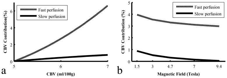
Cerebral blood flow, cerebral blood volume (CBV), and water permeability through blood-brain barrier are important hemodynamic parameters in brain physiology. Pulsed arterial spin labeling and vascular-space occupancy techniques have been used to measure regional cerebral blood flow and CBV, respectively. However, these techniques generally ignore the effects of one hemodynamic parameter on the measurement of others. For instance, the influences of CBV changes on arterial spin labeling or the permeability effects on vascular-space occupancy typically were not accounted for in the quantification of blood flow or volume. In the current work, the biophysical effects of CBV on pulsed arterial spin labeling and permeability on vascular-space occupancy signals are evaluated using a general two-compartment model. The dependence of these effects on the T(1) at various field strengths is also assessed by simulations. Results indicate that CBV has negligible to small influences on pulsed arterial spin labeling signal (<6.6% at 3 T) and permeability effects are negligible on vascular-space occupancy signal (<0.1% at 3 T) under normal physiologic conditions. In addition, CBV effect on pulsed arterial spin labeling is further diminished at high field strengths, but residual blood contamination in vascular-space occupancy signal may be enhanced at high fields due to the reduced difference between extra- and intravascular T(1) values.
(c) 2010 Wiley-Liss, Inc.
Figures


Similar articles
-
On the measurement of absolute cerebral blood volume (CBV) using vascular-space-occupancy (VASO) MRI.Magn Reson Med. 2009 Mar;61(3):659-67. doi: 10.1002/mrm.21872. Magn Reson Med. 2009. PMID: 19097238 Free PMC article.
-
Three-dimensional acquisition of cerebral blood volume and flow responses during functional stimulation in a single scan.Neuroimage. 2014 Dec;103:533-541. doi: 10.1016/j.neuroimage.2014.08.025. Epub 2014 Aug 23. Neuroimage. 2014. PMID: 25152092 Free PMC article.
-
Improved cortical-layer specificity of vascular space occupancy fMRI with slab inversion relative to spin-echo BOLD at 9.4 T.Neuroimage. 2008 Mar 1;40(1):59-67. doi: 10.1016/j.neuroimage.2007.11.045. Epub 2007 Dec 8. Neuroimage. 2008. PMID: 18249010 Free PMC article.
-
Noninvasive functional imaging of cerebral blood volume with vascular-space-occupancy (VASO) MRI.NMR Biomed. 2013 Aug;26(8):932-48. doi: 10.1002/nbm.2905. Epub 2013 Jan 28. NMR Biomed. 2013. PMID: 23355392 Free PMC article. Review.
-
A review of the development of Vascular-Space-Occupancy (VASO) fMRI.Neuroimage. 2012 Aug 15;62(2):736-42. doi: 10.1016/j.neuroimage.2012.01.013. Epub 2012 Jan 8. Neuroimage. 2012. PMID: 22245650 Free PMC article. Review.
Cited by
-
Noise concerns and post-processing procedures in cerebral blood flow (CBF) and cerebral blood volume (CBV) functional magnetic resonance imaging.Neuroimage. 2017 Jul 1;154:43-58. doi: 10.1016/j.neuroimage.2016.09.007. Epub 2016 Sep 11. Neuroimage. 2017. PMID: 27622397 Free PMC article. Review.
-
Robust Multi-TE ASL-Based Blood-Brain Barrier Integrity Measurements.Front Neurosci. 2021 Dec 3;15:719676. doi: 10.3389/fnins.2021.719676. eCollection 2021. Front Neurosci. 2021. PMID: 34924924 Free PMC article.
-
Comparison of BOLD and CBV using 3D EPI and 3D GRASE for cortical layer functional MRI at 7 T.Magn Reson Med. 2020 Dec;84(6):3128-3145. doi: 10.1002/mrm.28347. Epub 2020 Jun 18. Magn Reson Med. 2020. PMID: 32557752 Free PMC article.
-
Characterisation of laminar and vascular spatiotemporal dynamics of CBV and BOLD signals using VASO and ME-GRE at 7T in humans.Imaging Neurosci (Camb). 2024 Aug 13;2:imag-2-00263. doi: 10.1162/imag_a_00263. eCollection 2024. Imaging Neurosci (Camb). 2024. PMID: 40800482 Free PMC article.
-
Characterisation of laminar and vascular spatiotemporal dynamics of CBV and BOLD signals using VASO and ME-GRE at 7T in humans.bioRxiv [Preprint]. 2024 Jan 26:2024.01.25.576050. doi: 10.1101/2024.01.25.576050. bioRxiv. 2024. Update in: Imaging Neurosci (Camb). 2024 Aug 13;2:imag-2-00263. doi: 10.1162/imag_a_00263. PMID: 38410457 Free PMC article. Updated. Preprint.
References
-
- Raichle ME. In: Handbook of physiology, The nervous system. Mountcastle VB, Plum F, editors. Bethesda: The American Physiological Society; 1987.
-
- Girouard H, Iadecola C. Neurovascular coupling in the normal brain and in hypertension, stroke, and Alzheimer disease. J Appl Physiol. 2006;100(1):328–335. - PubMed
-
- Wang J, Licht DJ. Pediatric perfusion MR imaging using arterial spin labeling. Neuroimaging Clin N Am. 2006;16(1):149–67. ix. - PubMed
-
- Desai BS, Monahan AJ, Carvey PM, Hendey B. Blood-brain barrier pathology in Alzheimer’s and Parkinson’s disease: implications for drug therapy. Cell Transplant. 2007;16(3):285–299. - PubMed
-
- Correale J, Villa A. The blood-brain-barrier in multiple sclerosis: functional roles and therapeutic targeting. Autoimmunity. 2007;40(2):148–160. - PubMed
Publication types
MeSH terms
Grants and funding
LinkOut - more resources
Full Text Sources
Medical
