. 2010 May;51(5):1702-11.
doi: 10.1002/hep.23510.

Deletion of CD39 on natural killer cells attenuates hepatic ischemia/reperfusion injury in mice

Affiliations

Deletion of CD39 on natural killer cells attenuates hepatic ischemia/reperfusion injury in mice

Guido Beldi et al. Hepatology. 2010 May.

Abstract

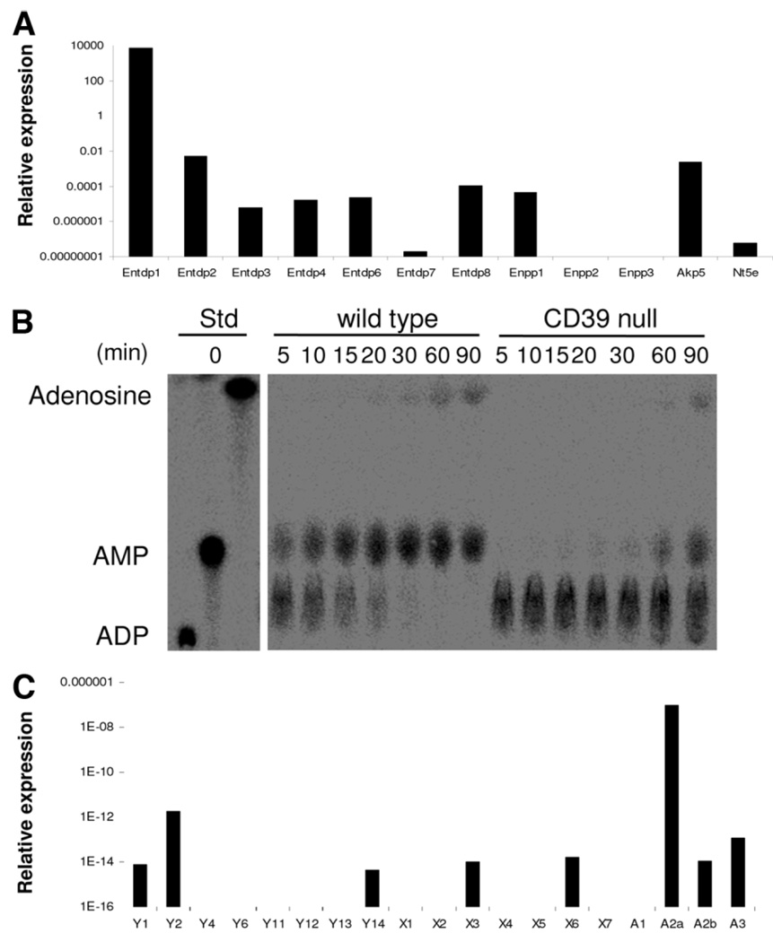
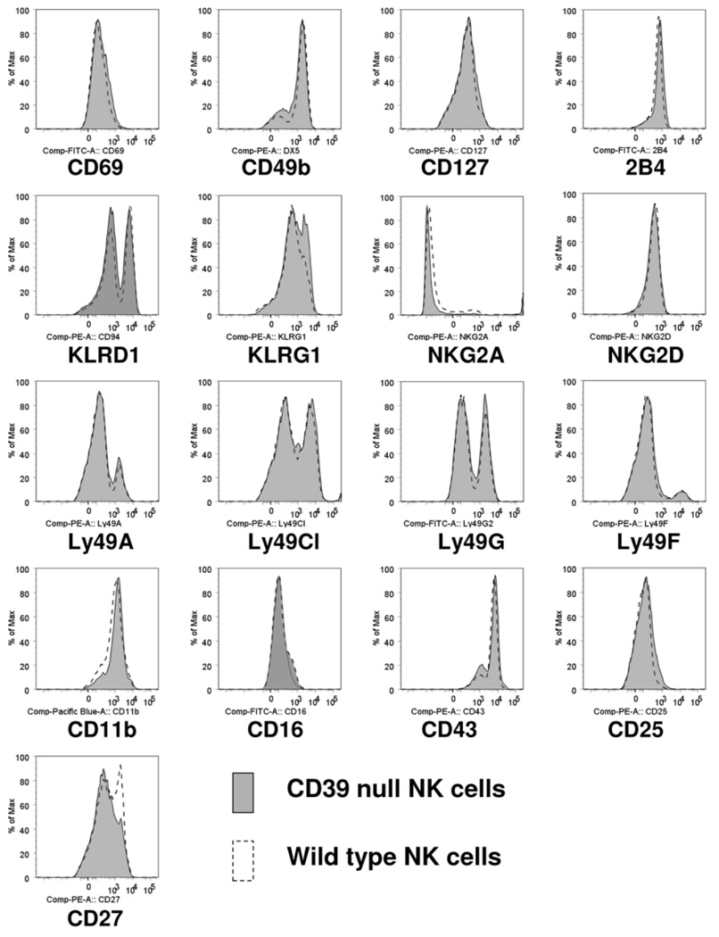
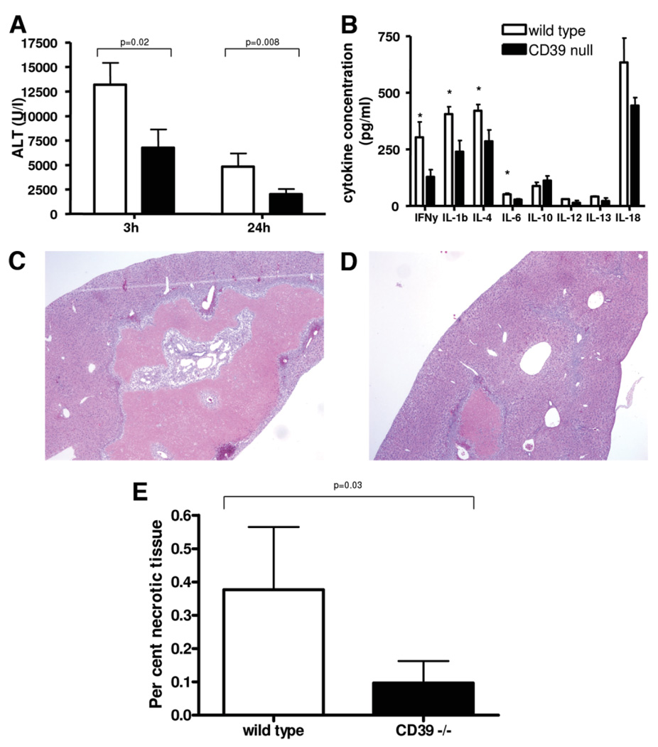
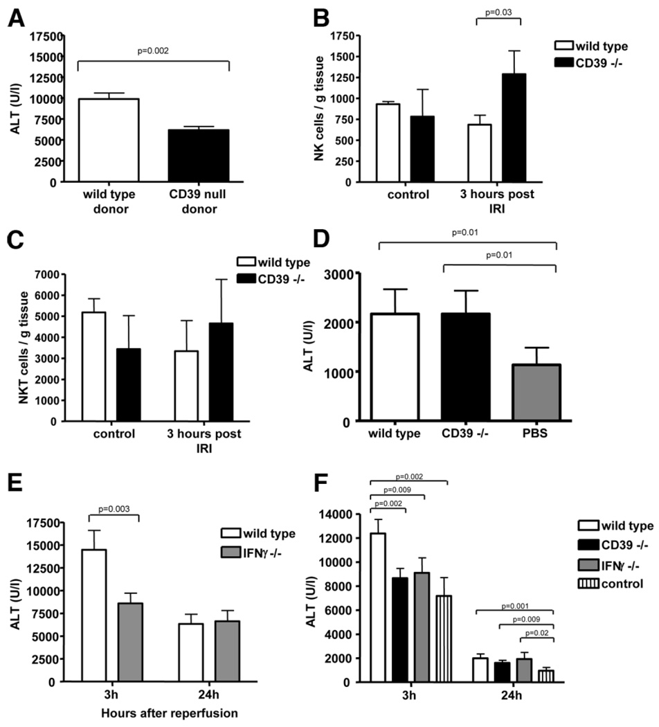
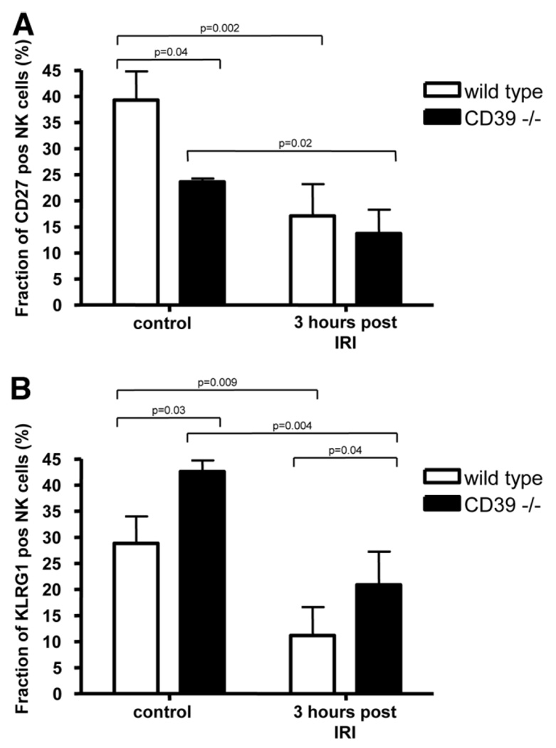
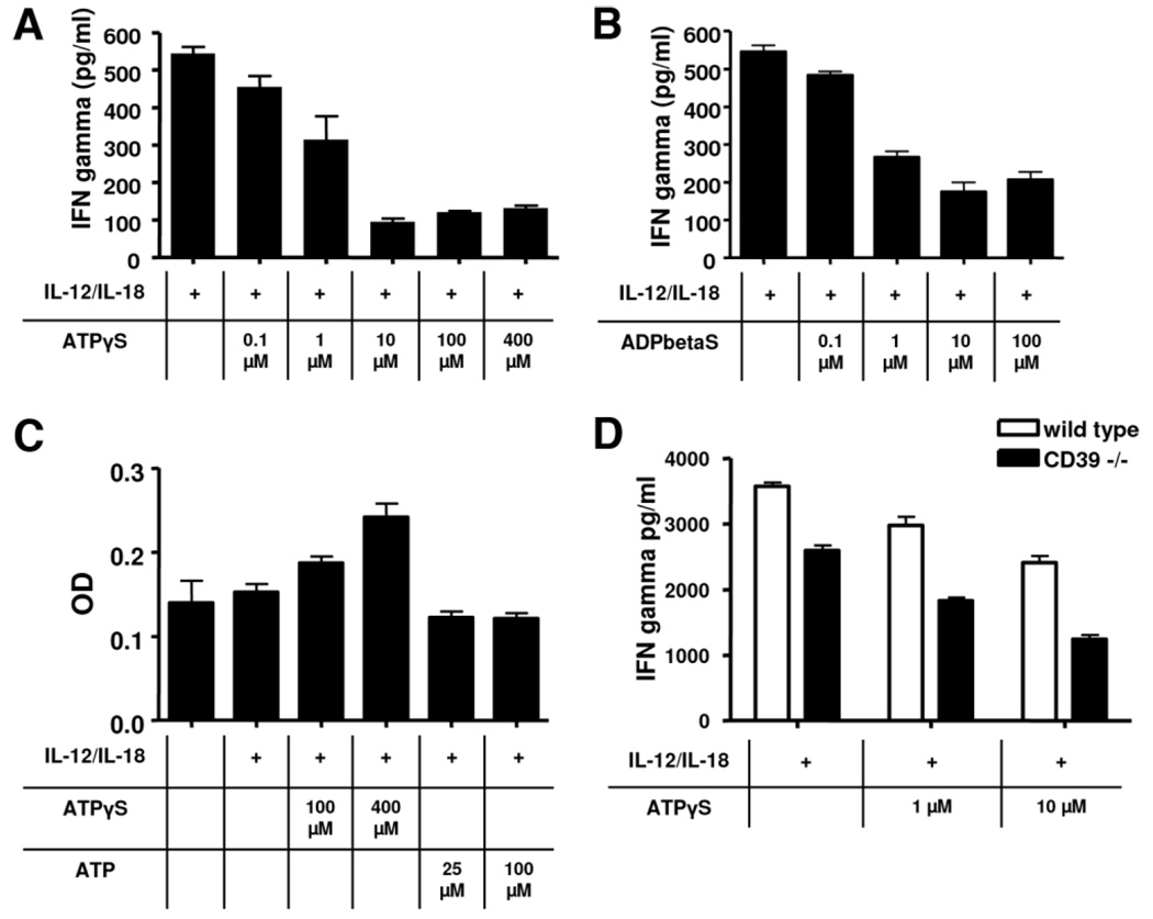
Natural killer (NK) cells play crucial roles in innate immunity and express CD39 (Ecto-nucleoside triphosphate diphosphohydrolase 1 [E-NTPD1]), a rate-limiting ectonucleotidase in the phosphohydrolysis of extracellular nucleotides to adenosine. We have studied the effects of CD39 gene deletion on NK cells in dictating outcomes after partial hepatic ischemia/reperfusion injury (IRI). We show in mice that gene deletion of CD39 is associated with marked decreases in phosphohydrolysis of adenosine triphosphate (ATP) and adenosine diphosphate to adenosine monophosphate on NK cells, thereby modulating the type-2 purinergic (P2) receptors demonstrated on these cells. We note that CD39-null mice are protected from acute vascular injury after single-lobe warm IRI, and, relative to control wild-type mice, display significantly less elevation of aminotransferases with less pronounced histopathological changes associated with IRI. Selective adoptive transfers of immune cells into Rag2/common gamma null mice (deficient in T cells, B cells, and NK/NKT cells) suggest that it is CD39 deletion on NK cells that provides end-organ protection, which is comparable to that seen in the absence of interferon gamma. Indeed, NK effector mechanisms such as interferon gamma secretion are inhibited by P2 receptor activation in vitro. Specifically, ATPgammaS (a nonhydrolyzable ATP analog) inhibits secretion of interferon gamma by NK cells in response to interleukin-12 and interleukin-18, providing a mechanistic link between CD39 deletion and altered cytokine secretion.

Conclusion: We propose that CD39 deficiency and changes in P2 receptor activation abrogate secretion of interferon gamma by NK cells in response to inflammatory mediators, thereby limiting tissue damage mediated by these innate immune cells during IRI.


Conflict of interest statement

Potential conflict of interest: Nothing to report.

Figures

Fig. 1
Fig. 1
Expression patterns of ectonucleotidases and purinergic receptors on NK cells. Isolated splenic NK (NK1.1-positive, CD49b-positive, CD3-negative) cells from Rag1−/− mice were studied by reverse transcription PCR. (A) Among all the ectonucleotidases tested, the highest expression was noted for CD39/E-NTPDase1. Expression levels of other ectonucleotidases, nucleotide pyrophosphatase (Enpp), and alkaline phosphatase (Akp5) were significantly lower and ecto-5′-ectonucleotidase/CD73 (Nt5e) was near absent. (B) Analysis of NTPDase activity on NK cells was done by TLC. The products of [C]ADP hydrolysis by purified hepatic NK cells are shown. Extracellular nucle-otides were efficiently hydrolyzed to AMP and adenosine by wild-type cells, whereas cells null for CD39 show substantially delayed hydrolysis of nucleotides and limited production of aden-osine. (C) Isolated NK cells expressed the P2 receptors P2Y1, P2Y2, P2Y14, P2X3, and P2X6; NK also expressed the P1 receptors A2A (at high levels), as well as A2B and A3 but not A1.
Fig. 2
Fig. 2
Expression of NK cell-specific surface markers on sorted NK cells. Isolated splenic NK (NK1.1-positive, CD49b-positive, CD3-negative) cells from wild-type mice and mice null for CD39 were characterized. Surface markers, in general, did not differ between wild-type and mutant NK cells, except KLRG1. Also, CD39-null NK cells expressed decreased levels of CD27 as compared to the wild-type.
Fig. 3
Fig. 3
Analysis of liver injury after hepatic ischemia with various periods of reperfusion. (A) Liver injury as assessed by alanine aminotrans-ferase (ALT) levels was significantly decreased in mice null for CD39, when compared to wild-type mice after 3 and 24 hours of reperfusion. (B) Cytokine profiling using arrays after 3 hours of reperfusion revealed decreased levels of circulating cytokines with the exception being IL-10. Representative liver sections of serial hematoxylin & eosin staining after 4 days of reperfusion; (C) wildtype and (D) CD39-null. (E) Results of morphometric analysis of area of necrosis revealed significantly increased area of necrosisin wild-type mice compared to mice null for CD39. Values are means ± standard deviation of at least four animals per time point. Levels of significance were assessed by unpaired t tests. P values are as indicated; asterisks indicate P < 0.05 in (B).
Fig. 4
Fig. 4
CD39 on NK cells modulate hepatic IRI. (A) To exclude confounding influences of the hepatic and systemic endothelium, adoptive transfer of wild-type and CD39 bone marrow-derived cells was performed after total body irradiation of wild-type mice. After transfer of CD39-null bone marrow, liver injury was significantly decreased in comparison to transfer of wild-type bone marrow after 3 hours of reperfusion. (B, C) NK cells but not NKT cells expand at 3 hours after reperfusion injury. (D) Performing adoptive transfer of NKT cells into Rag1 null mice revealed decreased injury in the control group and no difference between transfer of wild-type versus CD39-null NKT cells after 24 hours of reperfusion. (E) IRI in mice null for IFNγ was decreased after 3 hours but not after 24 hours. (F) Adoptive transfer of purified NK cells from wild-type mice and mice null for CD39 and null for IFNγ into Rag2/common gamma-null mice. Hepatic injury was significantly decreased after transfer of CD39-null and IFNγ null NK cells after 3 hours of reperfusion as compared to transfer of wild-type NK cells. Values are mean ± standard deviation of at least four animals per time point. Levels of significance were assessed by unpaired t tests. P values are as indicated.
Fig. 5
Fig. 5
Differential numbers of NK cell subsets after IRI in vivo. Hepatic mononuclear cells were isolated in control animals from wild-type and CD39-null mice at baseline (n = 3 each) and at 3 hours after hepatic IRI (n = 4 each). These were gated for NK1.1-positive, CD3-negative, (A) CD27-positive, and (B) KLRG1-positive cells. Significantly decreased levels of CD27-positive cells were observed in CD39-null murine livers prior to injury. After IRI, numbers of CD27-positive NK cells significantly decreased in wild-type and mutant mice. Conversely, levels of KLRG1-positive cells were significantly increased in mutant mice, both under basal conditions and after IRI. Values are mean ± standard deviation. Levels of significance were assessed by unpaired t tests. P values are as indicated.
Fig. 6
Fig. 6
Secrrtion of INFγ by NK cells in vitro. Splenic NK cells were isolated from mice null for CD39 and matched the wild-types. Secretion of IFNγ was induced by administration of IL-12 and IL-18 for 24 hours. (A,B) Addition of ATPγS or ADPβS (nonhydrolyzable ATP/ADP analogs) significantly decreased secretion of IFNγ in a dose-dependent manner. (C) Analysis of cell viability after 36 hours of incubation using a MTT assay: Administration of ATPγS resulted in a dose-dependent increase of optical density. (D) Comparison of IFNγ secretion of wildtype versus CD39-null NK cells. Deletion of CD39 is associated with significantly reduced secretion of IFNγ. Graphs are representative of at least five experiments. Data are given as mean ± standard deviation.

Similar articles

Cited by

References

    1. Lappas CM, Day YJ, Marshall MA, Engelhard VH, Linden J. Adenosine A2A receptor activation reduces hepatic ischemia reperfusion injury by inhibiting CD1d-dependent NKT cell activation. J Exp Med. 2006;203:2639–2648. - PMC - PubMed
    1. Feng L, Cheng F, Ye Z, Li S, He Y, Yao X, et al. The effect of renal ischemia-reperfusion injury on expression of RAE-1 and H60 in mice kidney. Transplant Proc. 2006;38:2195–2198. - PubMed
    1. Kato A, Graul-Layman A, Edwards MJ, Lentsch AB. Promotion of hepatic ischemia/reperfusion injury by IL-12 is independent of STAT4. Transplantation. 2002;73:1142–1145. - PubMed
    1. Takeuchi D, Yoshidome H, Kato A, Ito H, Kimura F, Shimizu H, et al. Interleukin 18 causes hepatic ischemia/reperfusion injury by suppressing anti-inflammatory cytokine expression in mice. HEPATOLOGY. 2004;39:699–710. - PubMed
    1. Friedman DJ, Rennke HG, Csizmadia E, Enjyoji K, Robson SC. The vascular ectonucleotidase ENTPD1 is a novel renoprotective factor in diabetic nephropathy. Diabetes. 2007;56:2371–2379. - PubMed

Publication types

MeSH terms