. 2010 Mar;51(3):881-90.
doi: 10.1002/hep.23381.

MicroRNA-dependent regulation of DNA methyltransferase-1 and tumor suppressor gene expression by interleukin-6 in human malignant cholangiocytes

Affiliations

MicroRNA-dependent regulation of DNA methyltransferase-1 and tumor suppressor gene expression by interleukin-6 in human malignant cholangiocytes

Chiara Braconi et al. Hepatology. 2010 Mar.

Abstract

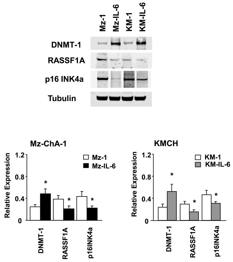
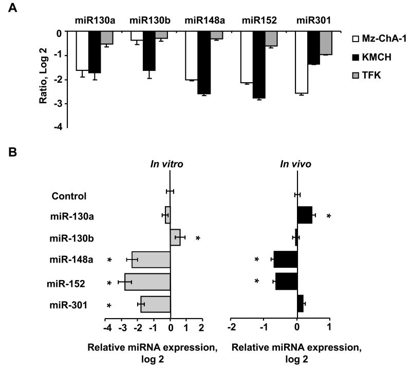
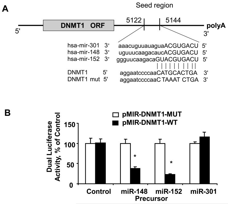
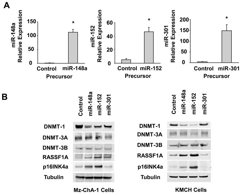
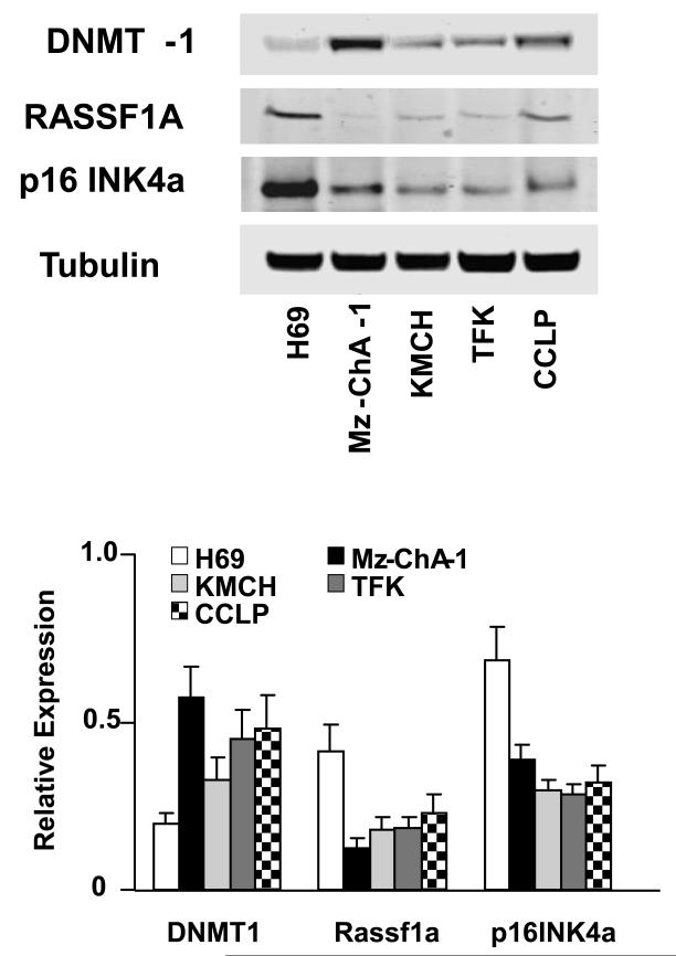
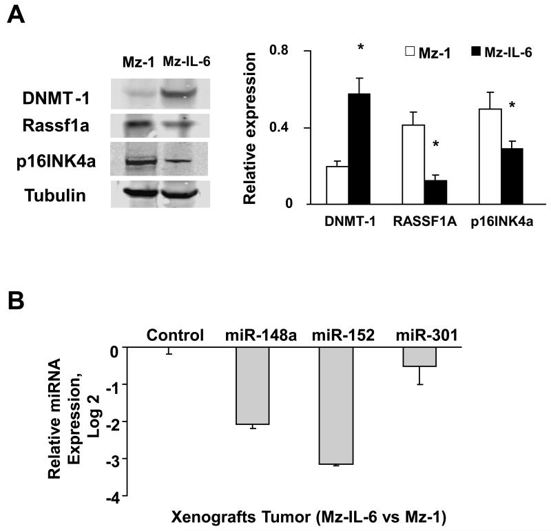
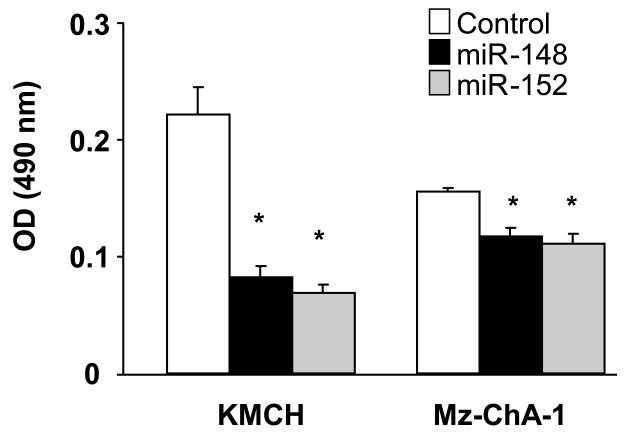
Although the inflammation-associated cytokine interleukin-6 (IL-6) has been implicated in cholangiocarcinoma growth, the relationship between IL-6 and oncogenic changes is unknown. IL-6 can increase expression of DNA methyltransferase-1 (DNMT-1) and epigenetically regulate the expression of several genes, including microRNAs (miRNAs). DNMT-1 up-regulation occurs in hepatobiliary cancers and is associated with a poor prognosis. To understand the potential regulation of DNMT-1 by IL-6-dependent miRNAs, we examined the expression of a group of miRNAs which have sequence complementarity to the 3'-untranslated region of DNMT-1, namely miR-148a, miR-152, and miR-301. The expression of these miRNAs was decreased in cholangiocarcinoma cells. Moreover, the expression of all three miRNAs was decreased in IL-6-overexpressing malignant cholangiocytes in vitro and in tumor cell xenografts. There was a concomitant decrease in expression of the methylation-sensitive tumor suppressor genes Rassf1a and p16INK4a. Using luciferase reporter constructs, DNMT-1 was verified as a target for miR-148a and miR-152. Precursors to miR-148a and miR-152 decreased DNMT-1 protein expression, increased Rassf1a and p16INK4a expression, and reduced cell proliferation.

Conclusion: These data indicate that IL-6 can regulate the activity of DNMT-1 and expression of methylation-dependent tumor suppressor genes by modulation of miR-148a and miR-152, and provide a link between this inflammation-associated cytokine and oncogenesis in cholangiocarcinoma.


Figures

Figure 1
Figure 1. IL-6 alters DNMT-1 protein expression in malignant cholangiocytes
Cell lysates were obtained from Mz-ChA-1 or KMCH cells stably transfected to over-express IL-6 or their respective controls. Immunoblot analysis was performed to assess DNMT-1, p16INK4a, and Rassf1a protein expression. The blots were stripped and reprobed for α-tubulin as a loading control and for quantitation. Representative immunoblots are shown along with quantitative data that show the mean ± standard error from 4 separate blots. * p < 0.05 when compared with respective controls.
Figure 2
Figure 2. Expression of potential DNMT-1 targeting miRNAs in cholangiocarcinoma
miRNA was isolated and profiling performed by hybridization to miRNA-specific probes on epoxy-coated slides. Panel A: Samples from KMCH-1, Mz-ChA-1 or TFK-1 malignant cholangiocytes were labeled with Cy5 whereas samples from H69 nonmalignant cholangiocytes were labeled with Cy3. The data in the panel represent the average ± standard errors of the log2 of the relative expression (ratio of Cy5/Cy3 fluorescence intensity) for each specific miRNA. Panel B: miRNA was isolated and microarray analysis performed from IL-6-overexpressing Mz-ChA-1 cells (Mz-IL-6) and their respective control (Mz-1) cells grown in cell culture in vitro or as tumor cell xenografts in immunodeficient mice. Compared to controls, IL-6 over-expressing Mz-ChA-1 cells (Mz-IL-6) showed a reduced expression of miR-148a and miR-152 in vitro (left panel) as well as in tumor xenografts in vivo (right panel) whereas the reduction of miR-301 was only seen in vitro. Data represents the mean and standard error of the ratio Mz-IL6/Mz-1 in four determinations performed in triplicate. * p < 0.05 when compared with respective controls.
Figure 3
Figure 3. Targeting of DNMT-1 3′-UTR by miRNAs
Panel A: The location of target sites of miR-148a, miR-152 and miR-301 in the DNMT-1 3′-UTR are shown. The sequence of the mutated DNMT-1 3-UTR construct with mutations to disrupt base pairing at shared putative microRNA binding sites is also displayed. Panel B: Mz-ChA-1 cells were transfected with the Renilla luciferase expression construct pRL-tk, the luciferase construct pMIR-DNMT1-WT-luc or pMIR-DNMT1-MUT-luc (in which mutations were introduced in the DNMT-1 target site) and miR-148a, miR-152, miR-301 or control precursor. After 24 h, Dual-Luciferase assays were performed. The decrease in relative firefly luciferase activity was present with miR-148a and miR-152 and the pMIR-DNMT1-WT-luc (black bars), while no effect was detected with the pMIR-DNMT1-MUT-luc construct (white bars), confirming that the 3′-UTR of DNMT-1 is a direct target of modulation by miR-148a and miR-152. The data represent the mean and standard deviations from six determinations from three independent transfections. * p < 0.05 compared to the respective controls.
Figure 4
Figure 4. Increased expression of miR 148 and, miR-152 alters expression of DNMT-1 and selected tumor suppressor genes
Panel A: Mz-ChA-1 cells were transfected with either miR-148a, miR-152, miR-301 or control precursor. After 24 h, real-time PCR assays were performed using TaqMan Human MicroRNA Assay kit and the expression of miRs was normalized to that of the small nuclear RNA U6B (RNU6B). A significant increase in microRNA expression was observed following transfection with precursor miR-148a and miR-152 compared to control miRNA precursors. The data represent the mean and standard errors from four independent transfections. * p < 0.05 relative to respective controls. Panel B: Mz-ChA-1 and KMCH cells were transfected with either miR-148a, miR-152, miR-301 or control precursor. After 72 hours, cell lysates were obtained for Western blot analysis of DNMTs, Rassf1a, and p16INK4a. The blots were stripped and re-probed for α-tubulin as a loading control.
Figure 5
Figure 5. Expression of selected genes in normal and malignant cholangiocytes
Cell lysates were obtained from H69 non-malignant cholangiocytes and KMCH, CC-LP, TFK and MzChA-1 cholangiocarcinoma cell lines. Immunoblot analysis was performed for DNMT-1 and for the tumor suppressor genes RASSF1a, and p16INK4a using antigen specific antibodies. The blots were stripped and reprobed for α-tubulin as a loading control and for quantitation. Representative immunoblots are shown along with quantitative data showing the mean ± standard error from 4 separate blots. For each cholangiocarcinoma cell line, DNMT expression was significantly increased and Rassf1a and p16INK4a expression significantly decreased compared with non-malignant H69 cells, with p<0.05.
Figure 6
Figure 6. Enforced expression of IL-6 alters expression of DNMT-1 targeting miRNA in vivo
IL-6 over-expressing (Mz-IL-6) or control (Mz-1) cells (3×10⁶) were injected subcutaneously into the flank of athymic BALB/c mice. Panel A: Western blot analysis was performed in tissue lysates from tumor cell xenografts using antibodies to DNMT-1, Rassf1a, p16INK4a, and α-tubulin. A representative blot image is shown on left panel whereas the densitometric analysis is shown on right panel. Panel B: miRNA was extracted from xenograft tissues and miR-148a, miR-152 and miR-301 expression was assessed by quantitative real-time PCR. Potential DNMT-1 targeting miRNA expressions were normalized to expression of RNU6B. The data represent results from three experiments performed in triplicate. *p<0.05 when compared with miRNA expression in control cell xenografts.
Figure 7
Figure 7. Effect of potential DNMT-1 targeting miRNAs on cell growth in cholangiocarcinoma cells
Mz-ChA-1 and KMCH cells (5 × 10⁴/well) were plated in 96-well plates after transfection with miRNA-specific precursors to miR-148a and miR-152 or with control precursors (100 nM). Cell proliferation was assessed after 24 hours using a viable cell assay and the proliferation index derived. The mean ± standard errors from 4 separate experiments are illustrated. *p < 0.05 when compared with controls.

References

    1. Patel T. Cholangiocarcinoma. Nat Clin Pract Gastroenterol Hepatol. 2006 Jan;3(1):33–42. - PubMed
    1. Park J, Tadlock L, Gores GJ, Patel T. Inhibition of interleukin 6-mediated mitogen-activated protein kinase activation attenuates growth of a cholangiocarcinoma cell line. Hepatology. 1999 Nov;30(5):1128–1133. - PubMed
    1. Meng F, Yamagiwa Y, Ueno Y, Patel T. Over-expression of interleukin-6 enhances cell survival and transformed cell growth in human malignant cholangiocytes. J Hepatol. 2006 Jun;44(6):1055–1065. - PMC - PubMed
    1. Blechacz B, Gores GJ. Cholangiocarcinoma: Advances in pathogenesis, diagnosis, and treatment. Hepatology. 2008 Mar 3;48(1):308–321. - PMC - PubMed
    1. Hodge DR, Xiao W, Clausen PA, Heidecker G, Szyf M, Farrar WL. Interleukin-6 regulation of the human DNA methyltransferase (HDNMT) gene in human erythroleukemia cells. J Biol Chem. 2001 Oct 26;276(43):39508–39511. - PubMed

Publication types

MeSH terms

Substances