

Multiple-mouse MRI with multiple arrays of receive coils
- PMID: 20146352
- PMCID: PMC2901989
- DOI: 10.1002/mrm.22236
Multiple-mouse MRI with multiple arrays of receive coils
Abstract
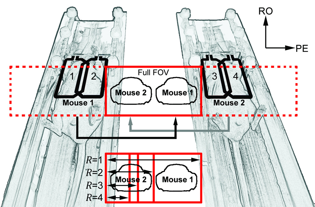
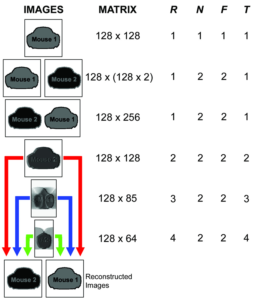
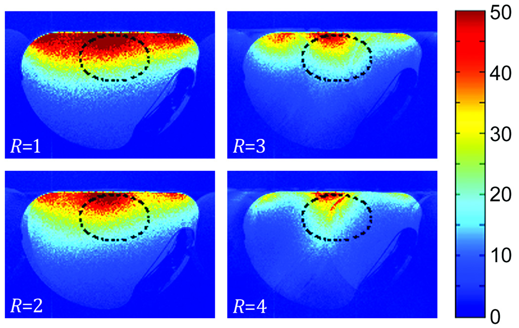
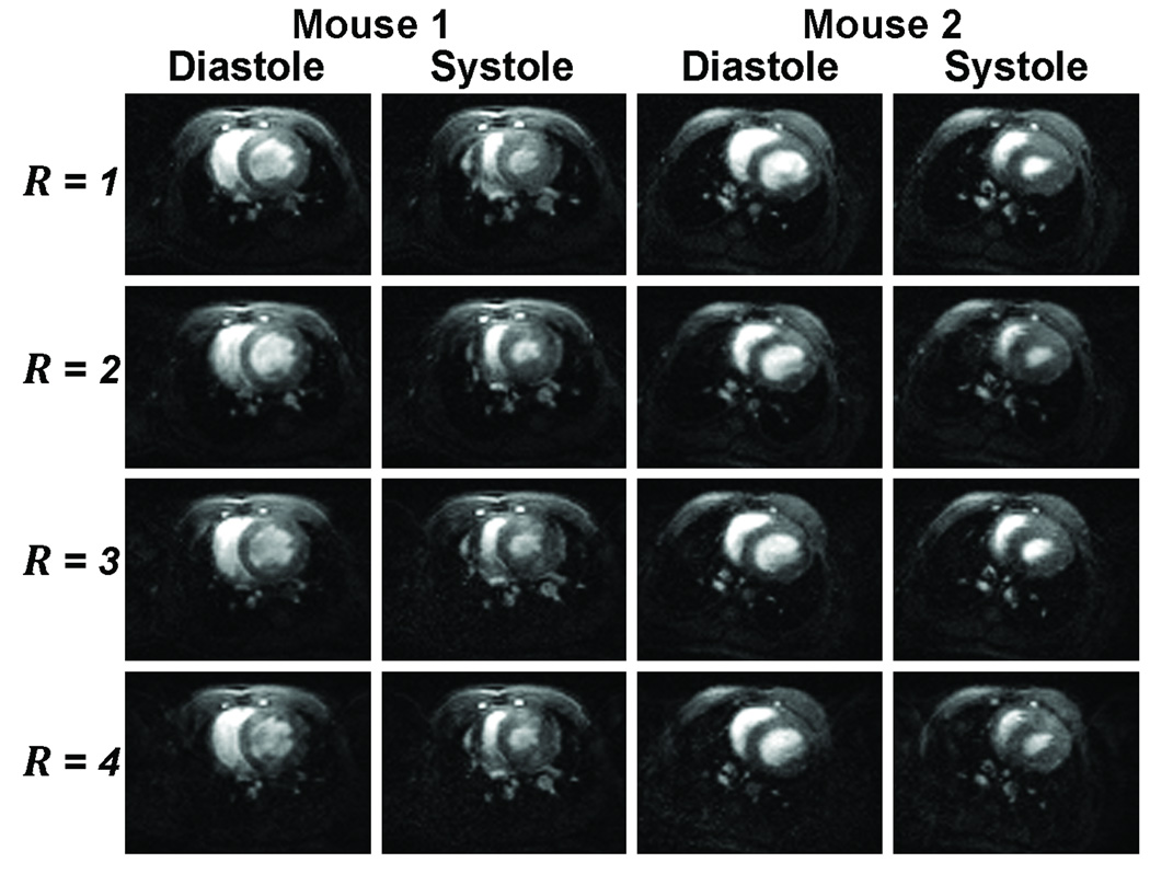
Compared to traditional single-animal imaging methods, multiple-mouse MRI has been shown to dramatically improve imaging throughput and reduce the potentially prohibitive cost for instrument access. To date, up to a single radiofrequency coil has been dedicated to each animal being simultaneously scanned, thus limiting the sensitivity, flexibility, and ultimate throughput. The purpose of this study was to investigate the feasibility of multiple-mouse MRI with a phased-array coil dedicated to each animal. A dual-mouse imaging system, consisting of a pair of two-element phased-array coils, was developed and used to achieve acceleration factors greater than the number of animals scanned at once. By simultaneously scanning two mice with a retrospectively gated cardiac cine MRI sequence, a 3-fold acceleration was achieved with signal-to-noise ratio in the heart that is equivalent to that achieved with an unaccelerated scan using a commercial mouse birdcage coil.
(c) 2010 Wiley-Liss, Inc.
Figures
Similar articles
-
A throughput-optimized array system for multiple-mouse MRI.NMR Biomed. 2013 Mar;26(3):237-47. doi: 10.1002/nbm.2841. Epub 2012 Aug 10. NMR Biomed. 2013. PMID: 22887122 Free PMC article.
-
A 20-channel receive-only mouse array coil for a 3 T clinical MRI system.Magn Reson Med. 2011 Aug;66(2):584-95. doi: 10.1002/mrm.22791. Epub 2011 Mar 23. Magn Reson Med. 2011. PMID: 21433066 Free PMC article.
-
A semiflexible 64-channel receive-only phased array for pediatric body MRI at 3T.Magn Reson Med. 2016 Sep;76(3):1015-21. doi: 10.1002/mrm.25999. Epub 2015 Sep 29. Magn Reson Med. 2016. PMID: 26418283 Free PMC article.
-
Characterization of the impact to PET quantification and image quality of an anterior array surface coil for PET/MR imaging.MAGMA. 2014 Apr;27(2):149-59. doi: 10.1007/s10334-013-0388-1. Epub 2013 Jun 26. MAGMA. 2014. PMID: 23800803 Review.
-
Mouse morphological phenotyping with magnetic resonance imaging.Methods Mol Med. 2006;124:103-27. doi: 10.1385/1-59745-010-3:103. Methods Mol Med. 2006. PMID: 16506419 Review.
Cited by
-
Feasibility of multianimal hyperpolarized (13) C MRS.Magn Reson Med. 2015 May;73(5):1726-32. doi: 10.1002/mrm.25307. Epub 2014 Jun 5. Magn Reson Med. 2015. PMID: 24903532 Free PMC article.
-
Accelerated cardiac magnetic resonance imaging in the mouse using an eight-channel array at 9.4 Tesla.Magn Reson Med. 2011 Jan;65(1):60-70. doi: 10.1002/mrm.22605. Magn Reson Med. 2011. PMID: 20740650 Free PMC article.
-
A throughput-optimized array system for multiple-mouse MRI.NMR Biomed. 2013 Mar;26(3):237-47. doi: 10.1002/nbm.2841. Epub 2012 Aug 10. NMR Biomed. 2013. PMID: 22887122 Free PMC article.
-
Functional and morphological cardiac magnetic resonance imaging of mice using a cryogenic quadrature radiofrequency coil.PLoS One. 2012;7(8):e42383. doi: 10.1371/journal.pone.0042383. Epub 2012 Aug 1. PLoS One. 2012. PMID: 22870323 Free PMC article.
-
Mouse Cardiovascular Imaging.Curr Protoc. 2024 Sep;4(9):e1116. doi: 10.1002/cpz1.1116. Curr Protoc. 2024. PMID: 39222027 Review.
References
-
- Roemer PB, Edelstein WA, Hayes CE, Souza SP, Mueller OM. The NMR phased array. Magn Reson Med. 1990;16(2):192–225. - PubMed
-
- Pruessmann KP, Weiger M, Scheidegger MB, Boesiger P. SENSE: sensitivity encoding for fast MRI. Magn Reson Med. 1999;42(5):952–962. - PubMed
-
- Sodickson DK, Manning WJ. Simultaneous acquisition of spatial harmonics (SMASH): fast imaging with radiofrequency coil arrays. Magn Reson Med. 1997;38(4):591–603. - PubMed
-
- Griswold MA, Jakob PM, Heidemann RM, Nittka M, Jellus V, Wang J, Kiefer B, Haase A. Generalized autocalibrating partially parallel acquisitions (GRAPPA) Magn Reson Med. 2002;47(6):1202–1210. - PubMed
-
- Griswold MA, Jakob PM, Nittka M, Goldfarb JW, Haase A. Partially parallel imaging with localized sensitivities (PILS) Magn Reson Med. 2000;44(4):602–609. - PubMed
Publication types
MeSH terms
Grants and funding
LinkOut - more resources
Full Text Sources
Medical
