Review
. 2010 May;16(14):1568-81.
doi: 10.2174/138161210791164180.

In vivo beta-cell imaging with VMAT 2 ligands--current state-of-the-art and future perspective

Affiliations
Review

In vivo beta-cell imaging with VMAT 2 ligands--current state-of-the-art and future perspective

Rajakrishnan Veluthakal et al. Curr Pharm Des. 2010 May.

Abstract

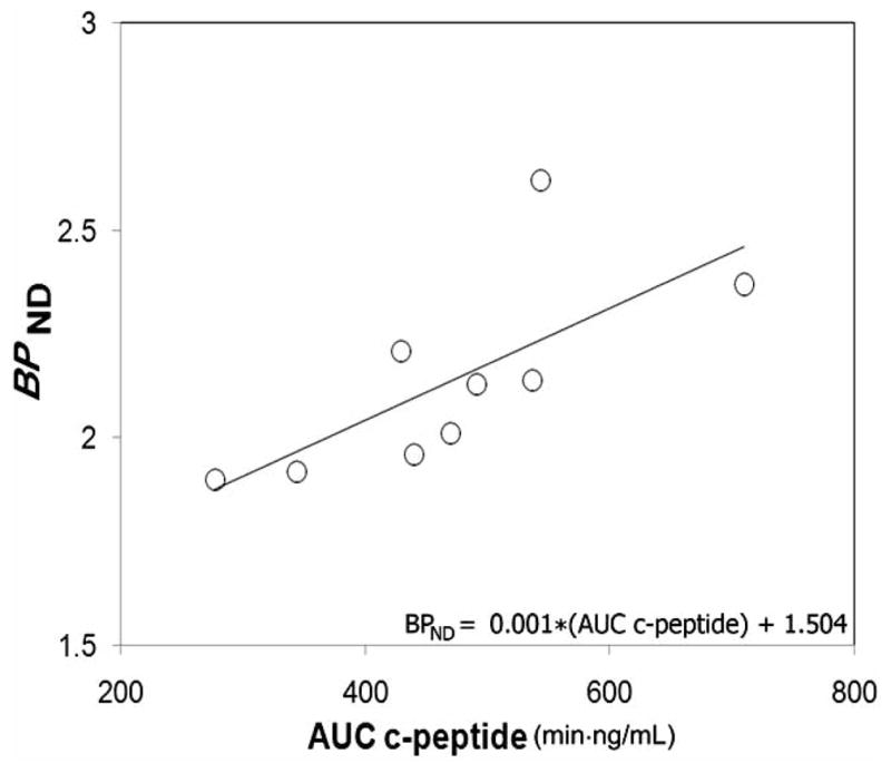
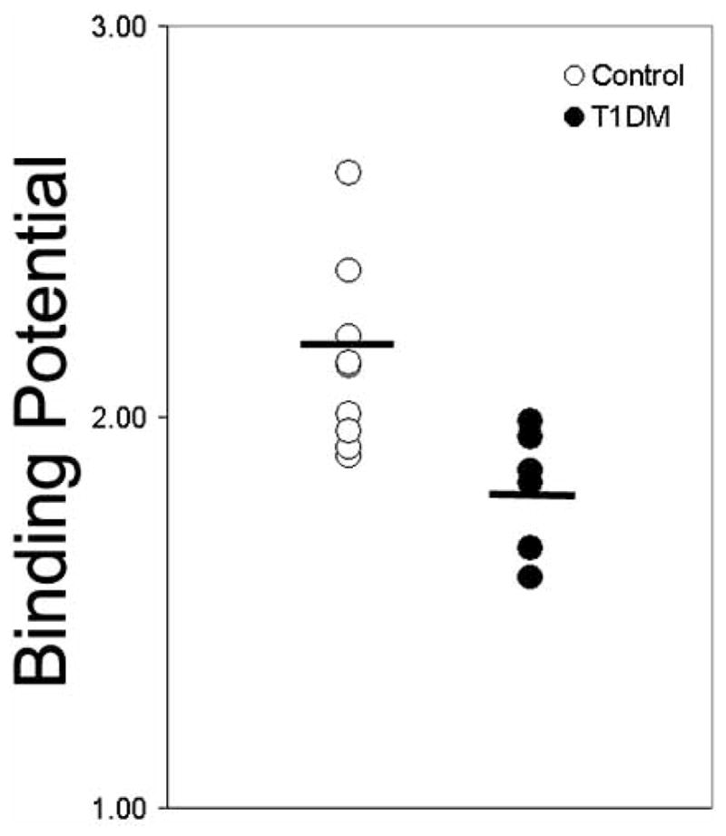
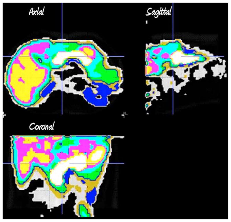
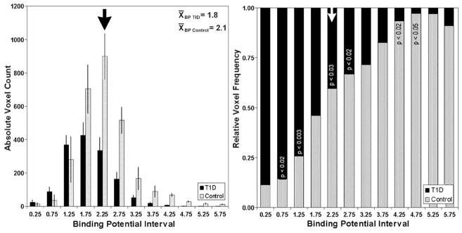
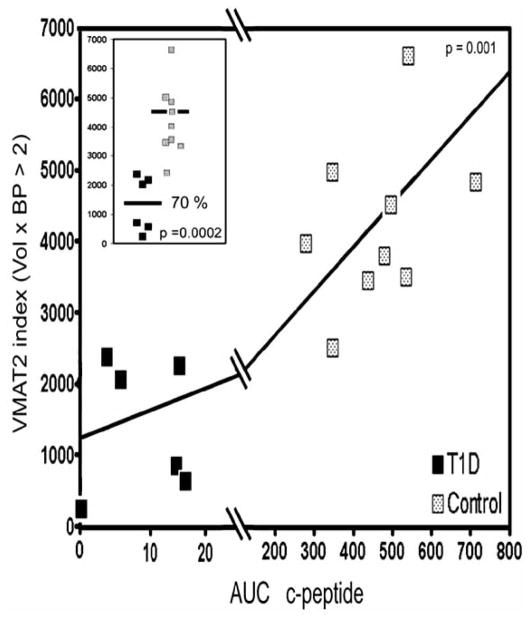
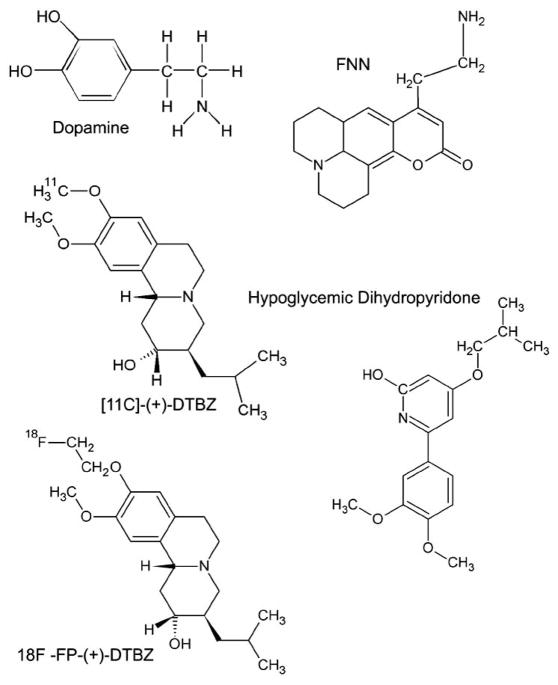
In diabetic disease, blood glucose, HbA1c and insulin levels qualify as biomarkers reflecting endocrine pancreas function, but their shortfall in being truly useful predictors or surrogate endpoints of "abnormal processes or disease" lies in that alteration in their levels are dependent on a variety comorbidities and occur too late in the disease process to be useful sentinels. Non invasive imaging of molecular targets within the beta cell carry the promise of revealing quantitative information about beta-cell mass that can, at least theoretically, be used to monitor, in real-time, the natural history of T1DM progression, assess novel therapies designed to drive the proliferation and differentiation of endogenous beta cell progenitors, appraise methods of preserving mature beta cell mass as well as to track the function and viability of transplanted cells and tissues. In this article, we review and deconstruct available information regarding the methodology of making non invasive measurements of VMAT2 in the pancreas and the validity of these measurements to estimate beta cell mass in vivo.


Figures

Fig. 1
Fig. 1
Association between binding potential and glucose-stimulated insulin secretion in control population. Average pancreatic BPND values and AUC c-peptide measures for each healthy control individual were evaluated for strength of association by linear correlation. The regression line BPND = 0.001 * AUC c-peptide + 1.504 yielded a correlation coefficient of r2 = 0.50 and p =0.03. The X and Y intercepts were −1504 and 1.504, respectively. Using non parametric statistics and analyzing the data from T1D patients and controls together, we found a significant correlation (r = 0.82, p =0.001) between insulin secretion, measured as AUC c-peptide, and binding potential (BPND).
Fig. 2
Fig. 2
Comparison of Binding Potentials in a population of healthy euglycemic individuals and a weight age matched population of patients with long-term type 1 diabetes. The average non thresholded binding potential is shown by the black bar. The p-value for the difference in the means was 0.014.
Fig. 3
Fig. 3
Tissue segmentation by clustering of voxel kinetic data. Axial, sagittal and coronal sections of the clustered kinetic data accurate define the different abdominal tissues. Following clustering each kinetic family of voxel data is assigned a different color.
Fig. 4
Fig. 4
Histogram summary of voxel BPND in control and T1D pancreata. Left panel. The average voxel count at each BPND interval is displayed for controls (n=9) in gray and patients with T1D (n=6) in black. The error bars represent the S.E.M. Right panel. The voxel counts at each BPND interval were normalized to the total number of voxels for controls in gray and patients with T1D in black. The significance of the difference at each BP interval is shown in the stacked columns.
Fig. 5
Fig. 5
Relationship between thresholded VMAT2 index (BPnd ≥ 2.5) and insulin secretory capacity in healthy controls (grey squares) and patients with longstanding type 1 diabetes (filled squares). The strength and significance of the relationship between VMAT2 index and AUC C-peptide was calculated using the Spearman Rank nonparametric method (r =0.82, p < 0.002). Inset: the mean values of the VMAT2 index for healthy subjects and patients with T1DM are shown (black bars) as well as the individual values. The method of Student was used to calculate the two tailed p value.
Fig. 6
Fig. 6
Examination of the Population Variability in VMAT2 in a cohort of BB rats. Young euglycemic BB rats ( > 7 weeks old) and BB rats with frank diabetes (blood glucose > 350 mg/dL and >12 weeks old) were scanned with [11C] (+) DTBZ and the VMAT2 quantitatied as max SUV. The p value for the difference between the population means was less than <0.001.
Fig. 7
Fig. 7. Comparison of radioligands, VMAT2 quantitation and blocking studies
Rodents were anesthetized and imaged dynamically with about 7–11 MBq of [11C]DTBZ (left panel) or [18F]FP DTBZ (right panel). In some studies cold DTBZ was given before or after hot ligand. From reconstructions of the dynamic image data, pancreas (Pan A–C) and kidney (Kidney A–C) regions of interest were identified to obtain time activity curves (TACs) corrected for dose and animal weight (displayed as SUV TACs). The extent to which excess cold ligand is able to block the radioactivity taken up by the pancreas is shown in the % block column. Comparing this value across left and right panels shows that [18F]-FP-DTBZ is more readily displaced relative to [11C] (+) DTBZ by cold DTBZ (2 mg/Kg) as shown by the higher % blocking value. SUV or standardized uptake value is a measure of radioligand uptake that compares the concentration of radioligand in tissue of interest to what it would hypothetically be where the dose to have been dispersed in a glass of water of equal volume/mass. In each case it can be seen that [18F]-FP-DTBZ gives a higher signal.
Fig. 8
Fig. 8
Dopamine tissue concentrations in saline and AMPT-treated rat brains and pancreata. Data are mean +/− S.E.M. for N = 6 animals.
Fig. 9
Fig. 9
Structures for compounds discussed in article.

Similar articles

Cited by

References

    1. BDW Group. Biomarkers and surrogate endpoints: preferred definitions and conceptual framework. Clin Pharmacol Ther. 2001;69:89–95. - PubMed
    1. http://www.cancer.gov/dictionary/?CdrID=45618.

    1. Gepts W. Pathologic anatomy of the pancreas in juvenile diabetes mellitus. Diabetes. 1965;14:619–33. - PubMed
    1. Gepts W, Lecompte PM. The pancreatic islets in diabetes. Am J Med. 1981;70:105–15. - PubMed
    1. Saito K, Yaginuma N, Takahashi T. Differential volumetry of A, B and D cells in the pancreatic islets of diabetic and nondiabetic subjects. Tohoku J Exp Med. 1979;129:273–83. - PubMed

Publication types

MeSH terms

LinkOut - more resources