

Thymic stromal lymphopoietin
- PMID: 20146705
- PMCID: PMC2895428
- DOI: 10.1111/j.1749-6632.2009.05128.x
Thymic stromal lymphopoietin
Abstract
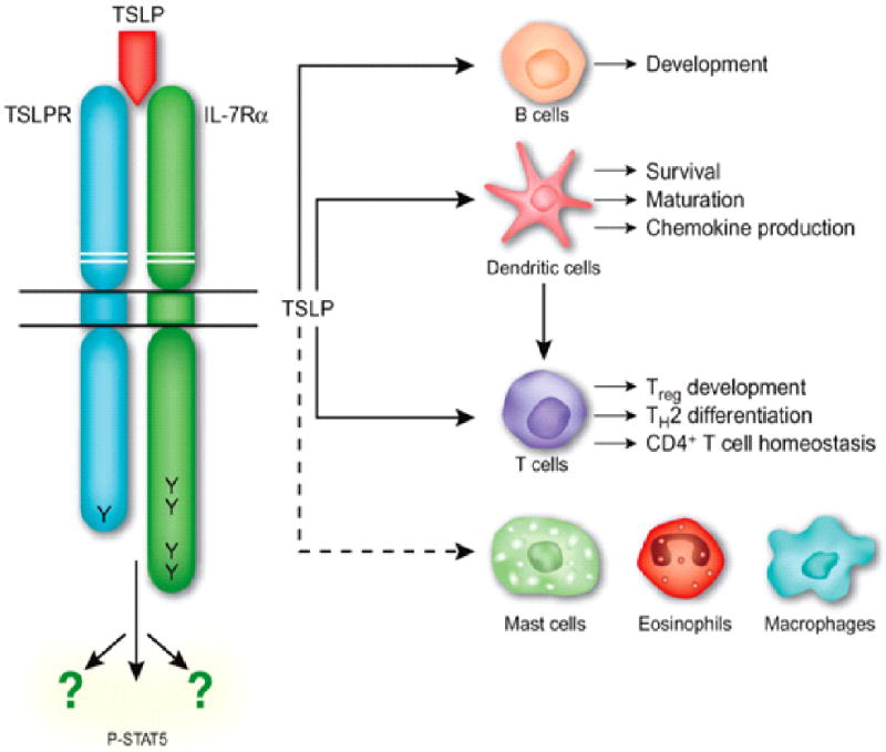
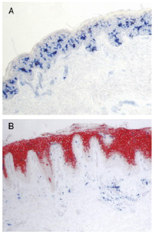
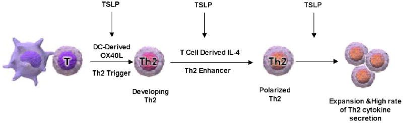
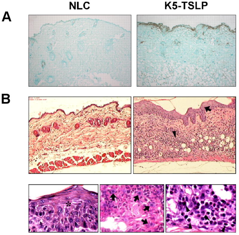
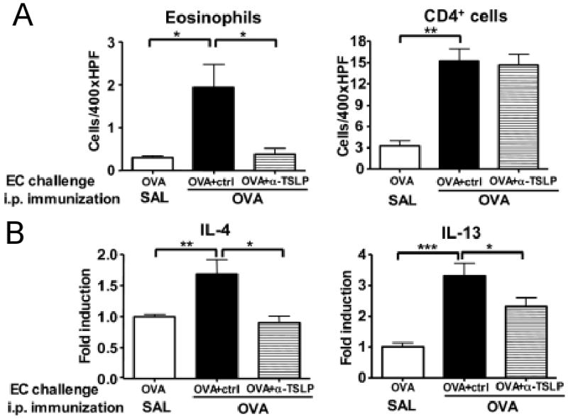
Thymic stromal lymphopoietin (TSLP) is an epithelial cell-derived cytokine expressed in skin, gut, lungs, and thymus. TSLP signals via a TSLP receptor (TSLPR), a heterodimer of the IL-7 receptor alpha chain and the TSLPR chain. The TSLPR chain is closely related to the common receptor gamma chain that is expressed on a wide range of cell types in the adaptive and innate immune system. TSLP exerts a profound influence on the polarization of dendritic cells to drive T helper (Th) 2 cytokine production. TSLP also directly promotes T-cell proliferation in response to T-cell receptor activation and Th2 cytokine production and supports B-cell expansion and differentiation. TSLP further amplifies Th2 cytokine production by mast cells and natural killer T cells. These properties confer on TSLP a critical role in driving Th2-mediated inflammation. This role is supported by the finding that TSLP expression is upregulated in keratinocytes of atopic dermatitis skin lesions and in bronchial epithelial cells in asthma.
Figures
Similar articles
-
Expression and Regulation of Thymic Stromal Lymphopoietin and Thymic Stromal Lymphopoietin Receptor Heterocomplex in the Innate-Adaptive Immunity of Pediatric Asthma.Int J Mol Sci. 2018 Apr 18;19(4):1231. doi: 10.3390/ijms19041231. Int J Mol Sci. 2018. PMID: 29670037 Free PMC article. Review.
-
Myeloid dendritic cells are primed in allergic asthma for thymic stromal lymphopoietin-mediated induction of Th2 and Th9 responses.Allergy. 2014 Aug;69(8):1068-76. doi: 10.1111/all.12435. Epub 2014 Jun 3. Allergy. 2014. PMID: 24888572
-
Thymic stromal lymphopoietin receptor blockade reduces allergic inflammation in a cynomolgus monkey model of asthma.J Allergy Clin Immunol. 2013 Aug;132(2):455-62. doi: 10.1016/j.jaci.2013.05.011. Epub 2013 Jun 26. J Allergy Clin Immunol. 2013. PMID: 23810153
-
Early production of thymic stromal lymphopoietin precedes infiltration of dendritic cells expressing its receptor in allergen-induced late phase cutaneous responses in atopic subjects.Allergy. 2009 Jul;64(7):1014-22. doi: 10.1111/j.1398-9995.2009.01947.x. Epub 2009 Feb 2. Allergy. 2009. PMID: 19187393
-
The role of thymic stromal lymphopoietin in allergic inflammation and chronic obstructive pulmonary disease.Arch Immunol Ther Exp (Warsz). 2010 Apr;58(2):81-90. doi: 10.1007/s00005-010-0064-3. Epub 2010 Feb 9. Arch Immunol Ther Exp (Warsz). 2010. PMID: 20143171 Review.
Cited by
-
Acute exposure of ozone induced pulmonary injury and the protective role of vitamin E through the Nrf2 pathway in Balb/c mice.Toxicol Res (Camb). 2015 Nov 10;5(1):268-277. doi: 10.1039/c5tx00259a. eCollection 2016 Jan 1. Toxicol Res (Camb). 2015. PMID: 30090343 Free PMC article.
-
Cutaneous Neuroimmune Interactions of TSLP and TRPV4 Play Pivotal Roles in Dry Skin-Induced Pruritus.Front Immunol. 2021 Dec 2;12:772941. doi: 10.3389/fimmu.2021.772941. eCollection 2021. Front Immunol. 2021. PMID: 34925342 Free PMC article.
-
The importance of TSLP in allergic disease and its role as a potential therapeutic target.Expert Rev Clin Immunol. 2014 Nov;10(11):1463-74. doi: 10.1586/1744666X.2014.967684. Expert Rev Clin Immunol. 2014. PMID: 25340427 Free PMC article. Review.
-
Aberrant T cell immunity triggered by human Respiratory Syncytial Virus and human Metapneumovirus infection.Virulence. 2017 Aug 18;8(6):685-704. doi: 10.1080/21505594.2016.1265725. Epub 2016 Dec 2. Virulence. 2017. PMID: 27911218 Free PMC article. Review.
-
Skin-derived TSLP systemically expands regulatory T cells.J Autoimmun. 2017 May;79:39-52. doi: 10.1016/j.jaut.2017.01.003. Epub 2017 Jan 23. J Autoimmun. 2017. PMID: 28126203 Free PMC article.
References
-
- Friend SL, Hosier S, Nelson A, et al. Exp Hematol. 1994;22(3):321. - PubMed
-
- Quentmeier H, Drexler HG, Fleckenstein D, et al. Leukemia. 2001;15(8):1286. - PubMed
-
- Soumelis V, Reche PA, Kanzler H, et al. Nature immunology. 2002;3(7):673. - PubMed
-
- Rimoldi M, Chieppa M, Salucci V, et al. Nature immunology. 2005;6(5):507. - PubMed
Publication types
MeSH terms
Substances
Grants and funding
LinkOut - more resources
Full Text Sources
Other Literature Sources
Medical
Molecular Biology Databases
