

Behavior of the electrocardiographic T peak to end interval in childhood
- PMID: 20146777
- PMCID: PMC6932471
- DOI: 10.1111/j.1542-474X.2009.00334.x
Behavior of the electrocardiographic T peak to end interval in childhood
Abstract
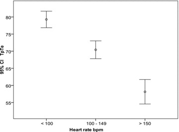
Background: The T-wave peak to T-wave end (TpTe) interval reflects spatial and transmural dispersion in repolarization and serves as an arrhythmogenic index. Normal TpTe interval data in children are lacking. We evaluated the effects of age, gender, heart rate, leads (II and V(5)) on TpTe and T-wave voltage.
Methods: Four hundred healthy children (age 4 days to 16.7 years) were enrolled. From a resting 12-lead digital ECG, TpTe, RR, QT, JT intervals, and T amplitude were measured (leads II and V(5)). Bazett and Fridericia formulas were applied to TpTe for heart rate correction and TpTe/QT and TpTe/JT were calculated. Descriptive and analytical statistics were applied, significance level set at P < or = 0.05.
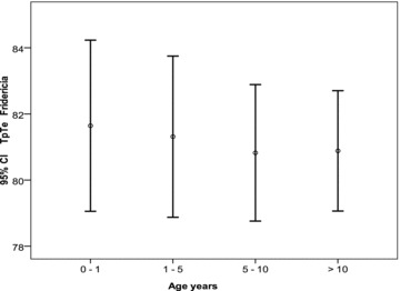
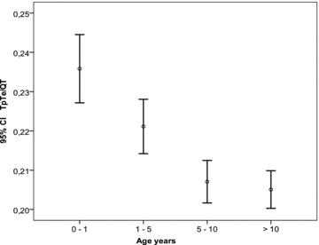
Results: TpTe in leads II and V(5) correlate well. Contrary to adults, no gender differences in TpTe were observed in childhood. TpTe lengthens with increasing age, and is inversely related to heart rate. TpTe 98th percentile is 85 msec in first 5 years, increasing to 92 msec in adolescence. TpTe Fridericia is a good correction formula in childhood; TpTe Bazett overcorrects in the younger age. TpTe/QT and TpTe/JT are longer in younger subjects due to greater QT shortening than the TpTe interval at higher heart rates.
Conclusions: In children, TpTe in lead II and V(5) correlate well. The TpTe interval lengthens with advancing age as heart rate diminishes. TpTe Fridericia is a good correction formula in children. Younger subjects have higher TpTe/QT and TpTe/JT indices compared to older children. T-wave voltage increases with age, tallest in the 5-10-year-age group particularly in V(5).
Figures
References
-
- Xue J, Gao W, Chen Y, et al Study of repolarization heterogeneity and electrocardiographic morphology with a modeling approach. J Electrocardiol 2008;41:581–587. - PubMed
-
- Yan GX, Antzelevitch C. Cellular basis for the normal T wave and the electrocardiographic manifestations of the long‐QT syndrome. Circulation 1998;98:1928–1936. - PubMed
-
- Yan GX, Shimizu W, Antzelevitch C. The characteristics and distribution of M cells in arterially‐perfused canine left ventricular wedge preparations. Circulation 1998;98:1921–1927. - PubMed
MeSH terms
LinkOut - more resources
Full Text Sources
Other Literature Sources
