. 2010 Feb 11:11:13.
doi: 10.1186/1471-2121-11-13.

Role of apoptosis-inducing factor (AIF) in programmed nuclear death during conjugation in Tetrahymena thermophila

Affiliations

Role of apoptosis-inducing factor (AIF) in programmed nuclear death during conjugation in Tetrahymena thermophila

Takahiko Akematsu et al. BMC Cell Biol. .

Abstract

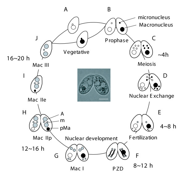
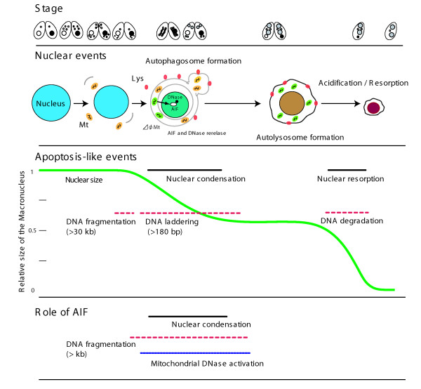
Background: Programmed nuclear death (PND), which is also referred to as nuclear apoptosis, is a remarkable process that occurs in ciliates during sexual reproduction (conjugation). In Tetrahymena thermophila, when the new macronucleus differentiates, the parental macronucleus is selectively eliminated from the cytoplasm of the progeny, concomitant with apoptotic nuclear events. However, the molecular mechanisms underlying these events are not well understood. The parental macronucleus is engulfed by a large autophagosome, which contains numerous mitochondria that have lost their membrane potential. In animals, mitochondrial depolarization precedes apoptotic cell death, which involves DNA fragmentation and subsequent nuclear degradation.

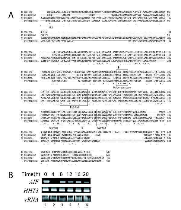
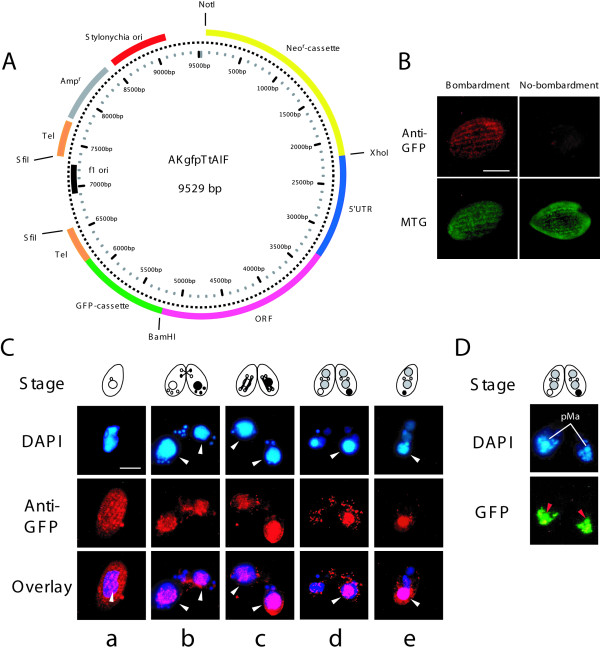
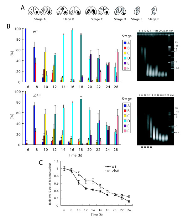
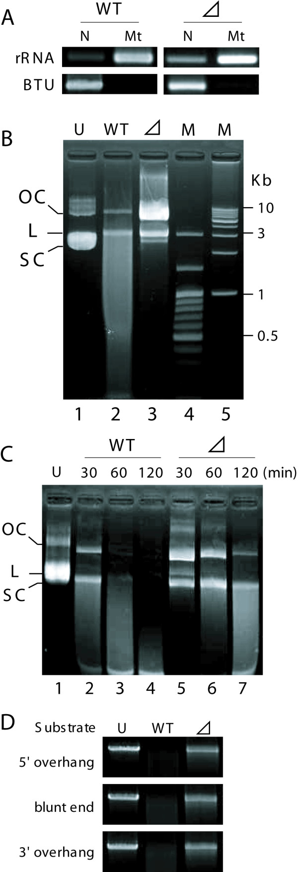
Results: We focused on the role of mitochondrial apoptosis-inducing factor (AIF) during PND in Tetrahymena. The disruption of AIF delays the normal progression of PND, specifically, nuclear condensation and kilobase-size DNA fragmentation. AIF is localized in Tetrahymena mitochondria and is released into the macronucleus prior to nuclear condensation. In addition, AIF associates and co-operates with the mitochondrial DNase to facilitate the degradation of kilobase-size DNA, which is followed by oligonucleosome-size DNA laddering.

Conclusions: Our results suggest that Tetrahymena AIF plays an important role in the degradation of DNA at an early stage of PND, which supports the notion that the mitochondrion-initiated apoptotic DNA degradation pathway is widely conserved among eukaryotes.


Figures

Figure 1
Figure 1
Nuclear events during conjugation of Tetrahymena thermophila. Conjugation in T. thermophila is a complicated process that is initiated by interaction between complementary mating types, which form a conjugating pair. A. Vegetative phase. B. Meiotic prophase. C. Meiosis. D. Nuclear exchange. One of four meiotic products mitotically divides, forming two pronucei. Subsequently, one of the pronuclei is reciprocally exchanged between mating partners. E. Fertilization (synkaryon formation). F. PZD (postzygotic division). Fertilized nucleus successively divides twice. G. Mac I. Anteriorly-located nuclei differentiate into the new macronuclei, while posterior nuclei remain the micronuclei. H. Mac IIp. The parental macronucleus migrates posteriorly and begins to degenerate. I. Mac IIe. Pair separates (exconjugants). J. Mac III. One of two micronuclei is eliminated. Progeny of T. thermophila do not undergo conjugation during the first ~100 vegetative fissions after conjugation called "immature." A: macronuclear anlagen. m: micronuclei. pMa: parental macronucleus. The scale bar in photograph indicates 10 μm.
Figure 2
Figure 2
Sequence alignment of AIF proteins and analysis of AIF expression. A. CLUSTAL-W was used to generate AIF sequence alignment, including Homo sapiens, Dictyostelium discoideum, Caenorhabditis elegans and Tetrahymena thermophila. Boxes indicate the NAD/FAD binding domain and oxidoreductase domain. MLS in N-terminal portion of T.thermohila indicates mitochondrial localization sequence. Asterisks indicate identical amino acids. Colons and semicolons indicate amino acid similarity. Arrowheads indicate a potential DNA binding site of human AIF. B. RT-PCR analysis of AIF transcript during conjugation. Histone h3 (HHT3) was used as a control. SSU rRNA was used as a loading control.
Figure 3
Figure 3
Translocation of AIF from mitochondria to parental macronucleus. A. Map of expression vector, named AKgfpTtAIF, with neomycin resistance cassette (Neor-cassette), 5'UTR and ORF of AIF, codon-optimized GFP (GFP-cassette), replication origin derived from Stylonychia lemnae and telomeres from Tetrahymena. Neomycin resistance is expressed under control of β-tubulin promoter. Before biolistic bombardment, the plasmid was digested with SfiI to expose telomere sequences on both ends. B. After biolistic bombardment, cytoplasmic localization of AIF::GFP was detected using α-GFP. This fluorescent pattern was coincided with MitoTracker Green (MTG). No-bombardment indicates non-transformed wild-type cell. Scale bar in photograph indicates 10 μm. C. Translocation of AIF::GFP was visualized with α-GFP antibodies. White arrows indicate parental macronucleus. Overlay represents fusion image of blue (nuclei) and red (AIF::GFP) fluorescence. Scale bar in photograph indicates 10 μm. D. Fluorescence microscopy of living cells expressing AIF::GFP at the stage of MacIIp. Red arrows indicate AIF::GFP in parental macronuclei. pMa denotes parental macronucleus.
Figure 4
Figure 4
Construction of AIF-deficient strain. A. Map of expression vector, pKoTtAIF, with neomycin resistance cassette (Neor-cassette). It consists of 5' and 3' UTR sequences of AIF and part of its 3' ORF. The neomycin resistance is expressed under the control of β-tubulin promoter. Before biolistic bombardment, the plasmid was digested with BamHI. B. Schematic representation of the wild-type (WT) and mutant locus of the AIF together with the targeting plasmid. Replacement of AIF to neomycin-resistant gene (Neor) in the macronucleus was confirmed by PCR using 10 ng of genomic DNA from isolated macronuclei as template. Small triangles located in the gene loci indicate specific primer pairs for the PCR amplification. C. Cell growth curve of CU428. Circles and squares indicate cell density of wild-type strain and ΔAIF, respectively. Points and attached bars correspond to the means of four identical measurements and standard deviations. The inset indicates RT-PCR analysis of the expression levels of AIF and Neor. β-tubulin (BTU) was used as a control.
Figure 5
Figure 5
Progression of PND by disruption of AIF. A. Nuclear events during conjugation were divided into 6 stages (stage A ~stage F). B. Time course analysis of progression of the nuclear events in wild-type and ΔAIF between 6 h and 28 h after initiation of conjugation. The percentages of the nuclear stages were counted, and were expressed as a percentage of the total number of tested cells (300-400 cells). Columns and attached bars correspond to the means of four identical measurements and standard deviations. Fragmental DNA was isolated from the strains every 2 h during conjugation. The black stars between 8-12 h in ΔAIF indicate delay of kb-sized DNA fragmentation. M denotes kbp-ladder size marker (left) and 100-bp ladder size marker (right). C. Changes in size of parental macronuclei between 6 h and 28 h after initiation of conjugation. Columns (points) and attached bars correspond to the means of four identical measurements (80-100 cells) and standard deviations.
Figure 6
Figure 6
Mitochondrial nuclease activity. A. Fractionation PCR. A partial fragment of the mitochondrial large subunit ribosomal RNA (rRNA) or β-tubulin (BTU) was amplified by PCR, using fraction samples from wild-type and ΔAIF that contained equal amounts of DNA. N and Mt indicate nuclei/unbroken cell fraction and mitochondrial fraction, respectively. No contamination of nuclear DNA was detected in mitochondrial fraction. B. Purified mitochondria (2 μg protein) from wild-type (lane 2) and ΔAIF (lane 3) were incubated with 2 μg substrate plasmid DNA with a circular form for 30 min at 37°C in 30 μl reaction buffer containing 20 mM KCl and 50 mM MOPS (pH 6.5). Lane 4 (M) and 5 (M) indicate 100-bp ladder size marker and λHindIII-digest, respectively. The substrate DNA appears in the nicked open circular (OC), linear (L), and supercoiled (SC) forms. C. The nuclease assay was performed under various incubation times. Lane 2-4, substrate DNA was coincubated with wild-type mitochondria. Lane 5-7, substrate DNA was coincubated with ΔAIF mitochondria. Undigested sample is seen in lane 1. D. Substrate specificity of the activities. End forms of linear plasmids with 5'- or 3'-overhang or with blunt ends are indicated at the left of gel.
Figure 7
Figure 7
Schematic representation of PND and a possible role of AIF, based on information described in [6]and from the present study.

Similar articles

Cited by

References

    1. Prescott DM. The DNA of ciliated protozoa. Microbiol Rev. 1994;58:233–267. - PMC - PubMed
    1. Mpoke S, Wolfe J. DNA digestion and chromatin condensation during nuclear death in Tetrahymena. Exp Cell Res. 1996;225:357–365. doi: 10.1006/excr.1996.0186. - DOI - PubMed
    1. Davis MC, Ward JG, Herrick G, Allis CD. Programmed nuclear death: apoptotic-like degradation of specific nuclei in conjugating Tetrahymena. Dev Biol. 1992;154:419–432. doi: 10.1016/0012-1606(92)90080-Z. - DOI - PubMed
    1. Kobayashi T, Endoh H. Caspase-like activity in programmed nuclear death during conjugation of Tetrahymena thermophila. Cell Death Differ. 2003;10:634–640. doi: 10.1038/sj.cdd.4401216. - DOI - PubMed
    1. Lu E, Wolfe J. Lysosomal enzymes in the macronucleus of Tetrahymena during its apoptosis-like degradation. Cell Death Differ. 2001;8:289–297. doi: 10.1038/sj.cdd.4400807. - DOI - PubMed

Publication types

MeSH terms

Substances

LinkOut - more resources