. 2010 Apr 2;285(14):10362-9.
doi: 10.1074/jbc.M109.092544. Epub 2010 Feb 9.

Human ELG1 regulates the level of ubiquitinated proliferating cell nuclear antigen (PCNA) through Its interactions with PCNA and USP1

Affiliations

Human ELG1 regulates the level of ubiquitinated proliferating cell nuclear antigen (PCNA) through Its interactions with PCNA and USP1

Kyoo-Young Lee et al. J Biol Chem. .

Abstract

The level of monoubiquitinated proliferating cell nuclear antigen (PCNA) is closely linked with DNA damage bypass to protect cells from a high level of mutagenesis. However, it remains unclear how the level of monoubiquitinated PCNA is regulated. Here, we demonstrate that human ELG1 protein, which comprises an alternative replication factor C (RFC) complex and plays an important role in preserving genomic stability, as an interacting partner for the USP1 (ubiquitin-specific protease 1)-UAF1 (USP1-associated factor 1) complex, a deubiquitinating enzyme complex for PCNA and FANCD2. ELG1 protein interacts with PCNAs that are localized at stalled replication forks. ELG1 knockdown specifically resulted in an increase in the level of PCNA monoubiquitination without affecting the level of FANCD2 ubiquitination. It is a novel function of ELG1 distinct from its role as an alternative RFC complex because knockdowns of any other RFC subunits or other alternative RFCs did not affect PCNA monoubiquitination. Lastly, we identified a highly conserved N-terminal domain in ELG1 that was responsible for the USP1-UAF1 interaction as well as the activity to down-regulate PCNA monoubiquitination. Taken together, ELG1 specifically directs USP1-UAF1 complex for PCNA deubiquitination.


Figures
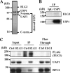
FIGURE 1.
FIGURE 1.
ELG1 interacts with USP1-UAF1 complex. A, the USP1 complex was purified from HeLa nuclear extracts and stained by Coomassie blue stain. Proteins indicated were identified by mass spectrometry. B, nuclear extracts from HeLa cells were immunoprecipitated (IP) with anti-USP1-antibody or IgG control antibody. The immunoprecipitated proteins were identified with the antibodies indicated. C, HEK293T cells were transected with FLAG-ELG1 expression vectors or control vectors (Ctrl). Total extracts from the transfected cells were IP after 48 h with anti-FLAG-antibody. The immunoprecipitated proteins were identified with the antibodies indicated.
FIGURE 2.
FIGURE 2.
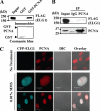
ELG1 interacts with PCNA. A, purified GST or GST-PCNA proteins were mixed with protein extracts containing FLAG-ELG1 and pulled down for immunoblot analysis. B, HEK293T cells were transected with FLAG-ELG1 expression vectors. Immunoprecipitations were carried out on nuclear extracts using an anti-PCNA antibody, and the eluate was then subjected to immunoblot analysis. C, ELG1 and PCNA co-localize in response to DNA damage. RPE cells stably expressing CFP-ELG1 protein were treated with 0.01% MMS for 1 h and incubated in fresh medium for another 12 h. Cells were then fixed, permeabilized, and stained with anti-PCNA antibody for confocal microscopy analysis. The co-localization was shown by an overlay view of the blue and red channels in the same field. DIC, differential interference contrast image. Bar, 10 (top) and 20 μm (bottom).
FIGURE 3.
FIGURE 3.
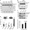
ELG1 knockdown increases the level of monoubiquitinated PCNA in chromatin. A, HEK293T cells were transfected with various siRNAs (si-ELG1) targeting the coding region of the ELG1 mRNA or 3′-untranslated region (UTR) as indicated. Histone H4 protein was used as a loading control. PCNA-Ub, monoubiquitinated PCNA. B, nuclear extracts were prepared 48 h after transfection of HEK293T cells with siRNA targeting ELG1 or control siRNA and then subjected to immunoprecipitation with anti-PCNA antibody. Monoubiquitinated PCNA was identified with anti-ubiquitin (P4D1) antibody. An arrowhead indicates monoubiquitinated PCNA. C, the increase in PCNA monoubiquitination following ELG1 knockdown was not an off-target effect. HEK293T cells were cotransfected with the combination of siRNA targeting the 3′-UTR of ELG1 (#2 siRNA in A) and a FLAG-ELG1 expression vector as indicated. D, ELG1 knockdown does not affect cell cycle profile. HEK293T cells were transfected with siRNA targeting ELG1 or control siRNA twice with an interval of 24 h. Cells were harvested after 48 h from the second transfection, and their cell cycle profiles were determined by flow cytometry analysis. The x and y axes represent DNA content as measured by 7-amino-actinomycin D (7-AAD) staining and cells undergoing DNA replication as measured by bromodeoxyuridine incorporation, respectively. Each rectangle encompasses cells in G1, S, and G2/M phases of the cell cycle clockwise from the lower left. The numbers indicate the percentage of cells in the rectangle out of total cells. E, an increase in PCNA monoubiquitination was not observed following canonical RFC subunit (RFC1 or RFC4) knockdown. F, knockdown of other alternative RFC large subunits (CTF18 or RAD17) did not cause any significant change in PCNA monoubiquitination. The chromatin-bound fractions were prepared 72 h after transfection of HEK293T cells with the indicated siRNAs or a plasmid expressing FLAG-tagged ELG1 and then subjected to immunoblot analyses. The #3 siRNA in A that targets the coding region of ELG1 was used for ELG1 knockdown in B, E, and F.
FIGURE 4.
FIGURE 4.
ELG1 facilitates USP1-mediated PCNA deubiquitination. A, overexpression of ELG1 and USP1 decreased UV-induced PCNA monoubiquitination (PCNA-Ub). HEK293T cells were transected with FLAG-ELG1 or Myc-USP1 expression vectors. Cells were irradiated with 20 J/m2 UV after 48 h and incubated further for 6 h. The soluble (S) and chromatin-bound (B) fractions were isolated and subjected to immunoblot analysis. Histone H3 and tubulin proteins were used as loading controls for chromatin-bound and soluble fractions, respectively. B, the increase in PCNA monoubiquitination following USP1 knockdown depends on ELG1. HEK293T cells were cotransfected with different combinations of control (Ctrl) or ELG1 siRNA with a Myc-USP1 expression vector as indicated. The chromatin-bound fractions were analyzed by immunoblotting. C, ELG1 knockdown did not affect FANCD2 monoubiquitination. The chromatin-bound fractions were prepared 72 h after transfection of HEK293T cells with the indicated siRNAs and then subjected to immunoblot analyses. D, the I-SceI-induced HR assay was performed as described previously (20) after knockdown of USP1 or ELG1. BLM depletion was used as a positive control. The inset shows the structure of the recombination substrate DR-green fluorescent protein and the location of the I-SceI recognition site. Error bars, S.D. of triplicate experiments. E, SupF mutation frequencies for UV-irradiated plasmids are indicated for control siRNA and ELG1 siRNA-treated cells. Error bars, S.D. of triplicate experiments. The number in the graph indicates probability (p value) calculated by Student's t tests.
FIGURE 5.
FIGURE 5.
N terminus of ELG1 is important for the down-regulation of PCNA monoubiquitination and UAF1 interaction. A, amino acids 368–384 of ELG1 are important for the down-regulation of PCNA monoubiquitination (PCNA-Ub). HEK293T cells were co-transfected with the combination of siRNA targeting the 3′-UTR of ELG1 and vectors expressing various sizes of N terminus of ELG1 as indicated. The chromatin-bound fractions were analyzed to monitor PCNA ubiquitination. B, amino acids (a.a.) 368–384 of the ELG1 protein are important for its interaction with UAF1. HEK293T cells were transfected with a plasmids expressing different sizes of the FLAG-tagged N terminus of ELG1 and a plasmid expressing full-length Myc-UAF1-FLAG. Immunoprecipitations (IP) were carried out on nuclear extracts using an anti-Myc antibody. The immunoprecipitated proteins were analyzed by immunoblotting to detect the presence of the ELG1 protein. C, the sequence of the human ELG1 between amino acids 368 and 384 is evolutionarily conserved. The location and peptide sequences of amino acids 368–384 are shown in the diagram. The deleted amino acids (368–372 and 375–380) to generate a UAF1 interaction mutant of ELG1 (ELG1 Mut) are shown at the bottom as dotted lines. D, HEK293T cells were transected with vectors expressing wild type (WT) or deletion mutant (Mut) of ELG1 along with a Myc-UAF1-FLAG expression vector. Nuclear extracts were immunoprecipitated with anti-Myc or anti-RFC4 antibodies and analyzed by immunoblotting. E, HEK293T cells were transected with vectors expressing wild type or deletion mutant of ELG1. Nuclear extracts were immunoprecipitated with anti-USP1 antibody and analyzed by immunoblotting. F, HEK293T cells were cotransfected with the combination of ELG1 siRNA and vectors expressing wild type or deletion mutant of ELG1. The nuclear extracts were analyzed to monitor PCNA ubiquitination. The #2 siRNA in Fig. 3A that targets the 3′-UTR region of ELG1 was used for ELG1 knockdown in A and F.

Similar articles

Cited by

References

    1. Moldovan G. L., Pfander B., Jentsch S. (2007) Cell 129, 665–679 - PubMed
    1. Hoege C., Pfander B., Moldovan G. L., Pyrowolakis G., Jentsch S. (2002) Nature 419, 135–141 - PubMed
    1. Wang S. C., Nakajima Y., Yu Y. L., Xia W., Chen C. T., Yang C. C., McIntush E. W., Li L. Y., Hawke D. H., Kobayashi R., Hung M. C. (2006) Nat. Cell Biol. 8, 1359–1368 - PubMed
    1. Lee K. Y., Myung K. (2008) Mol. Cells 26, 5–11 - PMC - PubMed
    1. Watanabe K., Tateishi S., Kawasuji M., Tsurimoto T., Inoue H., Yamaizumi M. (2004) EMBO J. 23, 3886–3896 - PMC - PubMed

Publication types

MeSH terms