. 2010 Mar;137(5):725-34.
doi: 10.1242/dev.040568.

COUP-TFs regulate eye development by controlling factors essential for optic vesicle morphogenesis

Affiliations

COUP-TFs regulate eye development by controlling factors essential for optic vesicle morphogenesis

Ke Tang et al. Development. 2010 Mar.

Abstract

Transcriptional networks, which are initiated by secreted proteins, cooperate with each other to orchestrate eye development. The establishment of dorsal/ventral polarity, especially dorsal specification in the optic vesicle, is poorly understood at a molecular and cellular level. Here, we show that COUP-TFI (Nr2f1) and COUP-TFII (Nr2f2) are highly expressed in the progenitor cells in the developing murine eye. Phenotype analysis of COUP-TFI and COUP-TFII single-gene conditional knockout mouse models suggests that COUP-TFs compensate for each other to maintain morphogenesis of the eye. However, in eye-specific COUP-TFI/TFII double-knockout mice, progenitor cells at the dorso-distal optic vesicle fail to differentiate appropriately, causing the retinal pigmented epithelium cells to adopt a neural retina fate and abnormal differentiation of the dorsal optic stalk; the development of proximo-ventral identities, neural retina and ventral optic stalk is also compromised. These cellular defects in turn lead to congenital ocular colobomata and microphthalmia. Immunohistochemical and in situ hybridization assays reveal that the expression of several regulatory genes essential for early optic vesicle development, including Pax6, Otx2, Mitf, Pax2 and Vax1/2, is altered in the corresponding compartments of the mutant eye. Using ChIP assay, siRNA treatment and transient transfection in ARPE-19 cells in vitro, we demonstrate that Pax6 and Otx2 are directly regulated by COUP-TFs. Taken together, our findings reveal novel and distinct cell-intrinsic mechanisms mediated by COUP-TF genes to direct the specification and differentiation of progenitor cells, and that COUP-TFs are crucial for dorsalization of the eye.


Figures

Fig. 1.
Fig. 1.
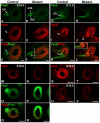
Expression of COUP-TFI and COUP-TFII in the developing mouse eye and generation of the COUP-TFI-floxed mouse. (A,B) The expression of COUP-TFI and COUP-TFII in the optic vesicle at E9.5. Frontal sections. (C,D) In the COUP-TF eye-specific double-mutant mouse at E9.5, the expression of COUP-TFI and COUP-TFII is drastically reduced. Frontal sections. (E-H) Expression of COUP-TFI and COUP-TFII in the optic vesicle at E9.5. Sagittal sections. (E,F) The distal plate. (G,H) The proximal plate. Arrow, temporal optic vesicle. Arrowhead, dorsal optic vesicle. (I-L) The expression of COUP-TFI and COUP-TFII in the optic cup at E11.5. Sagittal sections. (I,J) The proximal OS region. (K,L) The distal NR and RPE regions. (M) Generation of the COUP-TFI-floxed mouse. H, HindIII; S, Sau3AI. (N-Q) Expression of COUP-TFI and COUP-TFII in the eye of COUP-TFI (COUP-TFIΔ/Δ) and COUP-TFII (COUP-TFIΔ/Δ) single-mutant mice at E11.5. Sagittal sections. Arrowhead, COUP-TFI expression in RPE cells. In A-L and N-Q dorsal is to the top. Scale bars: 50 μm.
Fig. 2.
Fig. 2.
Coloboma, microphthalmia, secondary neural retina and extended neural retina in the COUP-TFI/TFII double-mutant mouse. (A) The optic fissure is closed completely at E14.5 in the control. (B,C) Three-allele-deleted compound mutants display the open optic fissure (asterisk) characteristic of a coloboma phenotype at E14.5. (D) The `complete' double-mutant mouse exhibits a severe coloboma phenotype at E14.5. (E) Eye from a control mouse at post-natal day (P) 18. (F) Microphthalmia in a double-mutant mouse at P18. (G,I) H&E staining of the control at E14.5. Arrow indicates the OD. (H,J) H&E staining of the double mutant at E14.5. Arrowheads indicate the ectopic NR-like structure in the ventral and proximal-dorsal prospective RPE region. G-J are frontal sections. Scale bars: 500 μm in G,H; 50 μm in I,J.
Fig. 3.
Fig. 3.
Extension of the NR into vOS territory and abnormal expression of Pax2 and Pax6 in the COUP-TFI/TFII double mutant. (A-F) The expression of Pax2 and Pax6 in the eye of control and COUP-TFI/TFII double mutant at E14.5. Arrow indicates the OD in the control. Arrowhead indicates the boundary between Pax2-positive and Pax6-positive domains in the mutant. The dashed line indicates the location at which the boundary would normally be. (G-L) The expression of Pax2 and Pax6 in the eye of the control and mutant at E11.5. Arrowhead indicates Pax6 expression in the vOS in the mutant. The inset in L shows Pax2/Pax6 double-positive cells in the vOS. (M-R) The expression of Pax2 and Pax6 at the OS in the control and mutant at E10.5. (S,T) Expression of Vax1 at the OS in the control and mutant at E10.5. (U,V) Expression of Vax2 in the optic cup in the control and mutant at E10.5. A-L and U,V are frontal sections, M-T sagittal sections. Scale bars: 100 μm.
Fig. 4.
Fig. 4.
Dorsal optic stalk cells fail to differentiate properly in the COUP-TFI/TFII double-knockout mouse. (A,B,D,E) H&E staining of control mice at E12.5 and E14.5. (C,F) H&E staining of the double-mutant at E12.5 and E14.5. Arrowheads indicate the opened optic fissure in the mutant. (G,I,K,M,O) The expression of Pax2 at the OS in the control at E11.5, E12.5 and E14.5. The arrow in G indicates the dOS. (H,J,L,N,P) The expression of Pax2 at the OS in the double mutant at E11.5, E12.5 and E14.5. Arrowhead indicates the prospective dOS in the mutants. A-P are frontal sections. Scale bars: 50 μm.
Fig. 5.
Fig. 5.
Transformation of the RPE into neural retina and misexpression of RPE regulatory genes in the COUP-TFI/TFII double mutant. (A-D) The expression of Chx10 (A,B) and Mitf (C,D) in the eye of the control and mutant at E14.5. (E,F) H&E staining of the control and mutant at E10.5. (G,H) The expression of Otx2 in the eye of the control and mutant at E10.5. (I-N) The expression of Mitf and Chx10 in the eye of the control and mutant at E10.5. Arrowheads in L and N indicate Chx10-expressing cells in the prospective RPE region in the mutant. The arrow in M and N indicates the junction between Mitf- and Chx10-expressing domains. (O-R) Expression of Pax6 protein (O,P) and transcripts (Q,R) in the eye of the control and mutant at E10.5. (S,T) The expression of Chx10 in the eye of the control and mutant at E11.5. Arrowheads indicate Chx10-expressing cells at the ventral and proximal-dorsal prospective RPE. All images are of frontal sections. Scale bars: 100 μm.
Fig. 6.
Fig. 6.
COUP-TFs directly regulate Pax6 and Otx2 transcription. (A) Real-time PCR analysis of RPE marker genes in human ARPE-19 cells. RNAs were isolated from siCON-treated cells (white) and siCOUP-TFI/TFII-treated cells (black). Expression levels of each gene were normalized to that of 18S rRNA. Data indicate mean ± s.e.m. *, P<0.05; , P<0.001. (B) Immunocytochemical assays with COUP-TFI, COUP-TFII and Pax6 antibodies in ARPE-19 cells transiently transfected with plasmids pCXN2-vector, pCXN2-COUP-TFI or pCXN2-COUP-TFII. When COUP-TFI (middle) or COUP-TFII (bottom) was overexpressed, the expression of Pax6 was repressed (arrowheads). The bar chart to the right shows the percentage of cells in which Pax6 expression was abolished or less/not affected in pCXN2-vector-transfected (367 cells), COUP-TFI-transfected (143 cells) or COUP-TFII-transfected (268 cells) cells. Error bars indicate s.e.m. (C,D) ChIP assays in ARPE-19 cells. COUP-TFII is recruited to the COUP-TF DR1 binding site in the 3′-UTR of the Pax6 gene in siCON-treated samples. COUP-TFII and Sp1 are recruited to the Sp1 binding site in the third intron of the Otx2 gene in siCON-treated samples. Lanes 1-3, siCON treatment; lanes 4-6, siCOUP-TFII or siSp1 treatment; lanes 1 and 4, input; lanes 2 and 5, control IgG antibody-treated samples; lanes 3 and 6, COUP-TFII antibody- or Sp1 antibody-treated samples. (E) Luciferase assays with pGL4-DR1X3-minP-Luc and pGL4-minP-Luc in siRNA-treated ARPE-19 cells. *, P<0.05. Scale bars: 50 μm.
Fig. 7.
Fig. 7.
Cell-intrinsic mechanisms mediated by COUP-TFI/TFII genes during morphogenesis of the murine eye. (A) COUP-TFI/TFII genes regulate RPE versus NR fates. (B) COUP-TFI/TFII genes program the NR and vOS identities to establish the proper boundary. See text for details.

References

    1. Azuma N., Tadokoro K., Asaka A., Yamada M., Yamaguchi Y., Handa H., Matsushima S., Watanabe T., Kida Y., Ogura T., et al. (2005). Transdifferentiation of the retinal pigment epithelia to the neural retina by transfer of the Pax6 transcriptional factor. Hum. Mol. Genet. 14, 1059-1068 - PubMed
    1. Baumer N., Marquardt T., Stoykova A., Spieler D., Treichel D., Ashery-Padan R., Gruss P. (2003). Retinal pigmented epithelium determination requires the redundant activities of Pax2 and Pax6. Development 130, 2903-2915 - PubMed
    1. Bertuzzi S., Hindges R., Mui S. H., O'Leary D., Lemke G. (1999). The homeodomain protein Vax1 is required for axon guidance and major tract formation in the developing forebrain. Genes Dev. 13, 3092-3105 - PMC - PubMed
    1. Bharti K., Liu W., Csermely T., Bertuzzi S., Arnheiter H. (2008). Alternative promoter use in eye development: the complex role and regulation of the transcription factor MITF. Development 135, 1169-1178 - PMC - PubMed
    1. Bramblett D., Pennesi M., Wu S., Tsai M. (2004). The transcription factor Bhlhb4 is required for rod bipolar cell maturation. Neuron 43, 779-793 - PubMed

Publication types

MeSH terms