. 2010 Mar;137(5):805-14.
doi: 10.1242/dev.042457.

Cadherin-7 and cadherin-6B differentially regulate the growth, branching and guidance of cranial motor axons

Affiliations

Cadherin-7 and cadherin-6B differentially regulate the growth, branching and guidance of cranial motor axons

Sarah H Barnes et al. Development. 2010 Mar.

Abstract

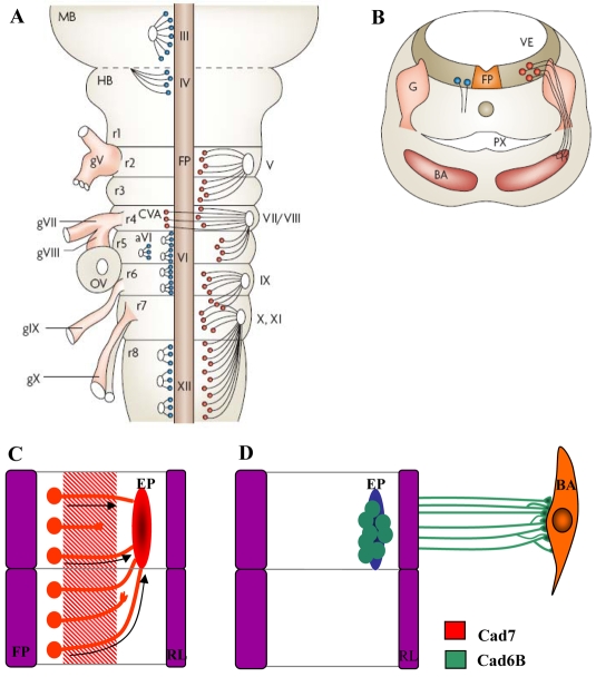
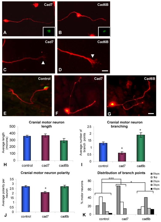
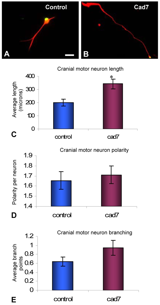
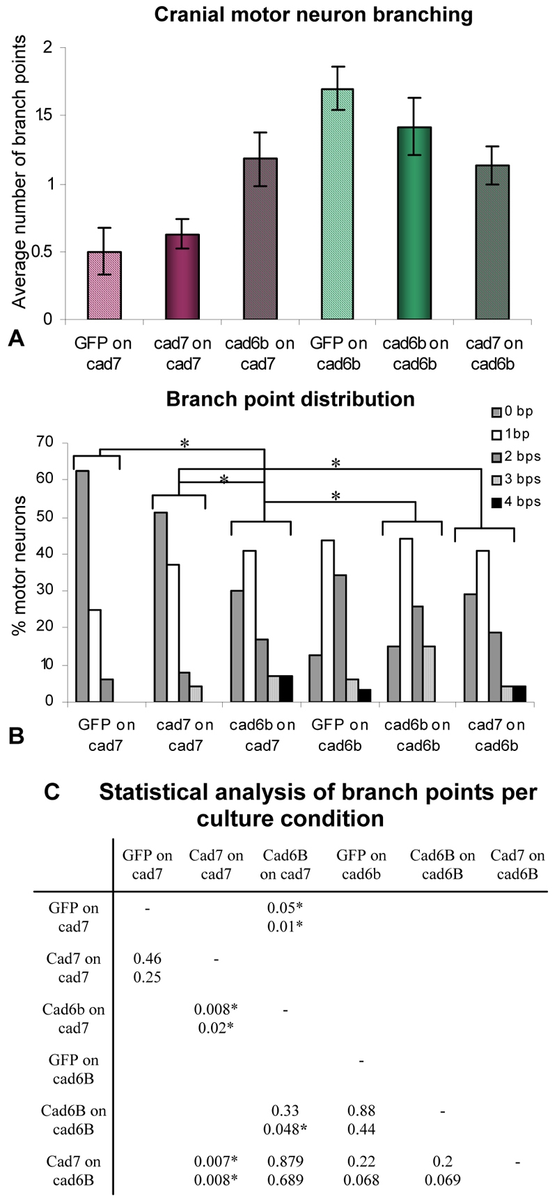
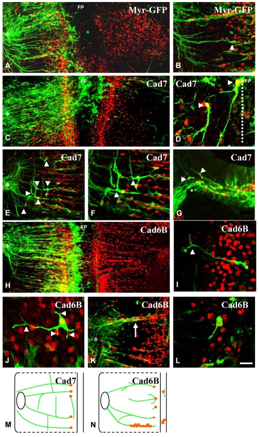
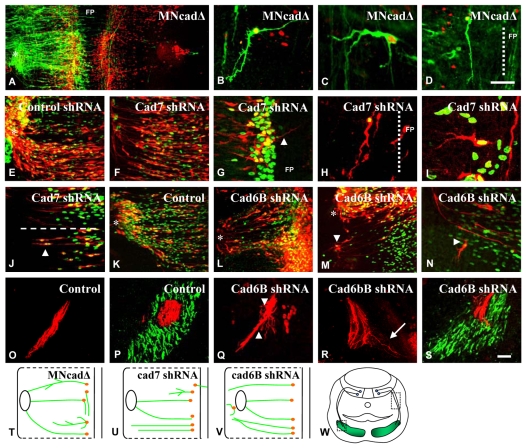
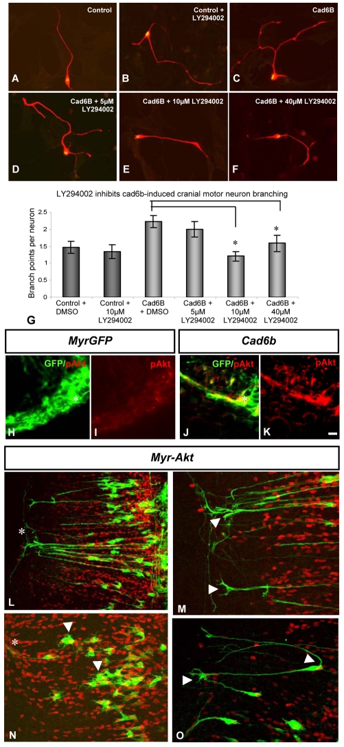
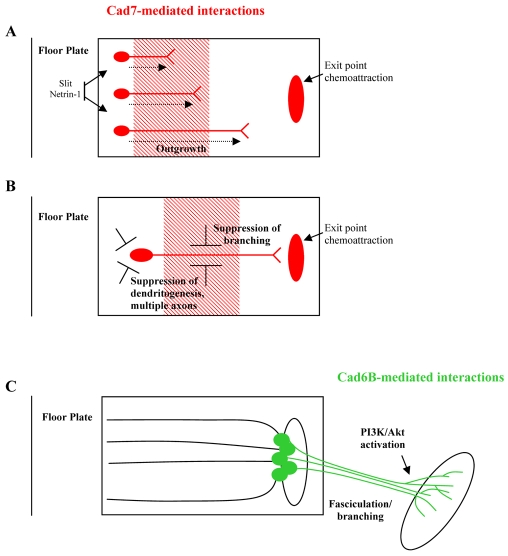
Cadherin-7 (Cad7) and cadherin-6B (Cad6B) are expressed in early and late phases of cranial motoneuron development, respectively. Cad7 is expressed by cranial motoneurons soon after they are generated, as well as in the environment through which their axons extend. By contrast, Cad6B is expressed by mature cranial motoneurons. We demonstrate in chick that these cadherins play distinct roles in cranial motor axon morphology, branching and projection. Using in vitro approaches, we show that Cad7 enhances motor axon outgrowth, suppresses the formation of multiple axons and restricts interstitial branching, thus promoting the development of a single unbranched axon characteristic of differentiating motoneurons. Conversely, Cad6B in vitro promotes motor axon branching, a characteristic of mature motoneurons. In vivo gain- and loss-of-function experiments for these cadherins yielded phenotypes consistent with this interpretation. In particular, a loss of cadherin-mediated interactions in vivo led to dysregulation of the cranial motoneuron normal branching programme and caused axon navigation defects. We also demonstrate that Cad6B functions via the phosphatidylinositol 3-kinase pathway. Together, these data show that Cad7 and Cad6B differentially regulate cranial motoneuron growth, branching and axon guidance.


Figures

Fig. 1.
Fig. 1.
Motoneuron development and Cad7 and Cad6B expression in the chick hindbrain. (A) Diagrams of flat-mounted brainstem showing the positions of cranial nerves and motor nuclei. Dorsally projecting branchiomotor (BM) neurons are shown in red, ventrally projecting somatic motoneurons in blue. Roman numerals indicate cranial nerves (III, oculomotor; IV, trochlear; V, trigeminal; VI, abducens; aVI, accessory abducens; VII/VII, facial/vestibuloacoustic; IX, glossopharyngeal; X, vagus; XI, cranial accessory; XII, hypoglossal; gV-X indicate sensory ganglia and cranial nerves as above). Rhombomeres are numbered. (B) Transverse section through the branchial region, showing hindbrain neuroepithelium and branchial arches (BA), with dorsally projecting BM (red) and ventrally projecting somatic motor (blue) motor axons. Adapted with permission from Guthrie (Guthrie, 2007). (C,D) Diagram of flat-mounted even- and odd-numbered rhombomere pairs at early (C) and late (D) developmental stages, with respect to BM axon outgrowth. (C) Cad7 (red) is expressed from E2-5 by the hindbrain neuroepithelium, extending cranial motoneurons and the boundary cap cells at the nerve exit point. (D) Cad6B (green) is expressed by mature motoneurons and their axons between E5 and E9. Arrows indicate the direction of migration of BM neuron somata. Expression data derived from Ju et al. (Ju et al., 2004). MB, midbrain; HB, hindbrain; FP, floor plate; OV, otic vesicle; BA, branchial arch; VE, ventricle; G, ganglion; PX, pharynx; EP, exit point; RL, rhombic lip.
Fig. 2.
Fig. 2.
Expression of cadherins and responses of cranial motoneurons to culture on cadherin-expressing substrata. (A-D) Immunohistochemistry using anti-Cad7 and anti-Cad6B antibodies in chick E5 cultured cranial motoneurons (red). Insets in A and B show Islet1/2 expression (green). (E-G) E5 cranial motoneurons cultured on control, Cad7- or Cad6B-expressing NIH3T3 cell monolayers. (H-K)The effects of cadherin-mediated interactions on cranial motoneuron morphology (length, polarity and branching). Significant differences from controls are indicated by asterisks (*, P<0.05; ***, P<0.001; Student's t-test in all cases, Xxxxxxx? test); n=90 neurons per condition. bps, branch points. Scale bars: in D, 20 μm for A,B and 10 μm for C,D; in G, 20 μm for E-G.
Fig. 3.
Fig. 3.
Effects of Cad7-mediated interactions on E3 cultured cranial motoneurons. (A,B) Chick E3 cranial motoneurons cultured on laminin and treated with control medium (A) or soluble Cad7 protein-containing medium (B). Immunostaining is with anti-Islet1/2 (green) and anti-neurofilament H (red) antibodies. (C-E) Effects of soluble Cad7 protein on E3 cranial motoneuron length, polarity and branching. Significant differences from controls are indicated by an asterisk (P≤0.05, Student's t-test); n=90 neurons per condition. Scale bar: in A, 40 μm for A and 50 μm for B.
Fig. 4.
Fig. 4.
The effects on chick E5 cranial motor axon branching of overexpression of Cad6B or Cad7 and culture on Cad7- or Cad6B-expressing substrata. (A) The effects of Cad7 or Cad6B overexpression by the motoneurons or by the culture environment (NIH3T3 cell monolayers) on the average number of axon branch points. (B) The percentage of motoneurons showing a particular number of branch points per culture condition. Significant differences from controls are indicated by an asterisk (P≤0.05; Mann-Whitney test). (C) Statistical analysis of motoneuron branching based on Student's t-test analysis (upper) of data in A and Mann-Whitney test analysis (lower) of data in B. Statistical differences are indicated by an asterisk (P≤0.05).
Fig. 5.
Fig. 5.
Effects of expression of full-length Cad7 and full-length Cad6b constructs in the chick hindbrain. (A-L) E4 hindbrains immunostained with anti-Islet1/2 (red) and anti-GFP (green) antibodies to detect electroporated neurons. Hindbrains were electroporated with myr-GFP control, Cad7 and Cad6B constructs as labelled. All panels show flat-mounts except G, which is a transverse section. Cranial motor axons project in a ventral-to-dorsal direction in controls (arrowhead in B). Medial is to the right and lateral to the left in all panels except A, C and H, which show both sides of the hindbrain. In Cad7-expressing hindbrains, axons project longitudinally (arrowheads in D-F). (F) A high-magnification image of E. In Cad6B-expressing hindbrains, cranial motor axons ectopically branch (I, arrowhead), extend multiple axons (J, arrowheads), aggregate at rhombomere boundaries (white arrow in K), and stall adjacent to the motor column (L). Asterisks indicate exit points. FP, floor plate. (M,N) Diagram of the behaviour of cranial motoneurons electroporated with full-length Cad7 (M) or full-length Cad6B (N). Scale bar: in L, 50 μm for A,C,E,H,K, 75 μm for B,F, 30 μm for G, and 15 μm for D,I,J,L.
Fig. 6.
Fig. 6.
Effects of expression of dominant-negative MNcadΔ, Cad7-shRNA and Cad6b-shRNA constructs in the chick hindbrain. (A-S) Hindbrains were fixed at E4 (A-J) or E5-6 (K-S) and immunostained with an anti-Islet1/2 antibody (red in A-D and green in E-N) to detect motoneurons, and with an anti-GFP antibody (green in A-D) or showing RFP natural fluorescence (red in E-S) to detect electroporated neurons (electroporation constructs as labelled). (A-N) MNcadΔ-expressing cranial motoneurons display morphological defects, such as an increase in branching (B) and ectopic axons from the cell body (C) and guidance defects, i.e. they project longitudinally (D). Dashed lines indicate the border of floor plate (FP). Cad7-shRNA-expressing axons project into floor plate (arrowhead in G), project longitudinally (H) or branch ectopically (I). Cad6B-shRNA-expressing axons display defects in the vicinity of the exit points, such as failing to turn rostrally (M,N; arrowheads). Asterisks indicate the exit point. (O-S) Transverse cryosections of embryos electroporated with Cad6B-shRNA or control (scrambled) Cad6B-shRNA, as labelled. (O,Q,R) Regions of the peripheral nerve are shown. Arrowheads in Q indicate defasciculation of the nerve and the arrow in R indicates an axon departing from the main axon bundle. (P,S) The muscle plate (boxed region in W) immunostained with MF20 antibody to label muscle (green). (T-V) Diagram of the behaviour of cranial motoneurons electroporated with MNcadΔ, Cad7-shRNA and Cad6B-shRNA plasmids. (W) Diagram of a transverse section through the hindbrain. Scale bars: 100 μm for A, in D, 60 μm for B-D,G-J, 20 μm for E,F; in S, 50 μm for K-S.
Fig. 7.
Fig. 7.
Evidence that Cad6B signals via the PI3K pathway. (A-F) Chick cranial motoneurons treated with DMSO (control) or with various concentrations of the PI3K inhibitor LY294002 grown on a Cad6B-expressing NIH3T3 cell monolayer. (G) The average number of branch points per neuron per condition. Significant differences from the control are indicated by an asterisk (P≤0.05, Student t-test); n=90 neurons per condition. (H-K) Cryosectioned embryos that have been electroporated with myr-GFP or with myr-GFP and full-length Cad6B, grown to E5-6 and immunostained with anti-pAkt (red) and anti-GFP (green) antibodies. pAkt is upregulated in Cad6B-overexpressing motoneurons (K). Asterisks indicate the ventral neural tube. (L-O) Effects of expression of a constitutively active myr-Akt construct in the hindbrain. Hindbrains were fixed at E5 and immunostained with anti-Islet1/2 (red) and anti-GFP (green) antibodies to detect electroporated motoneurons. Motor axons branch (L,M,O, arrowheads) and fail to extend a single axon, and in some cases project multiple short processes (arrowheads in N). Asterisks indicate ventral neural tube. Scale bar: in K, 20 μm for A-F,H-K, and 10 μm for L-O.
Fig. 8.
Fig. 8.
Model for the role of Cad7- and Cad6B-mediated interactions during BM neuron development in chick. (A-C) Flat-mount hindbrains showing the outgrowth of cranial BM neurons towards their exit points at ∼E3 (A) and ∼E4 (B) and their extension into the periphery at ∼E5 (C). (A) Cad7-mediated interactions (red) promote the early motor axon outgrowth. (B) Cad7-mediated interactions promote axon polarisation and suppress axon branching. (C) Cad6B-mediated interactions (green) promote branchiomotor/visceralmotor axon branching as they approach the branchial arch muscle plate, thus ensuring individual muscle masses are innervated. Cad6B branching is dependent upon the PI3K/Akt pathway.

Similar articles

Cited by

References

    1. Adler C. E., Fetter R. D., Bargmann C. I. (2006). UNC-6/Netrin induces neuronal asymmetry and defines the site of axon formation. Nat. Neurosci. 9, 511-518 - PMC - PubMed
    1. Andrews G. L., Mastick G. S. (2003). R-cadherin is a Pax6-regulated, growth-promoting cue for pioneer axons. J. Neurosci. 23, 9873-9880 - PMC - PubMed
    1. Boscher C., Mege R. M. (2008). Cadherin-11 interacts with the FGF receptor and induces neurite outgrowth through associated downstream signalling. Cell Signal. 20, 1061-1072 - PubMed
    1. Calautti E., Li J., Saoncella S., Brissette J. L., Goetinck P. F. (2005). Phosphoinositide 3-kinase signaling to Akt promotes keratinocyte differentiation versus death. J. Biol. Chem. 280, 32856-32865 - PubMed
    1. Caton A., Hacker A., Naeem A., Livet J., Maina F., Bladt F., Klein R., Birchmeier C., Guthrie S. (2000). The branchial arches and HGF are growth-promoting and chemoattractant for cranial motor axons. Development 127, 1751-1766 - PubMed

Publication types

MeSH terms