. 2010 May;84(9):4172-82.
doi: 10.1128/JVI.01567-09. Epub 2010 Feb 10.

Stimulation of HIV-1 replication in immature dendritic cells in contact with primary CD4 T or B lymphocytes

Affiliations

Stimulation of HIV-1 replication in immature dendritic cells in contact with primary CD4 T or B lymphocytes

Vincent Holl et al. J Virol. 2010 May.

Abstract

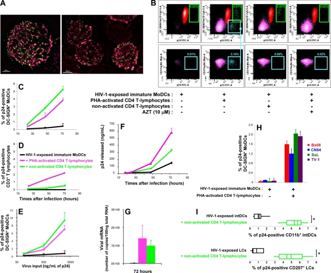
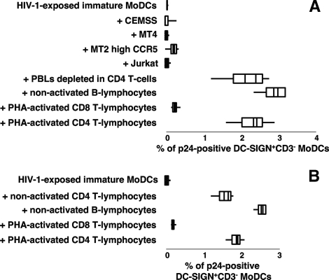
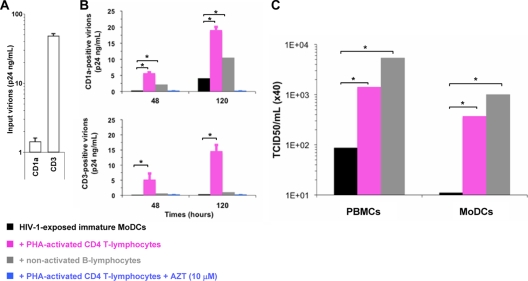
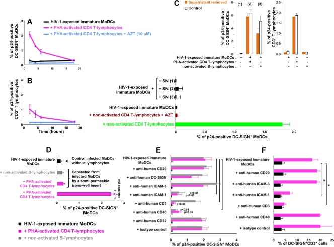
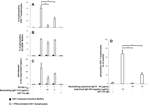
Sexual transmission is the major route of HIV-1 infection worldwide. Dendritic cells (DCs) from the mucosal layers are considered to be the initial targets of HIV-1 and probably play a crucial role in HIV-1 transmission. We investigated the role of cell-to-cell contact between HIV-1-exposed immature DCs and various lymphocyte subsets in the stimulation of HIV-1 replication. We found that HIV-1 replication and production in DCs were substantially enhanced by the coculture of DCs with primary CD4 T or nonpermissive B lymphocytes but not with primary activated CD8 T lymphocytes or human transformed CD4 T lymphocytes. Most of the new virions released by cocultures of HIV-1-exposed immature DCs and primary B lymphocytes expressed the DC-specific marker CD1a and were infectious for both immature DCs and peripheral blood mononuclear cells (PBMCs). Cocultured DCs thus produced large numbers of infectious viral particles under these experimental conditions. The soluble factors present in the supernatants of the cocultures were not sufficient to enhance HIV-1 replication in DCs, for which cell-to-cell contact was required. The neutralizing monoclonal antibody IgG1b12 and polyclonal anti-HIV-1 sera efficiently blocked HIV-1 transfer to CD4 T lymphocytes but did not prevent the increase in viral replication in DCs. Neutralizing antibodies thus proved to be more efficient at blocking HIV-1 transfer than previously thought. Our findings show that HIV-1 exploits DC-lymphocyte cross talk to upregulate replication within the DC reservoir. We provide evidence for a novel mechanism that may facilitate HIV-1 replication and transmission. This mechanism may favor HIV-1 pathogenesis, immune evasion, and persistence.


Figures

FIG. 1.
FIG. 1.
Increase in HIV-1 p24 antigen production in immature MoDCs in the presence of cocultured primary CD4 T lymphocytes. (A) Immunofluorescence analysis of the binding of HIV-1 pSG3EnvSF162 vpr-GFP pseudoparticles (green) to immature MoDCs 2 h postinfection. Membranes were stained with anti-human DC-SIGN IgG (red). Bars, 3 μm (left) and 10 μm (right). (B) Dot plots of intracellular p24-positive DC-SIGN+ MoDCs (green) and CD3+ CD4 T lymphocytes (purple) after 48 h of culture. The teal arrowhead indicates the percentage of infected CD4 T lymphocytes in the CD3-positive gate. PerCP Cy5.5; peridinin-chlorophyll cyanine 5.5; FITC, fluorescein isothiocyanate. (C and D) Immature MoDCs were exposed to HIV-1BaL for 2 h before the addition of uninfected autologous PHA-activated or nonactivated CD4 T lymphocytes. Percentages of MoDCs (C) and CD4 T lymphocytes (D) producing intracellular p24 antigen were determined by flow cytometry. Data are the means ± standard deviations (SD) of triplicate results. (E) Dose-dependent response curve obtained by plotting the amount of input virus loaded into immature DCs against the percentage of p24-positive DC-SIGN+ MoDCs detected after 48 h of coculture with autologous CD4 T lymphocytes. Data are the means ± SD of triplicate results. Data from one representative experiment of four are shown. (F) Cell-free supernatants were collected after various amounts of time, and extracellular viral p24 antigen levels were determined by ELISA. Data are the means ± SD of triplicate results. (G) Quantification of viral mRNA in cocultures of HIV-1-exposed MoDCs and activated or nonactivated CD4 T lymphocytes (n = 3) by RT-PCR. (H) Immature MoDCs were infected with various R5 HIV-1 primary isolates and cultured with or without activated CD4 T lymphocytes for 48 h (n = 3). (I) Box plot analyses of intracellular viral p24 antigen in cocultures (n = 5) of HIV-exposed LCs or intDCs and uninfected autologous nonactivated CD4 T lymphocytes after 3 days. Data are the means ± standard errors of the means (SEM) of results from n independent experiments. A two-tailed Mann-Whitney U test with Bonferroni's correction was used to assess differences between groups. A value of P of <0.025 was considered significant. *, P < 0.025 versus the control group.
FIG. 2.
FIG. 2.
Stimulation of HIV-1 replication in infected MoDCs cocultured with primary B lymphocytes but not with human CD4 T-cell lines. (A) Box plot analysis of the percentage of p24-positive DC-SIGN+ CD3 MoDCs following the coculture of HIV-1-exposed MoDCs and autologous CD4 T lymphocyte-depleted PBLs (n = 4), nonactivated B lymphocytes (n = 5), PHA-activated CD8 T lymphocytes (n = 7), PHA-activated CD4 T lymphocytes (n = 10), or various human CD4 T-cell lines (n = 4) for 48 h. (B) Box plot analysis of the percentage of infected DC-SIGN+ CD3 MoDCs in the presence of allogeneic T- or B-lymphocyte populations (n = 3) after 48 h of coculture. Data are the means ± SEM of results from n independent experiments.
FIG. 3.
FIG. 3.
The infectious HIV-1 particles released into the supernatant of the coculture are produced principally by infected MoDCs. (A and B) Virus particles positive for CD1a or CD3 were subjected to immunomagnetic separation and quantification by p24 ELISA (n = 4). Levels of these host cell surface markers detected on the envelopes of virions from input virus (A) or cell-free supernatants from cocultures (B) are shown. (B) Amounts of CD1a (top)- or CD3 (bottom)-positive virus particles collected after 2 and 5 days of culture (n = 4). The values plotted on all graphs are the means ± SEM of results from n independent experiments. (C) Infectivities (TCID50s) of the cell-free virus supernatants for PBMCs and immature MoDCs. Two-tailed Mann-Whitney U tests with Bonferroni's correction were used to assess differences between groups. A value of P of <0.017 was considered statistically significant. *, P < 0.017 versus controls.
FIG. 4.
FIG. 4.
Early cell-to-cell contact is required for the increase in HIV-1 replication in infected DCs. (A and B) CD4 T lymphocytes were added at various time points after the infection of immature MoDCs. The percentage of DC-SIGN+ DCs (A) or CD3+ CD4 T lymphocytes (B) expressing p24 was determined after 48 h. (C) Supernatants (SN) from HIV-1-exposed MoDCs, cultured alone [SN (1)] or cocultured with CD4 T lymphocytes [SN (2)] or B lymphocytes [SN (3)], were collected at 6 h and added to infected immature MoDCs. (Bottom) After 48 h, intracellular p24 levels were determined. (Top) In parallel, we used flow cytometry to measure intracellular p24 production at 48 h in cells from which supernatants (1, 2, and 3) had been removed (orange bars) or not removed (white bars). (D) HIV-1-exposed MoDCs and uninfected lymphocytes were left in contact or separated with a Transwell insert, and intracellular p24 levels were determined after 48 h. (E) PHA-activated CD4 T or B lymphocytes were pulsed with 20 μg/ml mouse monoclonal IgG2a directed against human CD3 (clone OKT3 [azide free]; Miltenyi Biotec) before being added to HIV-1-exposed immature MoDCs. We added 20 μg/ml mouse monoclonal IgG1 directed against CD40 (clone B-B20 [azide free]; Abcam), ICAM-3 (clone B-R1 [azide free]; Abcam), ICAM-1 (clone 1H4 [azide free]; Abcam), CD20 (clone MEM-97 [azide free]; Abcam), DC-SIGN (clone DCN46; BD Pharmingen), CD32 (clone 3D3; BD Pharmingen), or a mouse monoclonal IgG1 isotype (clone NCM1; Abcam) to the coculture at the same time as CD4 T lymphocytes. After 48 h of coculture, intracellular p24 levels were determined by flow cytometry. (F) In parallel, MoDC-CD4 T-cell conjugates were evaluated by determining the percentage of DC-SIGN+ CD3+ cells by flow cytometry. The values plotted on all graphs are means ± SD of triplicate results from a single representative experiment. A value of P of <0.05 was considered significant. *, P < 0.05 versus the corresponding control group.
FIG. 5.
FIG. 5.
Neutralizing activities of anti-HIV-1 monoclonal or polyclonaI IgGs toward the trans-infection of CD4 T lymphocytes. Monoclonal neutralizing IgG1b12 (A, B, and C) or polyclonal neutralizing IgG preparation no. 44 (D) was added, together with activated CD4 T lymphocytes, to HIV-1-exposed immature MoDCs. After 48 h of coculture, intracellular and extracellular p24 production levels were determined by flow cytometry and p24 ELISA, respectively. Shown are the percentages of p24-positive primary CD3+ CD4 T lymphocytes (A and D) and of p24-positive DC-SIGN+ MoDCs (B) and amounts of viral p24 released into the supernatant (C) when HIV-1-exposed immature MoDCs were cultured with or without activated CD4 T lymphocytes in the presence or absence of neutralizing IgGs or AZT. The values shown are the means ± SD of triplicate results. Purified neutralizing polyclonal IgGs were obtained from the serum of HIV-1-positive asymptomatic patient no. 44, and nonneutralizing polyclonal IgGs were obtained from a pool of sera from HIV-1-negative healthy donors. A value of P of <0.05 was considered significant. *, P < 0.05 versus the corresponding control group.

References

    1. Alvarez, C. P., F. Lasala, J. Carrillo, O. Muniz, A. L. Corbi, and R. Delgado. 2002. C-type lectins DC-SIGN and L-SIGN mediate cellular entry by Ebola virus in cis and in trans. J. Virol. 76:6841-6844. - PMC - PubMed
    1. Arrighi, J. F., M. Pion, E. Garcia, J. M. Escola, Y. van Kooyk, T. B. Geijtenbeek, and V. Piguet. 2004. DC-SIGN-mediated infectious synapse formation enhances X4 HIV-1 transmission from dendritic cells to T cells. J. Exp. Med. 200:1279-1288. - PMC - PubMed
    1. Balkow, S., F. Krux, K. Loser, J. U. Becker, S. Grabbe, and U. Dittmer. 2007. Friend retrovirus infection of myeloid dendritic cells impairs maturation, prolongs contact to naive T cells, and favors expansion of regulatory T cells. Blood 110:3949-3958. - PubMed
    1. Banchereau, J., and R. M. Steinman. 1998. Dendritic cells and the control of immunity. Nature 392:245-252. - PubMed
    1. Brossard, C., V. Feuillet, A. Schmitt, C. Randriamampita, M. Romao, G. Raposo, and A. Trautmann. 2005. Multifocal structure of the T cell-dendritic cell synapse. Eur. J. Immunol. 35:1741-1753. - PubMed

Publication types