. 2010 May;84(9):4183-93.
doi: 10.1128/JVI.02168-09. Epub 2010 Feb 10.

CD147/EMMPRIN acts as a functional entry receptor for measles virus on epithelial cells

Affiliations

CD147/EMMPRIN acts as a functional entry receptor for measles virus on epithelial cells

Akira Watanabe et al. J Virol. 2010 May.

Abstract

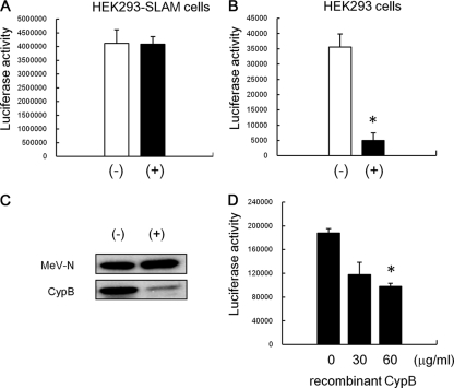
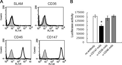
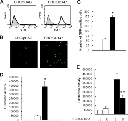
Measles is a highly contagious human disease caused by measles virus (MeV) and remains the leading cause of death in children, particularly in developing countries. Wild-type MeV preferentially infects lymphocytes by using signaling lymphocytic activation molecule (SLAM), whose expression is restricted to hematopoietic cells, as a receptor. MeV also infects other epithelial and neuronal cells that do not express SLAM and causes pneumonia and diarrhea and, sometimes, serious symptoms such as measles encephalitis and subacute sclerosing panencephalitis. The discrepancy between the tissue tropism of MeV and the distribution of SLAM-positive cells suggests that there are unknown receptors other than SLAM for MeV. Here we identified CD147/EMMPRIN (extracellular matrix metalloproteinase inducer), a transmembrane glycoprotein, which acts as a receptor for MeV on epithelial cells. Furthermore, we found the incorporation of cyclophilin B (CypB), a cellular ligand for CD147, in MeV virions, and showed that inhibition of CypB incorporation significantly attenuated SLAM-independent infection on epithelial cells, while it had no effect on SLAM-dependent infection. To date, MeV infection was considered to be triggered by binding of its hemagglutinin (H) protein and cellular receptors. Our present study, however, indicates that MeV infection also occurs via CD147 and virion-associated CypB, independently of MeV H. Since CD147 is expressed in a variety of cells, including epithelial and neuronal cells, this molecule possibly functions as an entry receptor for MeV in SLAM-negative cells. This is the first report among members of the Mononegavirales that CD147 is used as a virus entry receptor via incorporated CypB in the virions.


Figures

FIG. 1.
FIG. 1.
CypA and CypB are identified as MeV-N-interacting proteins by proteomic analysis. The C-terminal region of MeV-N, which is usually called the tail region, was used as a bait protein, and N_TAIL-binding proteins were pulled down from the COBL-a cell lysate. The binding proteins were trypsinized in solution without separation by SDS-PAGE and were subjected to MALDI-TOF MS. Detected ions were analyzed using the Mascot search engine. Amino acid sequences of CypA (A) and CypB (B) are shown. Peptides detected by MALDI-TOF MS are exhibited in red. Scores for each ion from CypA (C) and CypB (D) are shown, and individual ion scores of >36 indicate identity and extensive homology.
FIG. 2.
FIG. 2.
GST pull-down and coimmunoprecipitation (IP) assays showing direct binding of CypA and CypB to MeV-N. We incubated 2 μg of His-tagged MeV-N with 2 μg of GST-CypA (A), GST-CypB (B), or GST (A) for 30 min on ice, and then glutathione-Sepharose beads were added to each sample. After washing of the beads, trapped proteins were analyzed by SDS-PAGE followed by Western blotting (WB). To detect MeV-N, anti-His6 antibody was used as the primary antibody. For assays using CsA, a 5 μM final concentration was used for CypA and 5, 15, and 30 μM final concentrations were used for CypB. The numbers in the figure show the final concentration of CsA in each sample. (C) We incubated 2 μg of His-tagged MeV-N or His-tagged ENO1 with 2 μg of GST-CypA or GST-CypB. GST pull-down assays were performed according to the procedure described above. (D and E) We transiently transfected 1 μg of pCMV-myc-MeV-N with 1 μg of pCMV-HA-CypA (E) or pCMV-HA-CypB (D) into HEK293 cells (1.5 × 10⁶ cells). At 24 h posttransfection, the cells were lysed, and 0.5 μg of anti-myc monoclonal antibody and protein G Sepharose beads were added to the lysate. After washing of the beads, trapped proteins were analyzed by SDS-PAGE followed by Western blotting with anti-myc or anti-HA polyclonal antibody.
FIG. 3.
FIG. 3.
Evidence of incorporation of CypA and CypB into MeV particles. Culture medium of MeV-infected B95a cells was ultracentrifuged to pellet down the virus. The resultant pellet was resuspended with TN buffer and placed on a 20% to 60% sucrose layer (the volume of each sucrose layer was 1 ml). After ultracentrifugation, 600-μl fractions were collected sequentially from the bottom of the tube in the case of MeV-HL. (A) CypA, CypB, and MeV-N in each fraction of MeV-HL were detected by Western blotting. (B) The virus titer of each fraction was estimated using B95a cells. The virus titer and relative amounts of CypA, CypB, and MeV-N in each fraction of MeV-HL were graphed. (C) Trichloroacetic acid was added to fraction 2, and the resultant pellet was analyzed by SDS-PAGE and Coomassie brilliant blue staining. MeV-infected or mock-infected B95a cells were lysed with PBS containing 0.5% NP-40, and the cell lysates were placed on a 20% to 60% sucrose layer (the volume of each sucrose layer was 1 ml). After ultracentrifugation, 600-μl fractions were collected sequentially from the bottom of the tube. CypA, CypB, and MeV-N in each fraction of the MeV-infected cell lysate (D) and CypA and CypB in each fraction of the mock-infected cell lysate (E) were detected by Western blotting.
FIG. 4.
FIG. 4.
Incorporation of CypA or CypB into MeV. (A) MeV-HL, MeV-EGFP, and MeV-luc were purified by sucrose density gradient centrifugation. Virus particles were developed by SDS-PAGE and analyzed by Western blotting with anti-MeV-N, -CypA, and -CypB antibodies. (B) B95a cells were infected with MeV-luc at an MOI of 0.2. At 18 hpi, the cytoplasmic fraction of infected cells was extracted with hypotonic buffer. CypA, CypB, and MeV-N in the cytoplasmic fraction were detected by Western blotting. As a control, mock-infected B95a cells were used. (C and D) Localization of cyclophilins and MeV-N in MeV-luc-infected B95a cells was examined by immunofluorescence staining. CypB (C) and CypA (D) distributions (green; left panels), MeV-N distribution (red; second panels), their overlay (third panels), and phase-contrast images (right panels) are shown in striatal sections. Each panel represents sequential confocal scans of the same field. Arrows in the left panel indicate uninfected cells.
FIG. 5.
FIG. 5.
Effect of CsA treatment on MeV infectivity to epithelial cells. MeV-luc was propagated with and without 5 μg/ml CsA, and then the culture medium of infected cells was ultracentrifuged to pellet down viruses. The resultant pellets were rinsed and resuspended with fresh medium. The virus propagated without CsA was named MeV-luc (−), and that propagated with CsA was named MeV-luc (+). MeV-luc (−) and MeV-luc (+) were titrated using B95a cells. HEK293-SLAM cells (A) and HEK293 cells (B) were infected with the viruses at an MOI of 1.0. At 24 hpi, the luciferase activity of infected cells was measured. The white and black bars represent the infectivities of MeV-luc (−) and MeV-luc (+), respectively. Luciferase assays were performed in triplicate. Data are means plus standard errors of the means (SEM). *, P = 0.003 (Student's t test). (C) MeV-luc (−) and MeV-luc (+) were further purified by sucrose gradient density centrifugation and were analyzed by Western blotting with anti-MeV-N, -CypA, and -CypB antibodies. (D) HEK293 cells (6 × 10⁴ cells) were preincubated with 0, 30, or 60 μg/ml of recombinant CypB for 30 min at room temperature and then infected with MeV-luc at an MOI of 1.0 in the presence of 0, 30, or 60 μg/ml of recombinant CypB at 37°C for another hour. After incubation, cells were washed three times with medium containing fusion block peptide to stop infection and to remove viruses and recombinant CypB. The luciferase activity of infected cells was measured at 24 hpi. Luciferase assays were performed in triplicate. Data are means plus SEM. *, P = 0.0007 (Student's t test).
FIG. 6.
FIG. 6.
Contribution of CD147 to MeV infection of epithelial cells. (A) HEK293 cells (1 × 10⁶ cells) were incubated with 1 μg of anti-CD147, -CD36, -CD46, or -SLAM mouse monoclonal antibody for 1 hour. After being washed, cells were incubated with 0.5 μl of Alexa Fluor 468-conjugated goat anti-mouse IgG polyclonal antibody for 1 hour. After being washed, cells were analyzed by flow cytometry. (B) HEK293 cells (6 × 10⁴ cells) were preincubated with 50 μg/ml of anti-CD147, -CD36, or -CD46 antibody for 30 min at room temperature and then infected with MeV-luc at an MOI of 1.0 in the presence of 50 μg/ml of each antibody at 37°C for another hour. After incubation, cells were washed three times with medium containing fusion block peptide to stop infection and to remove viruses and antibodies. The luciferase activity of infected cells was measured at 24 hpi. Luciferase assays were performed in triplicate. Data are means plus SEM. *, P = 0.020 (Student's t test).
FIG. 7.
FIG. 7.
Effect of overexpression of CD147 on MeV infection in CHO-K1 cells. (A) Expression of human CD147 on CHO/CD147 cells was confirmed by flow cytometry with anti-human CD147 antibody. (B) CHO/CD147 and CHO/pCAG cells (3 × 10⁴ cells each) were incubated with MeV-EGFP at an MOI of 4.0 at 37°C for 1 hour. The MOI corresponded to that estimated for B95a cells. After the incubation, cells were washed three times with medium containing fusion block peptide. At 48 hpi, cells were observed by confocal fluorescence microscopy. (C) CHO/CD147 and CHO/pCAG cells (3 × 10⁴ cells each) were incubated with MeV-EGFP at an MOI of 1.0 at 37°C for 1 hour. After incubation, the cells were washed thrice with a medium containing fusion block peptide. At 48 hpi, we counted the number of GFP-positive cells by using a confocal fluorescence microscope. Data are means plus SEM. *, P = 0.0004 (Student's t test). (D) CHO/CD147 and CHO/pCAG cells (3 × 10⁴ cells each) were incubated with MeV-luc at an MOI of 4.0 at 37°C for 1 hour. After the incubation, cells were washed three times with medium containing fusion block peptide. The luciferase activity of infected cells was measured at 48 hpi. The white and black bars represent the infectivities to CHO/pCAG and CHO/CD147 cells, respectively. Luciferase assays were performed in triplicate. Data are means plus SEM. *, P = 0.022 (Student's t test). (E) CHO/CD147 and CHO/pCAG cells (3 × 10⁴ cells each) were incubated with MeV-luc in the presence of anti-CD147 antibody at an MOI of 4.0 at 37°C for 30 min. After the incubation, cells were washed three times with medium containing fusion block peptide. The luciferase activity of infected cells was measured at 48 hpi. The white and black bars represent the infectivities to CHO/pCAG and CHO/CD147 cells, respectively. (−), infection without antibody, as a negative control; (+), infection with anti-CD147 antibody. Luciferase assays were performed in triplicate. Data are means plus SEM. **, P = 0.035 (Student's t test).

Similar articles

Cited by

References

    1. Biswas, C., Y. Zhang, R. DeCastro, H. Guo, T. Nakamura, H. Kataoka, and K. Nabeshima. 1995. The human tumor cell-derived collagenase stimulatory factor (renamed EMMPRIN) is a member of the immunoglobulin superfamily. Cancer Res. 55:434-439. - PubMed
    1. Bose, S., M. Mathur, P. Baltes, N. Joshi, and A. K. Banerjee. 2003. Requirement for cyclophilin A for the replication of vesicular stomatitis virus New Jersey serotype. J. Gen. Virol. 84:1687-1699. - PubMed
    1. Braaten, D., E. K. Franke, and J. Luban. 1996. Cyclophilin A is required for an early step in the life cycle of human immunodeficiency virus type 1 before the initiation of reverse transcription. J. Virol. 70:3551-3560. - PMC - PubMed
    1. Bryce, J., C. Boschi-Pinto, K. Shibuya, and R. E. Black. 2005. WHO estimates of the causes of death in children. Lancet 365:1147-1152. - PubMed
    1. Cande, C., N. Vahsen, I. Kouranti, E. Schmitt, E. Daugas, C. Spahr, J. Luban, R. T. Kroemer, F. Giordanetto, C. Garrido, and J. M. Penninger. 2004. AIF and cyclophilin A cooperate in apoptosis-associated chromatinolysis. Oncogene 23:1514-1521. - PubMed

Publication types

LinkOut - more resources