. 2010 May;84(9):4769-81.
doi: 10.1128/JVI.02279-09. Epub 2010 Feb 10.

Generation of the pathogenic R5-tropic simian/human immunodeficiency virus SHIVAD8 by serial passaging in rhesus macaques

Affiliations

Generation of the pathogenic R5-tropic simian/human immunodeficiency virus SHIVAD8 by serial passaging in rhesus macaques

Yoshiaki Nishimura et al. J Virol. 2010 May.

Abstract

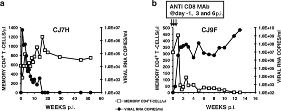
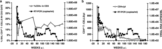
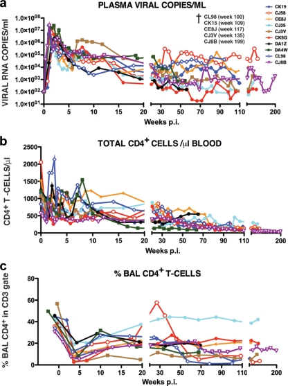
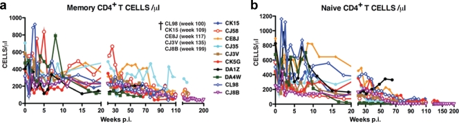
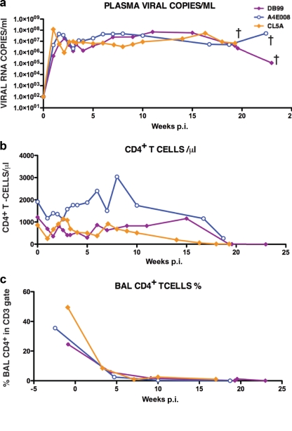
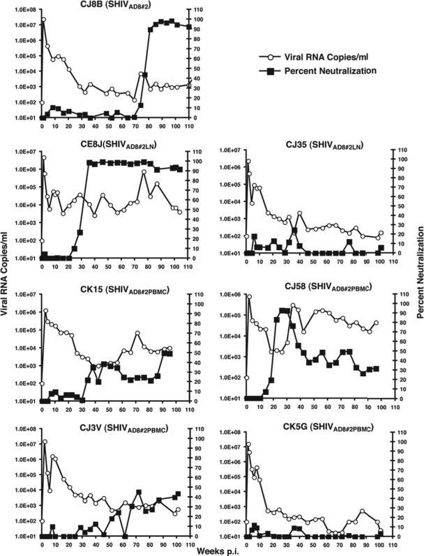
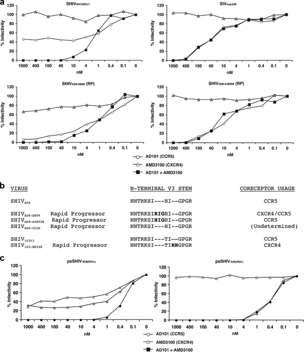
A new pathogenic R5-tropic simian/human immunodeficiency virus (SHIV) was generated following serial passaging in rhesus macaques. All 13 animals inoculated with SHIV(AD8) passaged lineages experienced marked depletions of CD4(+) T cells. Ten of these infected monkeys became normal progressors (NPs) and had gradual losses of both memory and naïve CD4(+) T lymphocytes, generated antiviral CD4(+) and CD8(+) T cell responses, and sustained chronic immune activation while maintaining variable levels of plasma viremia (10(2) to 10(5) RNA copies/ml for up to 3 years postinfection [p.i.]). To date, five NPs developed AIDS associated with opportunistic infections caused by Pneumocystis carinii, Mycobacterium avium, and Campylobacter coli that required euthanasia between weeks 100 and 199 p.i. Three other NPs have experienced marked depletions of circulating CD4(+) T lymphocytes (92 to 154 cells/microl) following 1 to 2 years of infection. When tested for coreceptor usage, the viruses isolated from four NPs at the time of their euthanasia remained R5 tropic. Three of the 13 SHIV(AD8)-inoculated macaques experienced a rapid-progressor syndrome characterized by sustained plasma viremia of >1 x 10(7) RNA copies/ml and rapid irreversible loss of memory CD4(+) T cells that required euthanasia between weeks 19 and 23 postinfection. The sustained viremia, associated depletion of CD4(+) T lymphocytes, and induction of AIDS make the SHIV(AD8) lineage of viruses a potentially valuable reagent for vaccine studies.


Figures

FIG. 1.
FIG. 1.
Infectivity of the original SHIVAD8 in rhesus monkeys. Macaques CJ7H (a) and CJ9F (b) were inoculated with 1 ml of undiluted SHIVAD8 by the i.v., i.p., and BM routes. Macaque CJ9F was treated with the depleting anti-CD8 MAb cM-T807 as indicated.
FIG. 2.
FIG. 2.
Serial animal-to-animal passage of SHIVAD8. (a) Passage history of SHIVAD8 and origin of SHIVAD8#2. (b) Rhesus monkey PBMC were infected with SHIVAD8 or the passaged SHIVAD8#2 virus stock, normalized for RT activity. CPM, counts per minute.
FIG. 3.
FIG. 3.
SHIVAD8#2 induces sustained plasma viremia and loss of CD4 T cells in an inoculated rhesus macaque. Plasma viremia and the percentage of BAL fluid CD4+ T cells (a) or the absolute numbers of circulating CD4+ T cells (b) in rhesus macaque CJ8B inoculated intravenously with SHIVAD8#2 are shown. RT-PCR, reverse transcription-PCR.
FIG. 4.
FIG. 4.
SHIVAD8#2 and its immediate derivatives cause immunodeficiency in rhesus macaques. The dashed arrows indicate virus transfer by blood transfusion. The thick arrows indicate LN or PBMC specimens used to generate virus stocks by coculturing with PBMC from uninfected donors. †, euthanized animals.
FIG. 5.
FIG. 5.
Total CD4+ T lymphocytes are gradually lost in normal progressors following infection with SHIVAD8#2 and its immediate derivatives. The levels of plasma viremia (a), absolute numbers of peripheral CD4+ T cells (b), and percentages of BAL fluid CD4+ T cells (c) are shown. The five normal progressors that developed AIDS and were euthanized are indicated (†).
FIG. 6.
FIG. 6.
Marked depletion of naïve and memory CD4+ T lymphocytes characterizes long-term SHIVAD8 infection in NP rhesus monkeys. Absolute numbers of memory CD4+ T cells (a) and naive CD4+ T cells (b) in 10 normal-progressor macaques during 200 weeks of SHIVAD8 infection are shown. †, euthanized animals.
FIG. 7.
FIG. 7.
Patterns of virus replication and CD4+T cell dynamics in SHIVAD8 rapid progressors. The levels of plasma viremia (a), absolute numbers of peripheral CD4+ T cells (b), and percentages of BAL fluid CD4+ T cells (c) are shown. †, euthanized animals.
FIG. 8.
FIG. 8.
Neutralizing-antibody activities detected in normal-progressor macaques following infection with SHIVAD8#2 or its immediate derivatives. Plasma samples (1:20 dilution) from the indicated SHIVAD8-infected macaques were incubated in quadruplicate for 1 h at 37°C with the virus isolates shown in parentheses and then used as an inoculum to infect TZM-bl cells. The luciferase activity present in cell lysates at 28 h p.i. was measured, and the average percent neutralization activity in plasma at each time point was determined. Prechallenge plasma samples served as negative controls and baselines for zero neutralizing-antibody activity.
FIG. 9.
FIG. 9.
Coreceptor utilization of SHIVAD8 derivatives isolated from rapid progressors. (a) TZM-bl cells were infected in quadruplicate with viruses (SHIVAD8-DB99 and SHIVAD8-A4E008) recovered from rapid progressors DB99 and A4E008, respectively, in the presence of the indicated amounts of the small-molecule coreceptor inhibitors AD101 (CCR5), AMD 3100 (CXCR4), or both. SIVmac239 and SHIVDH12RCL-7 were also analyzed as representative R5-tropic and dual-tropic viruses, respectively. The luciferase activities present in cell lysates 24 h p.i. were measured, and percent infectivities were determined in the absence or presence of coreceptor inhibitors. (b) gp120 sequences from the N-terminal V3 regions of SHIVAD8 variants, recovered from three RP animals, were aligned with the starting SHIVAD8 V3 loop. The V3 regions of the R5-tropic SHIVSF162P3 and its SHIVSF162-BR24N derivative, which also emerged in an RP, are included in the alignment. (c) Coreceptor utilization of virus, pseudotyped with Envs present in RP DB99 at the time of necropsy, containing or lacking the 3-aa RIG V3 loop insertion.

Similar articles

Cited by

References

    1. Amara, R. R., F. Villinger, J. D. Altman, S. L. Lydy, S. P. O'Neil, S. I. Staprans, D. C. Montefiori, Y. Xu, J. G. Herndon, L. S. Wyatt, M. A. Candido, N. L. Kozyr, P. L. Earl, J. M. Smith, H. L. Ma, B. D. Grimm, M. L. Hulsey, J. Miller, H. M. McClure, J. M. McNicholl, B. Moss, and H. L. Robinson. 2001. Control of a mucosal challenge and prevention of AIDS by a multiprotein DNA/MVA vaccine. Science 292:69-74. - PubMed
    1. Barnett, S. W., I. K. Srivastava, E. Kan, F. Zhou, A. Goodsell, A. D. Cristillo, M. G. Ferrai, D. E. Weiss, N. L. Letvin, D. Montefiori, R. Pal, and M. Vajdy. 2008. Protection of macaques against vaginal SHIV challenge by systemic or mucosal and systemic vaccinations with HIV-envelope. AIDS 22:339-348. - PubMed
    1. Barouch, D. H., S. Santra, J. E. Schmitz, M. J. Kuroda, T. M. Fu, W. Wagner, M. Bilska, A. Craiu, X. X. Zheng, G. R. Krivulka, K. Beaudry, M. A. Lifton, C. E. Nickerson, W. L. Trigona, K. Punt, D. C. Freed, L. Guan, S. Dubey, D. Casimiro, A. Simon, M. E. Davies, M. Chastain, T. B. Strom, R. S. Gelman, D. C. Montefiori, M. G. Lewis, E. A. Emini, J. W. Shiver, and N. L. Letvin. 2000. Control of viremia and prevention of clinical AIDS in rhesus monkeys by cytokine-augmented DNA vaccination. Science 290:486-492. - PubMed
    1. Beer, B. E., C. R. Brown, S. Whitted, S. Goldstein, R. Goeken, R. Plishka, A. Buckler-White, and V. M. Hirsch. 2005. Immunodeficiency in the absence of high viral load in pig-tailed macaques infected with simian immunodeficiency virus SIVsun or SIVlhoest. J. Virol. 79:14044-14056. - PMC - PubMed
    1. Brenchley, J. M., T. W. Schacker, L. E. Ruff, D. A. Price, J. H. Taylor, G. J. Beilman, P. L. Nguyen, A. Khoruts, M. Larson, A. T. Haase, and D. C. Douek. 2004. CD4+ T cell depletion during all stages of HIV disease occurs predominantly in the gastrointestinal tract. J. Exp. Med. 200:749-759. - PMC - PubMed

Publication types

MeSH terms

Substances

LinkOut - more resources