. 2010 Apr;84(8):4026-40.
doi: 10.1128/JVI.02597-09. Epub 2010 Feb 10.

Regulation of ICP0-null mutant herpes simplex virus type 1 infection by ND10 components ATRX and hDaxx

Affiliations

Regulation of ICP0-null mutant herpes simplex virus type 1 infection by ND10 components ATRX and hDaxx

Vera Lukashchuk et al. J Virol. 2010 Apr.

Abstract

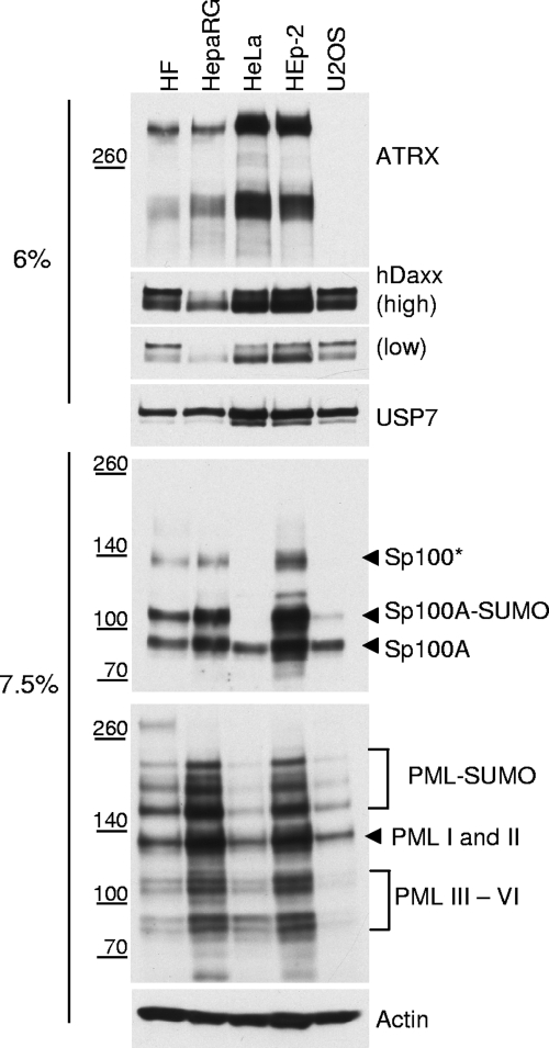
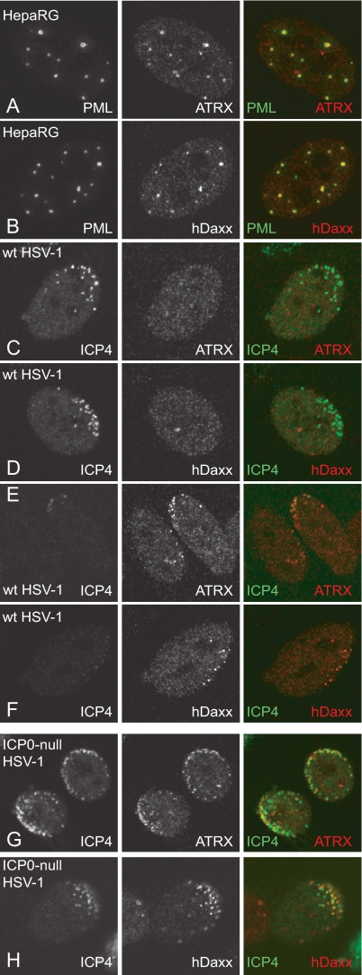
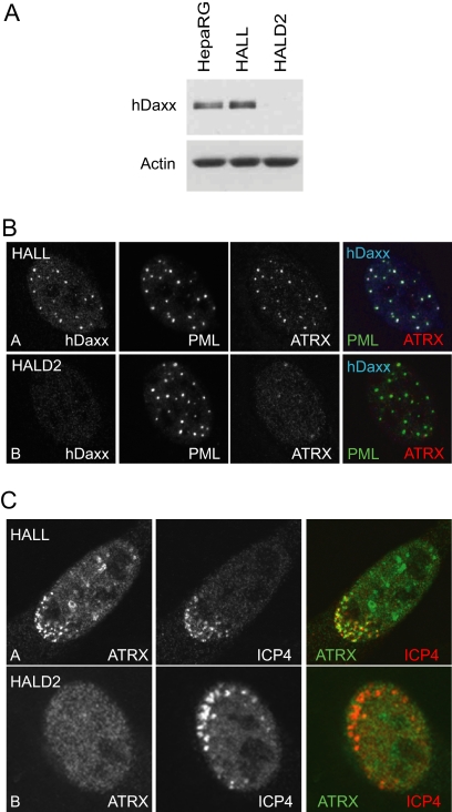
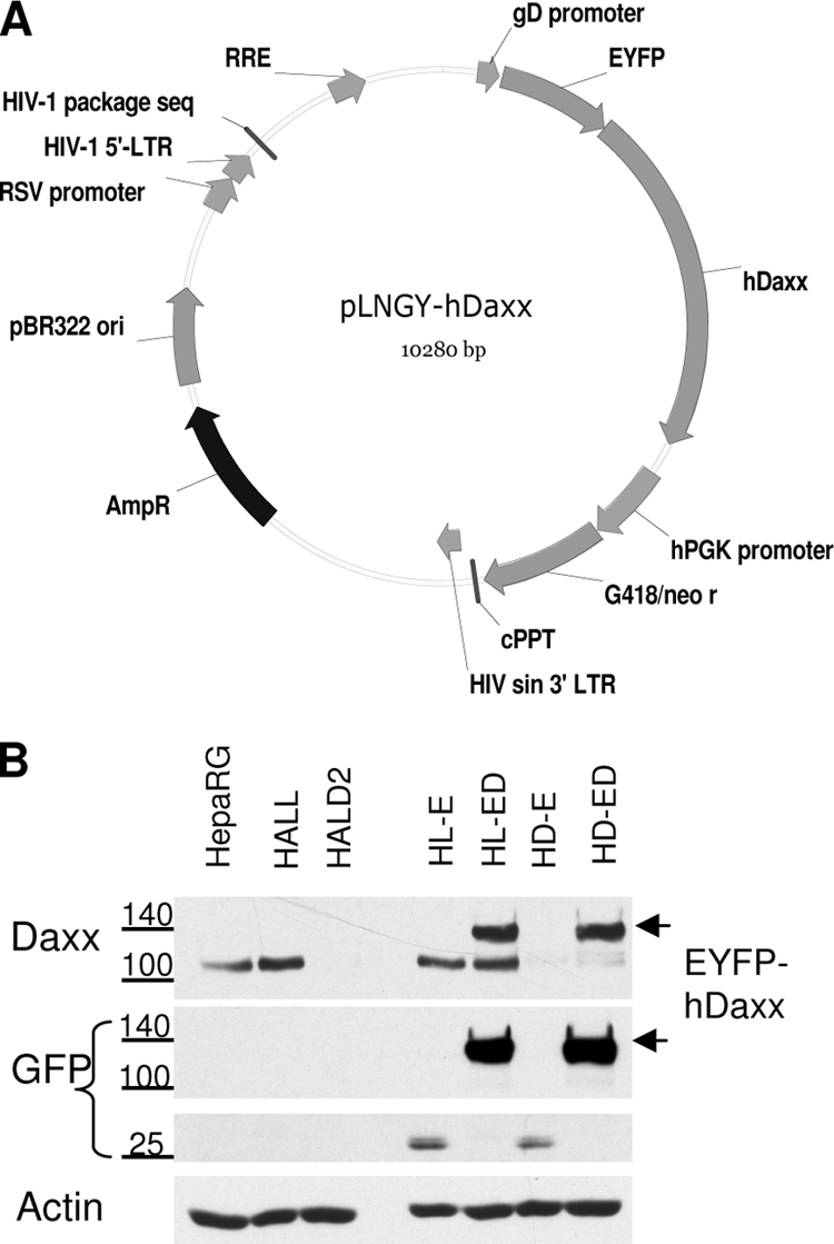
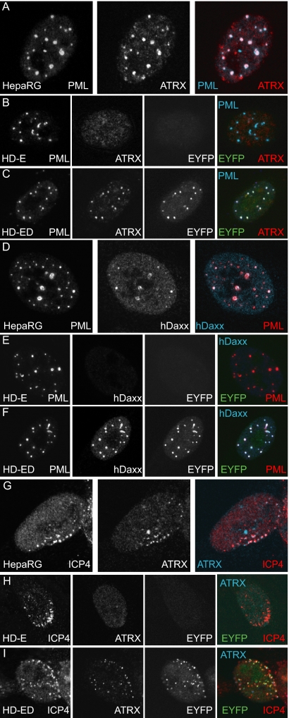
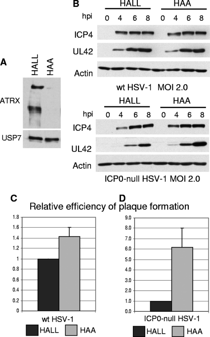
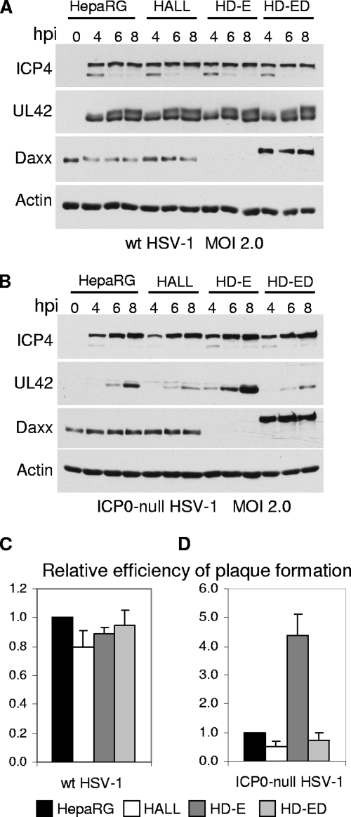
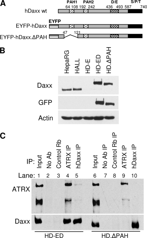
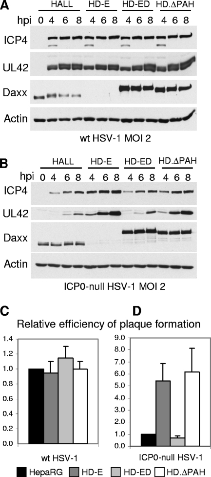
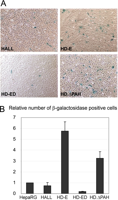
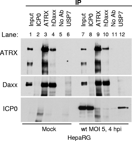
Herpes simplex virus type 1 (HSV-1) immediate-early gene product ICP0 activates lytic infection and relieves cell-mediated repression of viral gene expression. This repression is conferred by preexisting cellular proteins and is commonly referred to as intrinsic antiviral resistance or intrinsic defense. PML and Sp100, two core components of nuclear substructures known as ND10 or PML nuclear bodies, contribute to intrinsic resistance, but it is clear that other proteins must also be involved. We have tested the hypothesis that additional ND10 factors, particularly those that are involved in chromatin remodeling, may have roles in intrinsic resistance against HSV-1 infection. The two ND10 component proteins investigated in this report are ATRX and hDaxx, which are known to interact with each other and comprise components of a repressive chromatin-remodeling complex. We generated stable cell lines in which endogenous ATRX or hDaxx expression is severely suppressed by RNA interference. We found increases in both gene expression and plaque formation induced by ICP0-null mutant HSV-1 in both ATRX- and hDaxx-depleted cells. Reconstitution of wild-type hDaxx expression reversed the effects of hDaxx depletion, but reconstitution with a mutant form of hDaxx unable to interact with ATRX did not. Our results suggest that ATRX and hDaxx act as a complex that contributes to intrinsic antiviral resistance to HSV-1 infection, which is counteracted by ICP0.


Figures

FIG. 1.
FIG. 1.
Analysis of endogenous expression of ND10 proteins in cultured cell lines. Samples of the indicated cells were harvested, and whole-cell extracts derived from approximately 3 × 10⁴ cells were loaded onto 7.5% or 6% polyacrylamide gels. The antibodies used for Western blot detection were anti-PML 5E10, anti-ATRX 39F, anti-Daxx D7810, anti-Sp100 SpGH, anti-actin AL-40, and anti-USP7 BL851. USP7 and actin were used as loading controls for 6% and 7.5% gels, respectively. The differences in USP7 expression were reproducible.
FIG. 2.
FIG. 2.
ATRX and hDaxx are recruited to the sites of incoming HSV-1 genomes in HepaRG cells. Immunofluorescence images of uninfected HepaRG cells (A and B) and cells infected with wt HSV-1 (MOI, 0.001) (C to F) and ICP0-null mutant HSV-1 (MOI, 0.5) (G to H) at the edge of a developing plaque. Cells were processed for immunofluorescence analysis at 24 h postinfection and stained for the indicated proteins. Uninfected cells (A and B) were costained for PML (5E10) and ATRX (H300) or hDaxx (07-471). Infected cells were costained for ICP4 (58S) and ATRX or hDaxx. For panel H, Daxx was detected with anti-rabbit r1866 serum. The secondary antibodies were FITC-conjugated anti-mouse (green) (A to H), Alexa 555-conjugated anti-rabbit (red) (A to F), or Cy3-conjugated anti-rabbit (red) (G to H) IgGs.
FIG. 3.
FIG. 3.
ATRX is not recruited to sites associated with ICP0-null mutant HSV-1 genomes in hDaxx-depleted HepaRG cells. (A) hDaxx depletion. Cell extracts harvested from normal HepaRG, shLuci-expressing HALL, and shDaxx-expressing HALD2 cells and were resolved on a 7.5% polyacrylamide gel, and hDaxx expression was detected by Western blotting. Actin is a loading control. (B) Confocal immunofluorescence images demonstrating simultaneous detection of hDaxx, ATRX, and PML in HALL and HALD2 cells, using rabbit anti-Daxx (07-471), mouse anti-PML (5E10), and goat anti-ATRX (D19) antibodies. The secondary antibodies were FITC-conjugated anti-mouse, Alexa 647-conjugated anti-rabbit, and Alexa 555-conjugated anti-goat IgGs. (C) Immunofluorescence analysis of ATRX cells in developing ICP0-null mutant HSV-1 plaques in HALL and HALD2 cells. Cells were infected at an MOI of 0.5 (HALL) or 0.1 (HALD2) and costained the following day for ATRX (H300, anti-rabbit Alexa 488) and ICP4 (58S, anti-mouse Cy3).
FIG. 4.
FIG. 4.
Reintroduction of hDaxx into hDaxx-depleted cells. (A) Map of the pLNGY-hDaxx vector construct (Table 1). RRE, rev response element; hPGK promoter, human phosphoglycerate kinase promoter; cPPT, polypurine tract; LTR, long terminal repeats; sin, self-inactivating; RSV promoter, Rous sarcoma virus promoter; AmpR, ampicillin resistance gene; G418/neo, neomycin resistance gene. (B) Western blot analysis of hDaxx expression in the generated cell lines. The reintroduced fusion protein EYFP-hDaxx in HD-ED cells is indicated by arrowheads on the right. Antibodies used for detection of the proteins are indicated on the left: hDaxx and EYFP expression levels were detected by anti-hDaxx antibody (D7810) and anti-GFP antibody (ab290), respectively. The positions of relevant size markers are indicated.
FIG. 5.
FIG. 5.
Reintroduced hDaxx relocates ATRX to ND10 and to sites associated with incoming ICP0-null mutant HSV-1 genomes. Immunofluorescence analysis of PML, ATRX, and hDaxx distribution in uninfected (rows A to F) and ICP0-null HSV-1-infected (rows G to I) HepaRG, HD-E, and HD-ED cells (as indicated on the relevant panels). Cells on coverslips were costained for PML (r8) and ATRX (39F) (A to C) or PML (5E10) and hDaxx (07-471) (D to F). For analysis of the redistribution of the proteins during ICP0-null mutant virus infection, cells were infected at an MOI of 0.1 (HD-E) or 0.5 (HepaRG and HD-ED) and processed for immunofluorescence analysis the following day. Cells were costained for ICP4 (58S) and ATRX (H300). The secondary antibodies used throughout the experiment were Cy5-conjugated anti-rabbit (blue) and Alexa 555-conjugated anti-mouse (red) IgGs. Green fluorescence resulted from the autofluorescence of the EYFP tag.
FIG. 6.
FIG. 6.
Efficiency of HSV-1 infection in ATRX-depleted cells. (A) Detection of ATRX expression by Western blot analysis using 39F antibody, showing expression of both ATRX isoforms in HALL cells and barely detectable levels of expression in HAA cells. Samples were resolved on a 6% polyacrylamide gel. USP7 was used as a loading control. (B) Kinetics of viral protein expression in ATRX-depleted cells. HALL and HAA cells were infected with wt or ICP0-null HSV-1 at an MOI of 2, and samples were harvested for Western blot analysis at 0, 4, 6, and 8 h postinfection (hpi). Samples were resolved on a 7.5% polyacrylamide gel, and membranes were probed for ICP4 and UL42, using antibodies 58S and Z1F11, respectively. Actin was a loading control. (C) Efficiency of plaque formation of HSV-1 in ATRX-depleted cells. Cells were infected with wt HSV-1 strain in1863 and ICP0-null HSV-1 strain dl1403/CMVlacZ at sequential dilutions. After 24 h of infection, cells were fixed and stained for β-galactosidase expression. The plots represent the mean relative efficiencies of plaque formation as the numbers of plaques determined to occur in HAA cells in relation to the level for HALL cells from three independent experiments. Error bars represent standard error values.
FIG. 7.
FIG. 7.
Efficiency of HSV-1 infection in hDaxx-depleted cells. (A and B) Kinetics of ICP4 and UL42 expression in hDaxx-depleted and hDaxx-reconstituted cells. HepaRG, HALL, HD-E, and HD-ED cells were infected with wt (A) or ICP0-null mutant (B) HSV-1 at an MOI of 2, and samples were harvested for Western blot analysis at 0, 4, 6, and 8 h postinfection (hpi). Samples were resolved on a 7.5% polyacrylamide gel, and membranes were probed for ICP4, UL42, and hDaxx using antibodies 58S, Z1F11, and D7810, respectively. Actin was a loading control. (C and D) Efficiency of plaque formation of HSV-1 in hDaxx-depleted and hDaxx-reconstituted cells. Cells seeded into 24-well plates were infected with wt HSV-1 strain in1863 (C) or ICP0-null HSV-1 dl1403/CMVlacZ (D) at sequential dilutions and stained for β-galactosidase expression 24 h later. The plots represent mean values for plaque numbers relative to those in HepaRG cells, obtained from 4 independent experiments. Error bars represent standard error values.
FIG. 8.
FIG. 8.
Deletion of the ATRX interaction domain of hDaxx. (A) Schematic representation of wt hDaxx (based on that in reference 102), the EYFP-hDaxx construct, and the EYFP-hDaxxΔPAH1 mutant, from which amino acids 48 to 120 spanning the ATRX interacting domain PAH1 were removed by PCR-based splicing mutagenesis. PAH, paired amphipathic helices; D/E, Asp/Glu-rich motif; S/P/T, Ser/Pro/Thr-rich domain. (B) Western blot analysis of expression of hDaxx proteins in HepaRG, HALL, HD-E, HD-ED, and HD.ΔPAH cells. The membrane was probed with the indicated antibodies. Samples were resolved on a 7.5% polyacrylamide gel. (C) Immunoprecipitation analysis of ATRX and hDaxx interaction in HD-ED cells and HD.ΔPAH cells. Lanes 1 and 6 represent 2.5% input of the total extract sample incubated in each immunoprecipitation reaction. HD-ED and HD.ΔPAH cells extracts were immunoprecipitated with either anti-ATRX (antibody H300; lanes 4 and 9), anti-Daxx (antibody 07-471; lanes 5 and 10), control rabbit serum 201 raised against USP7 (lanes 3 and 8), or no antibody (lanes 2 and 7). Samples were analyzed on a 6% polyacrylamide gel and probed with anti-ATRX 39F and anti-hDaxx D7810 antibodies.
FIG. 9.
FIG. 9.
The ATRX interaction domain of hDaxx is essential for ATRX localization to ND10 and recruitment to HSV-1 early replication foci in the absence of ICP0. Panels A and B show uninfected HD.ΔPAH cells stained for the indicated proteins. Panels C and D show HD.ΔPAH cells located at the periphery of a developing plaque after infection with ICP0-null mutant HSV-1 at an MOI of 0.5 PFU/cell and after a 24-h infection period. ATRX, hDaxxΔPAH, PML, and ICP4 were detected with antibodies H300, 07-471, 5E10, and 58S, respectively.
FIG. 10.
FIG. 10.
The ATRX interaction region of hDaxx is required for fully efficient hDaxx-mediated repression of ICP0-null mutant HSV-1 gene expression (A and B) and plaque formation (C and D). (A and B) Cells were infected with the indicated viruses at an MOI of 2.0 PFU/cell, and samples were harvested at the indicated time points after infection. Samples were resolved on a 7.5% polyacrylamide gel. Membranes were probed for the expression of ICP4, UL42, and hDaxx using antibodies 58S, Z1F11, and D7810, respectively. Actin was a loading control. (C and D) Cells seeded into 24-well plates were infected with wt HSV-1 strain in1863 (C) or ICP0-null HSV-1 dl1403/CMVlacZ (D) at sequential dilutions and stained for β-galactosidase activity the following day to reveal the plaques. The plots represent mean values for plaque numbers relative to those in HepaRG cells, obtained from 3 independent experiments. Error bars represent standard error values.
FIG. 11.
FIG. 11.
Establishment of quiescent infections in hDaxx-depleted, EYFP-hDaxx-reconstituted, and EYFP-hDaxxΔPAH1-introduced cells. Cells were infected with HSV-1 strain in1374, incubated at the restrictive temperature of 38.5°C overnight, and processed for β-galactosidase staining the following day. (A) Images of HALL, HD-E, HD-ED, and HD.ΔPAH cells at 24 h after infection with in1374. (B) Relative cell numbers of β-galactosidase-expressing cells in the above-mentioned cell types, compared to those obtained in parallel in1374-infected HepaRG cells. The plots represent mean values obtained from 3 independent experiments. Error bars are standard error values.
FIG. 12.
FIG. 12.
ICP0 neither interacts with nor disrupts the ATRX/hDaxx complex. HepaRG cells were infected with wt HSV-1 at an MOI of 5, and cells were harvested in IP lysis buffer at 4 h postinfection. Immunoprecipitation was carried out using no antibody or anti-ICP0 190, anti-ATRX H300, anti-Daxx 07-471, and anti-USP7 210 antibodies, as indicated. Input lanes contained 2.5% of the sample used for immunoprecipitation. Western blot analysis was carried out using mouse anti-ATRX 39F, anti-Daxx D7810, and anti-ICP0 11060 antibodies.
FIG. 13.
FIG. 13.
Phosphorylation of hDaxx during wt HSV-1 infection. (A and B) Cells were seeded into 24-well plates at a density of 1 × 10⁵ cells per well and infected with wt (A and C) or ICP0-null (B and D) HSV-1 at an MOI of 5. Samples were harvested at the indicated time points (A and B) and resolved on 7.5% gels for ICP4, UL42, and actin and on 6% gels for hDaxx. For panels C and D, samples were harvested in IP lysis buffer (without EDTA), and then aliquots of the same samples were mock treated (left-hand sets of 3 lanes) or treated with λ-phosphatase (right-hand sets of 3 lanes) prior to Western blot analysis. M, mock.

Similar articles

Cited by

References

    1. Amin, H. M., S. Saeed, and S. Alkan. 2001. Histone deacetylase inhibitors induce caspase-dependent apoptosis and downregulation of daxx in acute promyelocytic leukaemia with t(15;17). Br. J. Haematol. 115:287-297. - PubMed
    1. Argentaro, A., J. C. Yang, L. Chapman, M. S. Kowalczyk, R. J. Gibbons, D. R. Higgs, D. Neuhaus, and D. Rhodes. 2007. Structural consequences of disease-causing mutations in the ATRX-DNMT3-DNMT3L (ADD) domain of the chromatin-associated protein ATRX. Proc. Natl. Acad. Sci. U. S. A. 104:11939-11944. - PMC - PubMed
    1. Baumann, C., A. Schmidtmann, K. Muegge, and R. De La Fuente. 2008. Association of ATRX with pericentric heterochromatin and the Y chromosome of neonatal mouse spermatogonia. BMC Mol. Biol. 9:29. - PMC - PubMed
    1. Boddy, M. N., K. Howe, L. D. Etkin, E. Solomon, and P. S. Freemont. 1996. PIC 1, a novel ubiquitin-like protein which interacts with the PML component of a multiprotein complex that is disrupted in acute promyelocytic leukaemia. Oncogene 13:971-982. - PubMed
    1. Boutell, C., A. Orr, and R. D. Everett. 2003. PML residue lysine 160 is required for the degradation of PML induced by herpes simplex virus type 1 regulatory protein ICP0. J. Virol. 77:8686-8694. - PMC - PubMed

Publication types

MeSH terms

LinkOut - more resources