. 2010 Apr;84(8):4013-25.
doi: 10.1128/JVI.02502-09. Epub 2010 Feb 10.

Herpes simplex virus glycoproteins H/L bind to cells independently of {alpha}V{beta}3 integrin and inhibit virus entry, and their constitutive expression restricts infection

Affiliations

Herpes simplex virus glycoproteins H/L bind to cells independently of {alpha}V{beta}3 integrin and inhibit virus entry, and their constitutive expression restricts infection

Tatiana Gianni et al. J Virol. 2010 Apr.

Abstract

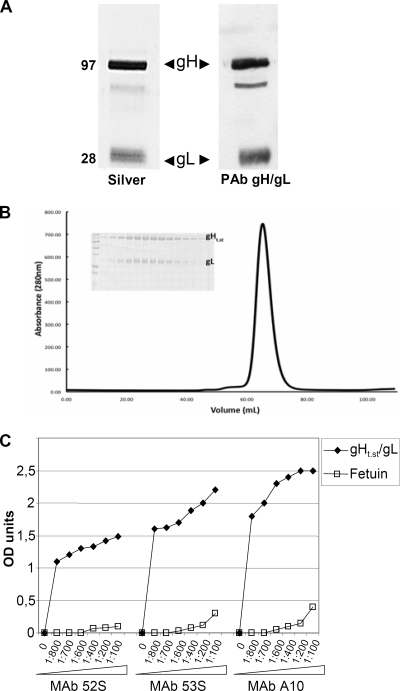
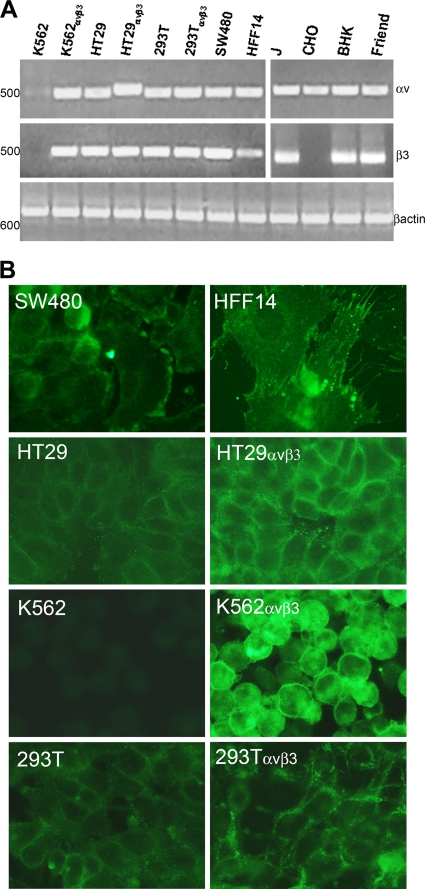
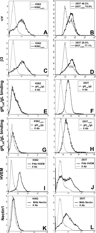
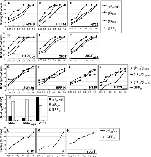
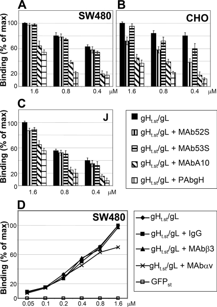
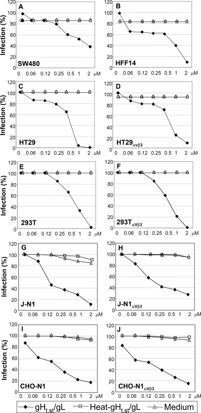
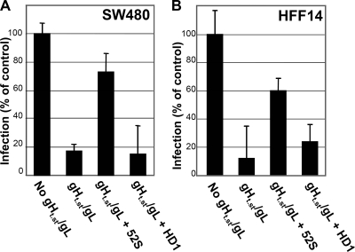
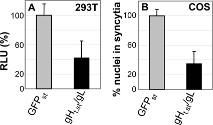
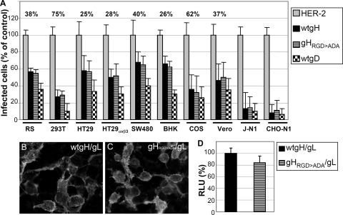
Herpes simplex virus (HSV) fusion with cells requires the gD, gB, and gH/gL glycoprotein quartet. gD serves as a receptor binding glycoprotein. gB and gH/gL execute fusion in an as-yet-unclear manner. To better understand the role of gH/gL in HSV entry, we produced a soluble version of gH/gL carrying a One-STrEP tag (gH(t.st)/gL). Previous findings implicated integrins as possible ligands to gH/gL (C. Parry et al., J. Gen. Virol. 86:7-10, 2005). We report that (i) gH(t.st)/gL bound a number of cells in a dose-dependent manner at concentrations similar to those required for the binding of soluble gB or gD. (ii) gH(t.st)/gL inhibited HSV entry at the same concentrations required for binding. It also inhibited cell-cell fusion in transfected cells. (iii) The absence of beta3 integrin did not prevent the binding of gH(t.st)/gL to CHO cells and infection inhibition. Conversely, integrin-negative K562 cells did not acquire the ability to bind gH(t.st)/gL when hyperexpressing alphaVbeta3 integrin. (iv) Constitutive expression of wild-type gH/gL (wt-gH/gL) restricted infection in all of the cell lines tested, a behavior typical of glycoproteins which bind cellular receptors. The extent of restriction broadly paralleled the efficiency of gH/gL transfection. RGD motif mutant gH/gL could not be differentiated from wt-gH with respect to restriction of infection. Cumulatively, the present results provide several lines of evidence that HSV gH/gL interacts with a cell surface cognate protein(s), that this protein is not necessarily an alphaVbeta3 integrin, and that this interaction is required for the process of virus entry/fusion.


Figures

FIG. 1.
FIG. 1.
Properties of purified gHt.st/gL. (A) Aliquots of gHt.st/gL made in insect cells and purified by means of Strep-Tactin resin according to the manufacturer protocol were separated by denaturing polyacrylamide gel electrophoresis and silver stained (Silver) (left lane) or reacted by Western blotting with a PAb to gH/gL (right lane). The values to the left are molecular sizes in kilodaltons. (B) Size exclusion chromatography on a Superdex 200 column (GE Healthcare) in 10 mM Tris (pH 8.0)-150 mM NaCl. Static light scattering was carried out over the gHt.st/gL peak during elution from the Superdex 200 column to determine the molecular weight of purified gHt.st/gL. The x axis is elution volume in ml, and the y axis is milliunits of absorbance at 280 nm (mAU). Insert, pattern of proteins as seen in denaturing gel electrophoresis with Coomassie blue staining. (C) ELISA reactivity of gHt.st/gL to neutralizing MAbs 52S, 53S, and A10 to gH/gL. gHt.st/gL or fetuin, as a negative control, was immobilized on 96-well plates and allowed to react with increasing dilutions of the MAbs. Reactivity was detected by means of anti-mouse antibodies conjugated to peroxidase, followed by o-phenylenediamine and reading of the optical density (OD) at 490 nm. Each point represents the average of triplicate measurements.
FIG. 2.
FIG. 2.
Expression of αV and β3 integrin subunits. (A) Detection of αV and β3 integrin transcripts by RT-PCR. The cDNA of the indicated cells was amplified by means of αV integrin or β3 integrin primers annealing to the human (left) or rodent (right) isoform. As a control for RT-PCR, all of the cells were checked for β-actin expression. The values on the left represent the migration positions of 500- or 600-bp markers. (B) IFA detection of αVβ3 integrin by means of MAb LM609. All pictures were taken with the same exposure time by means of a 63× objective. All of the inserts in panel A, except that relative to β3 on the right, were modified by Photoshop software as follows: −35% brightness, +30% contrast. HFF14 insert in panel B: brightness, −20%; contrast, +20%. All of panel B: brightness, +40%; contrast, +100%.
FIG. 3.
FIG. 3.
Expression of αV and β3 integrin and of HVEM and nectin1 receptors and gHt.st/gL binding as detected by flow cytometry. (A to D) The indicated cells were reacted with MAb L230 to αV integrin (A, B) or MAb AP3 to β3 integrin (C, D). (E to H) The indicated cells were reacted with gHt.st/gL, followed by MAb 52S. (I to L) The indicated cells were reacted with PAb to HVEM (I, J) or MAb R1.302 to nectin1 (K, L). Abscissa, fluorescence intensity.
FIG. 4.
FIG. 4.
CELISA binding of gHt.st/gL to cells. (A to F) gHt.st/gL, gDΔ290-299 (gD290t), gB730t, or GFPst was allowed to react with cells grown in 96-well plates. Binding was detected by means of appropriate antibodies (PAb R45 to gD, MAb H1817 to gB), followed by HRP-conjugated secondary antibodies or HRP-conjugated MAb to the One-STrEP tag (Strep-Tactin) for gHt.st/gL or GFPst and the o-phenylenediamine substrate. (G to J) gHt.st/gL or GFPst was reacted with the indicated cells in medium adjusted to pH 7, pH 6, or pH 5. (K) CELISA with K562, K562αVβ3, and 293T cells in suspension. gHt.st/gL, gB730t, or GFPst was reacted with cells as described above, except that the cells were in suspension. Binding was detected as in panels A to F. (L to N) Binding of gHt.st/gL to receptor-negative CHO and J cells or to heparan sulfate- and chondroitin sulfate-negative sog9 cells. Abscissa, μM concentrations.
FIG. 5.
FIG. 5.
Inhibition of gHt.st/gL binding to cells. (A to C) Inhibition of gHt.st/gL binding by Abs to gH/gL. gHt.st/gL at the indicated concentrations was preincubated with the indicated Abs for 1 h at room temperature and laid over the cells for 1 h of incubation at 4°C. Binding was detected as described in the legend to Fig. 4A to F. (D) Inhibition of gHt.st/gL binding to SW480 cells by Abs L230 and AP3 (7, 44) directed to αV and β3 integrins, respectively. Cells were exposed to antibodies for 1 h prior to the addition of gHt.st/gL and throughout gHt.st/gL binding. The amount of antibodies was kept constant (40 μg/ml). Mouse IgGs served as a negative control for antibodies. All details are as described in the legend to Fig. 4A to F. Each point or column represents the average of triplicate assays. Each assay was performed at least twice. Binding is expressed as a percentage of the highest value obtained for each panel. Bars denote standard deviations.
FIG. 6.
FIG. 6.
Inhibition of R8102 infection by gHt.st/gL. Cells were preincubated with gHt.st/gL at the indicated concentrations for 1 h. R8102 (3 PFU/cell) was added to the gHt.st/gL-containing medium for 90 min of incubation. The viral inoculum was removed, and cells were overlaid with gHt.st/gL and processed for β-Gal quantification at 8 h after infection. The negative controls consisted of incubation with heat-inactivated gHt.st/gL for 120 min at 80°C (heat-gHt.st/gL) or medium alone. A 100% infection value corresponds to infected cells not exposed to gHt.st/gL. 293T cells were either left untransfected or transfected with αV and β3 integrin plasmids (293TαVβ3). J and CHO cells were transfected either with nectin1 alone (J-N1 and CHO-N1) or with nectin1 plus αV and β3 integrin plasmids (J-N1αVβ3 and CHO-N1αVβ3). Each point represents the average of triplicate assays. Each experiment was performed at least two times. Infection is expressed as a percentage of the highest value obtained for each panel.
FIG. 7.
FIG. 7.
Preincubation of gHt.st/gL with MAb 52S restores R8102 infection. Cells were infected with R8102 (details are as in the legend to Fig. 6) in the absence (No gHt.st/gL) or in the presence of gHt.st/gL (gHt.st/gL). In the “gHt.st/gL + 52S” and “gHt.st/gL + HD1” samples, gHt.st/gL was preabsorbed to MAb 52S or HD1, respectively. Preincubation of gHt.st/gL with MAb 52S, but not MAb HD1, restored R8102 infection. Each column represents the average of triplicate assays. Bars represent standard deviations.
FIG. 8.
FIG. 8.
gHt.st/gL inhibition of cell-cell fusion in cells transiently expressing gD, gB, or gH/gL. (A) 293T cells were transfected with gD, gB, gH/gL, and T7 polymerase and seeded 24 h later at a 1:1 ratio with effector cells transfected with luciferase under the control of the T7 promoter. The mixed cell population was exposed to gHt.st/gL or GFPst (0.8 μM). The extent of cell-cell fusion was expressed as relative luciferase units (RLU). A value of 100% corresponds to cells treated with GFPst. Each column represents the average of triplicate assays. Bars represent standard deviations. (B) COS cells grown on glass coverslips were transfected with gD, gB, or gH/gL and exposed to gHt.st/gL or GFPst (0.8 μM) from 8 h after transfection until fixation at 48 h after transfection. Cells were stained by IFA with MAb H1817 to gB. The extent of fusion was expressed as the average number of nuclei present in syncytia. For each specimen, at least 100 nuclei were scored. A value of 100% corresponds to cells treated with GFPst. Bars denote standard deviations.
FIG. 9.
FIG. 9.
Restriction of R8102 infection by full-length gH/gL or by the gHRGD>ADA/gL and characterization of gHRGD>ADA. (A) Restriction of R8102 infection. The indicated cells were transfected with wt-gH or gHRGD>ADA (each plus gL) or with HER-2 as a negative control. As a positive control for restriction of infection, cells were transfected with wt-gD. In order to determine the number of transfected cells for each cell line, each transfection mixture included a YFPVenus plasmid. Twenty-four hours after transfection, cells were seeded into 96- or 24-well trays on glass coverslips and employed after a further 24 h. Cells in 96-well trays were infected with R8102 in triplicate; the number of infected cells was quantified as β-Gal activity and expressed as a percentage. A value of 100% represents cells transfected with HER-2. Each column represents the average of triplicate assays. Bars represent standard deviations. Cells on glass coverslips were employed to determine the efficiency of transfection. Specifically, for each cell line, four to eight microscopic fields were photographed and the number of YFPVenus-fluorescent cells was scored over the total number of cells. The percentage ratio of transfected over total cells represents the efficiency of transfection and is given for each cell line in the first row. J and CHO cells were transfected with nectin1 (J-N1 and CHO-N1) to render cells susceptible to R8102 infection simultaneously with gH/gL and YFPVenus. (B, C). Characterization of gHRGD>ADA. Cell surface expression of wt-gH/gL (B) or gHRGD>ADA (C). 293T cells were transfected with gHRGD>ADA or wt-gH plus gL. Cell surface expression was detected by IFA by means of MAb 53S in paraformaldehyde-fixed cells. (D) Cell-cell fusion of 293T cells transfected with gHRGD>ADA or wt-gH plus gL, gD, or gB. Cell-cell fusion was performed and quantified as described in the legend to Fig. 8. A value of 100% represents the value obtained with wt-gH. Each column represents the average of triplicate assays. Bars represent standard deviations. Panels B and C were modified by Photoshop as follows: −20% brightness, +20% contrast. RLU, relative luciferase units.

Similar articles

Cited by

References

    1. Akula, S. M., N. P. Pramod, F. Z. Wang, and B. Chandran. 2002. Integrin alpha3beta1 (CD 49c/29) is a cellular receptor for Kaposi's sarcoma-associated herpesvirus (KSHV/HHV-8) entry into the target cells. Cell 108:407-419. - PubMed
    1. Atanasiu, D., J. C. Whitbeck, T. M. Cairns, B. Reilly, G. H. Cohen, and R. J. Eisenberg. 2007. Bimolecular complementation reveals that glycoproteins gB and gH/gL of herpes simplex virus interact with each other during cell fusion. Proc. Natl. Acad. Sci. U. S. A. 104:18718-18723. - PMC - PubMed
    1. Avitabile, E., C. Forghieri, and G. Campadelli-Fiume. 2007. Complexes between herpes simplex virus glycoproteins gD, gB, and gH detected in cells by complementation of split enhanced green fluorescent protein. J. Virol. 81:11532-11537. - PMC - PubMed
    1. Avitabile, E., C. Forghieri, and G. Campadelli-Fiume. 2009. Cross talk among the glycoproteins involved in herpes simplex virus entry and fusion: the interaction between gB and gH/gL does not necessarily require gD. J. Virol. 83:10752-10760. - PMC - PubMed
    1. Avitabile, E., G. Lombardi, and G. Campadelli-Fiume. 2003. Herpes simplex virus glycoprotein K, but not its syncytial allele, inhibits cell-cell fusion mediated by the four fusogenic glycoproteins, gD, gB, gH,and gL. J. Virol. 77:6836-6844. - PMC - PubMed

Publication types

Substances

LinkOut - more resources