. 2010 May;84(9):4264-76.
doi: 10.1128/JVI.02214-09. Epub 2010 Feb 10.

Structure-based mutational analysis of the bovine papillomavirus E1 helicase domain identifies residues involved in the nonspecific DNA binding activity required for double trimer formation

Affiliations

Structure-based mutational analysis of the bovine papillomavirus E1 helicase domain identifies residues involved in the nonspecific DNA binding activity required for double trimer formation

Xiaofei Liu et al. J Virol. 2010 May.

Abstract

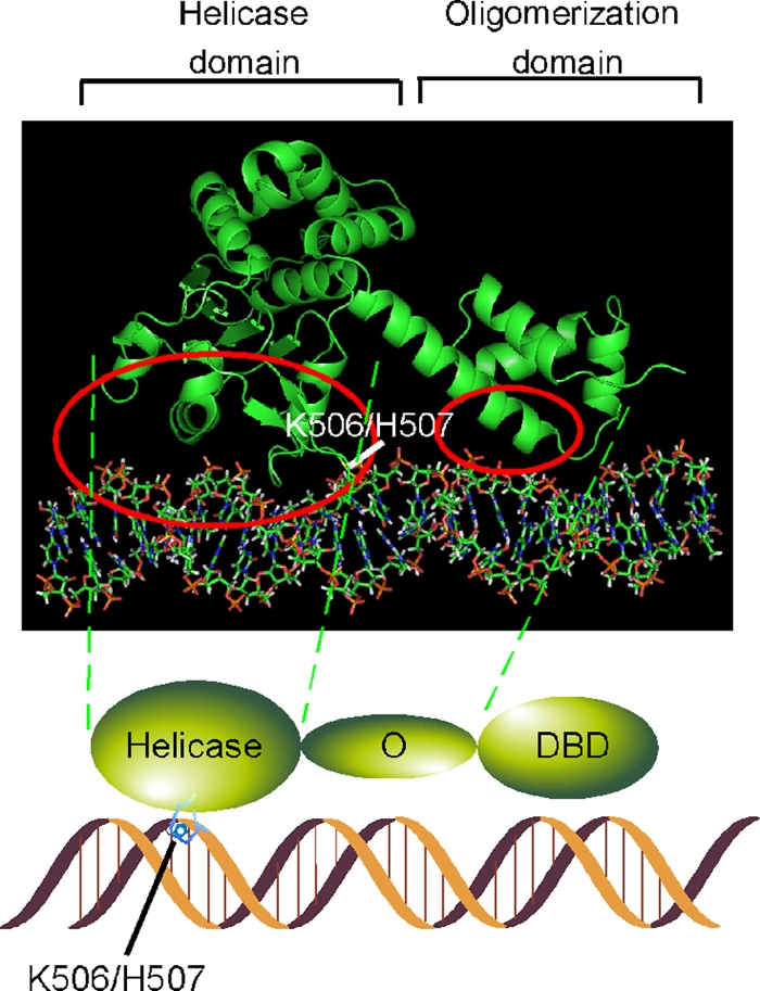
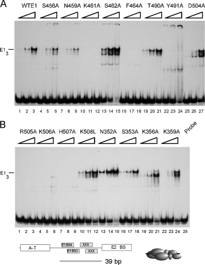
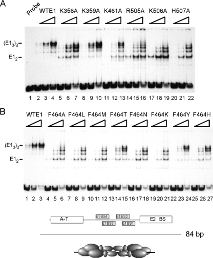
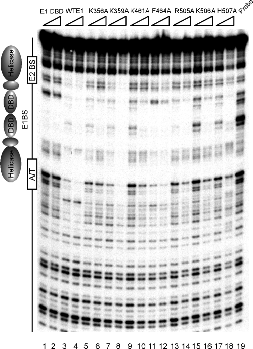
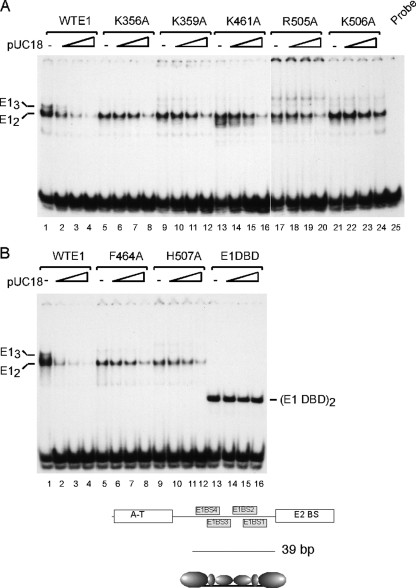
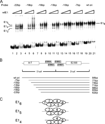
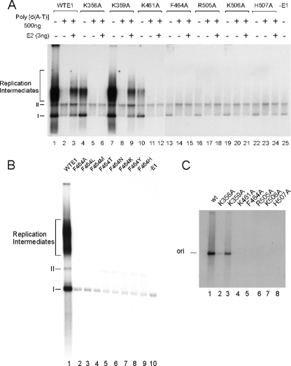
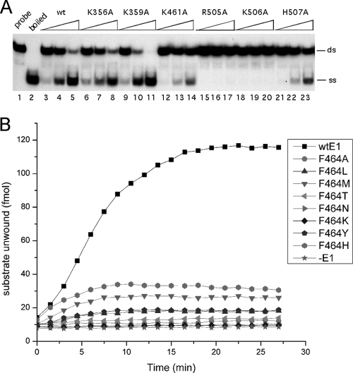
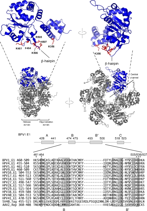
The papillomavirus E1 protein is a multifunctional initiator protein responsible for preparing the viral DNA template for initiation of DNA replication. The E1 protein encodes two DNA binding activities that are required for initiation of DNA replication. A well-characterized sequence-specific DNA binding activity resides in the E1 DBD and is used to tether E1 to the papillomavirus ori. A non-sequence-specific DNA binding activity is also required for formation of the E1 double trimer (DT) complex, which is responsible for the local template melting that precedes loading of the E1 helicase. This DNA binding activity is very poorly understood. We use a structure-based mutagenesis approach to identify residues in the E1 helicase domain that are required for the non-sequence-specific DNA binding and DT formation. We found that three groups of residues are involved in nonspecific DNA binding: the E1 beta-hairpin structure containing R505, K506, and H507; a hydrophobic loop containing F464; and a charged loop containing K461 together generate the binding surface involved in nonspecific DNA binding. These residues are well conserved in the T antigens from the polyomaviruses, indicating that the polyomaviruses share this nonspecific DNA binding activity.


Figures

FIG. 1.
FIG. 1.
Diagram depicting the regions of the E1 oligomerization and helicase domains predicted to be in close proximity to DNA when E1 is bound to dsDNA via its DBD. One subunit was extracted from the hexamer structure of the E1 oligomerization and helicase domains and oriented along the dsDNA in a linear arrangement, suggested by the arrangement of the E1 DBD directly adjacent to the oligomerization domain in the primary sequence. The oligomerization and helicase domains were oriented such that the β-hairpin residues K506 and H507, which are known to interact with DNA, are in contact with DNA. The regions circled in red contain candidate residues for interaction with dsDNA.
FIG. 2.
FIG. 2.
Trimer formation by wild-type E1 and E1 point mutants. Wild-type E1 and E1 point mutants were tested for trimer formation in the presence of ADP by EMSA. Three concentrations of E1 (6, 12, and 24 ng) were incubated with a 39-bp probe containing a part of the ori as illustrated below the panels. In panel A, S456A, N459A, K461A, S462A, F464A, T490A, Y491A, and D504A were tested. In panel B, R505A, K506A, H507A, K508L, N352A, S353A, K356A, and K359A were tested. Lane 25 in panel B contained probe alone. The migration of the E1 trimer (E13) is indicated.
FIG. 3.
FIG. 3.
DT formation by wild-type E1 and E1 point mutants. Wild-type E1 and E1 mutants were tested for DT formation in the presence of ADP by EMSA. Three concentrations of E1 (3, 6, and 12 ng) were incubated with the 84-bp ori probe containing the complete ori as illustrated below the panels. In panel A, wild-type E1, K356A, K359A, K461A, R505A, K506A, and H507A were tested. In panel B, wild-type E1, F464A, F464L, F464M, F464T, F464N, F464K, F464Y, and F464H were tested. The migration of the E1 dimer (E12) and DT (E13)2 are indicated.
FIG. 4.
FIG. 4.
E1 mutants with defects in trimer and DT formation fail to protect sequences flanking the E1 BS. DNase I footprinting was performed on the top strand of ori region comparing the protection observed with E1 DBD, wild-type E1, and E1 point mutants. E1 DBD (120 and 240 fmol, lanes 1 to 2) and 140 and 280 fmol of wild-type E1 and of the respective point mutants were used. Lane 19 contained no E1.
FIG. 5.
FIG. 5.
Residues on the E1 oligomerization and helicase domains are required for nonspecific DNA binding activity. EMSA was performed using a 39-bp ori probe where E1 binds as a dimer, as indicated below the panels. In each set of four, EMSA was performed in the absence or in the presence of 4, 8, or 16 ng of pUC 18 as nonspecific dsDNA competitor. In panel A, wild-type E1 (lanes 1 to 4), K356A (lanes 5 to 8), K359A (lanes 9 to 12), K461A (lanes 13 to 16), R505A (lanes 17 to 20), K506A (lanes 21 to 24) were tested. Lane 25 contained probe alone. In panel (B), wild-type E1 (lanes 1 to 4), F464A (lanes 5 to 8), H507A (lanes 9 to 12) and E1 DBD (lanes 13 to 16) were tested. The migration of E12 and E13 is indicated.
FIG. 6.
FIG. 6.
DNA sequences flanking the E1 BS are required for DT assembly. EMSA was performed using wild-type E1 and the 84-bp ori probe, and progressive deletions in this probe, as indicated in panel B. (A) Three quantities of E1 (3, 6, and 12 ng) were used for EMSA in the presence of ADP utilizing the seven probes indicated in panel B. The mobility of the different E1 complexes are indicated. (B) Schematic description of the 84-bp ori probe and the six progressive deletions used in panel A. (C) Diagram depicting the predicted arrangement of the E1 molecules in the E16 (E1 DT), E15, and E14 complexes.
FIG. 7.
FIG. 7.
Mutants defective for nonspecific DNA binding are defective for DNA replication in vitro and in vivo. (A) The abilities of wild-type E1 and E1 point mutants to support DNA replication in a cell-free replication system were compared. Replication of each mutant was tested under three different conditions. In the first lane for each mutant (lanes 1, 4, 7, 10, 13, 16, 19, and 22) the replication reactions were carried out in the presence of E1 and replication extract. In the second lane for each mutant (lanes 2, 5, 8, 11, 14, 17, 20, and 23) nonspecific competitor DNA [poly(dA-dT); 500 ng] was added to each replication reaction prior to the addition of E1. In the third lane in each set (lanes 3, 6, 9, 12, 15, 18, 21, and 23) both competitor DNA and E2 (3 ng) were added before the addition of E1. As described previously, addition of competitor DNA suppresses replication in the presence of wild-type E1 alone, whereas the addition of E2 restores significant replication even in the presence of competitor DNA. In lane 25, no E1 was added. (B) Wild-type E1 and the mutants F464A, L, M, N, K, Y, and H were tested for in vitro DNA replication under the standard conditions in the presence of E1 and replication extract. In lane 9 in panel A and lane 10 in panel B, no E1 was added. I and II refers to forms I and II of the replicated ori plasmid, respectively. (C) Wild-type E1 and seven E1 mutants (K356A, K359A, K461A, F464A, R505A, K506A, and H507A) were tested in a transient replication assay in CHO cells by cotransfection of a viral ori plasmid, an expression vector for E2, and expression vectors for the wild-type or mutant E1 proteins. DpnI-resistant replicated ori plasmid DNA was detected by Southern blotting and hybridization with an ori plasmid probe.
FIG. 8.
FIG. 8.
DNA helicase activity of E1 mutants. (A) Wild-type E1 (lanes 3 to 5) and the substitution mutants K356A (lanes 6 to 8), K359A (lanes 9 to 11), K461A (lanes 12 to 14), R505A (lanes 15 to 17), K506A (lanes 18 to 20), and H507A (lanes 21 to 23) were tested for DNA helicase activity using an oligonucleotide displacement assay. Three quantities (0.1, 0.2, and 0.4 pmol) of wild-type and mutant E1 were incubated with a partially single-stranded substrate for 30 min at 37°C. After termination of the reactions by the addition of SDS to 0.1%, the samples were analyzed by PAGE. In lane 1, probe alone was added; in lane 2, the boiled probe was added. The migration of dsDNA and ssDNA is indicated. (B) Eight different substitutions at F464 (F464A, L, M, T, N, K, Y, and H) were tested for DNA helicase activity using a time-resolved fluorescence based oligonucleotide displacement assay. A 1.5-pmol portion of wild-type E1 or the respective E1 mutants was incubated with 200 fmol of fluorescent substrate at 37°C, and fluorescence was measured every 2 min for 26 min.
FIG. 9.
FIG. 9.
Summary of the location of the residues in E1 that affect nonspecific DNA binding. (Top panels) Diagrams of a monomer of the E1 oligomerization and helicase domains based on the hexameric E1 structure determined by Enemark and Joshua-Tor (8). The two images are rotated 90° relative to each other. The residues affecting non-sequence-specific DNA binding (K356, K461, F464, R505, K506, and H507) are indicated. The center diagrams show the hexameric structures of the E1 oligomerization and helicase domains rotated 90° relative to each other. The bottom of the figure shows an alignment of the residues 460 to 509 from BPV E1 containing the highly conserved AAA+ elements B and B′ from 13 papillomavirus E1 sequences and the corresponding region from SV40 large T-ag and the Rep protein from adenoassociated virus 2 (AAV2). Invariant residues are highlighted, and the residues required for non-sequence-specific DNA binding are indicated by asterisks.

Similar articles

Cited by

References

    1. Abbate, E. A., J. M. Berger, and M. R. Botchan. 2004. The X-ray structure of the papillomavirus helicase in complex with its molecular matchmaker E2. Genes Dev. 18:1981-1996. - PMC - PubMed
    1. Castella, S., G. Bingham, and C. M. Sanders. 2006. Common determinants in DNA melting and helicase-catalyzed DNA unwinding by papillomavirus replication protein E1. Nucleic Acids Res. 34:3008-3019. - PMC - PubMed
    1. Chen, G., and A. Stenlund. 2002. Sequential and ordered assembly of E1 initiator complexes on the papillomavirus origin of DNA replication generates progressive structural changes related to melting. Mol. Cell. Biol. 22:7712-7720. - PMC - PubMed
    1. Chen, G., and A. Stenlund. 1998. Characterization of the DNA-binding domain of the bovine papillomavirus replication initiator E1. J. Virol. 72:2567-2576. - PMC - PubMed
    1. Chen, G., and A. Stenlund. 2001. The E1 initiator recognizes multiple overlapping sites in the papillomavirus origin of DNA replication. J. Virol. 75:292-302. - PMC - PubMed

Publication types

MeSH terms

LinkOut - more resources