. 2010 Jul;27(7):1612-20.
doi: 10.1093/molbev/msq045. Epub 2010 Feb 10.

Translocation of Y-linked genes to the dot chromosome in Drosophila pseudoobscura

Affiliations

Translocation of Y-linked genes to the dot chromosome in Drosophila pseudoobscura

Amanda M Larracuente et al. Mol Biol Evol. 2010 Jul.

Abstract

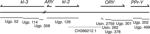
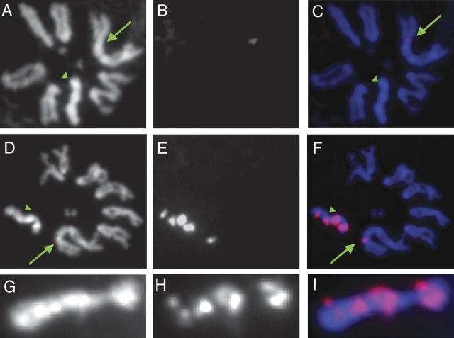
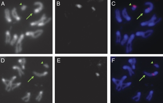
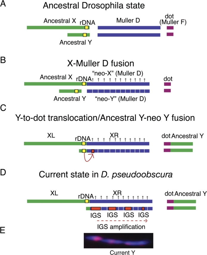
One of the most striking cases of sex chromosome reorganization in Drosophila occurred in the lineage ancestral to Drosophila pseudoobscura, where there was a translocation of Y-linked genes to an autosome. These genes went from being present only in males, never recombining, and having an effective population size of 0.5N to a state of autosomal linkage, where they are passed through both sexes, may recombine, and their effective population size has quadrupled. These genes appear to be functional, and they underwent a drastic reduction in intron size after the translocation. A Y-autosome translocation may pose problems in meiosis if the rDNA locus responsible for X-Y pairing had also moved to an autosome. In this study, we demonstrate that the Y-autosome translocation moved Y-linked genes onto the dot chromosome, a small, mainly heterochromatic autosome with some sex chromosome-like properties. The rDNA repeats occur exclusively on the X chromosome in D. pseudoobscura, but we found that the new Y chromosome of this species harbors four clusters bearing only the intergenic spacer region (IGS) of the rDNA repeats. This arrangement appears analogous to the situation in Drosophila simulans, where X-rDNA to Y-IGS pairing could be responsible for X-Y chromosome pairing. We postulate that the nascent D. pseudoobscura Y chromosome acquired and amplified copies of the IGS, suggesting a potential mechanism for X-Y pairing in D. pseudoobscura.


Figures

F<sc>IG</sc>. 1.
FIG. 1.
The five genes that were translocated from the Y chromosome to the dot chromosome are found on 10 scaffolds from the CAF1 assembly of the D. pseudoobscura genome (Drosophila 12 Genomes Consortium 2007). The scaffolds are represented by lines, and the length of the lines drawn are proportional to the length of the scaffolds. These scaffolds cover at least 158 kb, including estimated gap lengths, but this is likely to be a gross underestimate because it does not account for gaps between scaffolds. In Drosophila persimilis, this region spans approximately 312 kb including estimated gaps within the single scaffold in which these genes are found. The scaffolds are all unmapped (Ugp. = “unknown group” and Usin. = “unknown singleton”). The scaffold containing kl-3 does not overlap with the rest of the Y-to-dot genes in D. pseudoobscura, although in D. persimilis all of the genes, including kl-3, are contained on a single scaffold and are in the same order. ARY and kl-2 can be linked to the first exons of ORY using a scaffold (CH396212.1) from the CABA assembly (shown as dotted line).
F<sc>IG</sc>. 2.
FIG. 2.
Hybridization of the rDNA probes 18S and 28S and IGS probes to Drosophila pseudoobscura mitotic chromosomes from larval brains suggest that the rDNA repeats are exclusively X-linked in D. pseudoobscura and that the rDNA IGS spacer region is found on the X and in multiple clusters on the Y. The green arrow points to the X chromosome, and the green arrowhead points to the Y chromosome. (A) DAPI staining of D. pseudoobscura mitotic chromosomes. (B) Signal for the 18S and 28S probes. (C) Overlay of A and B with DAPI staining colored in blue and the probe staining colored in red. (D,E,F) DAPI DNA staining, probe hybridization, and overlay for the IGS probes (for the 226- and 267-bp subrepeats, combined), respectively. (G,H,I) DAPI DNA staining, IGS probe hybridization, and overlay for just the Y chromosome, respectively. The rDNA genes 18S and 28S map exclusively to the X chromosome (shown as green arrow in C); no signal is seen on the Y chromosome, supporting the mapping of bobbed (Sturtevant and Tan 1937; Sturtevant and Novitski 1941). The IGS subrepeats are present in at least four clusters on the Y chromosome (shown as green arrowhead in F) and in one cluster on the X chromosome (shown as green arrow in F). There is no signal from the dot chromosome.
F<sc>IG</sc>. 3.
FIG. 3.
FISH in Drosophila affinis and D. persimilis using D. pseudoobscura probe shows that the current Y chromosomes acquired rDNA genes and spacers. The green arrow points to the X chromosome, and the green arrowhead points to the Y chromosome. (A) DAPI staining of D. affinis mitotic chromosomes. (B) Signal for the 18S and 28S probes. (C) Overlay of B and C with DAPI staining colored in blue and the probe staining colored in red. (D,E,F) DAPI DNA staining of D. affinis mitotic chromosomes, probe hybridization, and the overlay for the IGS probes, respectively. Both the rDNA genes 18S and 28S and the IGS repeats map to the X and Y chromosomes in D. affinis (shown as a green arrow and a green arrowhead for the X and Y, respectively, in C and F). The IGS repeats are not tandemly repeated on the Y chromosome in D. affinis. (G) DAPI staining of D. persimilis mitotic chromosomes. (H) Signal for the 18S and 28S probes. (I) Overlay of G and H with DAPI staining colored in blue and the probe staining colored in red. Signal amplification (described in Materials and Methods) was required to detect the signal in D. persimilis panels H and I. (J,K,L) DAPI DNA staining of D. persimilis mitotic chromosomes, probe hybridization, and the overlay for the IGS probes, respectively. The rDNA genes 18S and 28S map to both the X chromosome and the current Y chromosome in D. persimilis (shown as a green arrow and a green arrowhead for the X and Y, respectively, in I). The IGS repeats occur in at least four clusters on the current D. persimilis Y chromosome, similar to D. pseudoobscura (L).
F<sc>IG</sc>. 4.
FIG. 4.
FISH in D. guanche using Drosophila pseudoobcura probes suggests that the ancestral locations of the rDNA for D. pseudoobscura were likely on the X and Y chromosomes. The green arrow points to the X chromosome and the green arrowhead points to the Y chromosome. (A) DAPI staining of D. guanche mitotic chromosomes. (B) Signal for the 18S and 28S probes. (C) Overlay of B and C with DAPI staining colored in blue and the probe staining colored in red. (D,E,F) DAPI DNA staining of D. guanche mitotic chromosomes, probe hybridization, and the overlay for the IGS probes, respectively. Both the rDNA genes 18S and 28S and the IGS repeats map to the X and Y chromosomes in D. guanche (shown as a green arrow and a green arrowhead for the X and Y, respectively, in C and F). The IGS repeats are not tandemly repeated on the Y chromosome in this species, indicating that the current Y-linked IGS arrays in D. pseudoobscura are derived.
F<sc>IG</sc>. 5.
FIG. 5.
The location of the rDNA in the melanogaster group, obscura-group and Drosophila hydei in the Drosophila subgenus suggest that the ancestral locations of the rDNA are on the X and Y chromosomes. D. simulans presents an exception where the ancestral Y-linked rDNA locus was lost after the amplification of acquired IGS repeats on the Y chromosome (Lohe and Roberts 1990). D. hydei has two clusters of rDNA repeats on the Y chromosome in addition to the X (Hennig et al. 1975). The three species that have the X-Muller D fusion (X–D) and the Y–A translocation (Y–A), Drosophila pseudoobscura, Drosophila persimilis, and Drosophila affinis, appear to have a more recent acquisition, followed by the amplification of the IGS repeats on the current Y chromosomes of D. pseudoobscura and D. persimilis (figs. 2 and 4). D. pseudoobscura appears to have lost its ancestral Y-linked rDNA locus (fig. 2), whereas D. persimilis and D. affinis retained rDNA on their current Y chromosomes.
F<sc>IG</sc>. 6.
FIG. 6.
We propose that there was a Y-to-dot translocation in D. pseudoobscura and that the current Y chromosome originated from an X–D fusion, followed by acquisition of IGS sequences. (A) The Drosophila ancestral state with respect to the sex chromosomes and rDNA repeats. (B) An X-Muller D fusion occurred between 11 and 18 Ma. The homolog of the fused element (neo-Y) was transmitted as a Y. The neo-Y eventually degenerates (represented by hatched lines) and, in response, the neo-X becomes dosage compensated (represented by upward arrows). (C) The IGS spacer is transferred to the neo-Y chromosome either from the ancestral X or as diagrammed: from the ancestral Y chromosome. Some fraction of the ancestral Y containing the rDNA may have remained free and fused with the current Y. (D) The ancestral Y chromosome translocated to the dot chromosome, and the current Y chromosome is a degenerated neo-Y chromosome that originated from the X–D fusion event. The IGS spacer acquired by the current Y chromosome is amplified, producing at least four clusters, and the ancestral Y-linked rDNA locus is lost. We illustrate this as a translocation of most of the ancestral Y to the dot with subsequent fusion of the remaining ancestral Y with the current Y rather than a complete fusion, although both scenarios are possible. The left arm of the X (XL) in D. pseudoobscura corresponds to the ancestral X chromosome and the right arm (XR) is the Muller D element. (E) The IGS spacer repeats (in red) occur in at least four large clusters on the Y chromosome (DAPI staining).

Similar articles

Cited by

References

    1. Ashburner M. Drosophila. a laboratory handbook. Cold Spring Harbor (NY): Cold Spring Harbor Laboratory Press; 1989.
    1. Ault JG, Rieder CL. Meiosis in Drosophila males. I. The question of separate conjunctive mechanisms for the XY and autosomal bivalents. Chromosoma. 1994;103:352–356. - PubMed
    1. Brianti MT, Ananina G, Recco-Pimentel SM, Klaczko LB. Comparative analysis of the chromosomal positions of rDNA genes in species of the tripunctata radiation of Drosophila. Cytogenet Genome Res. 2009;125:149–157. - PubMed
    1. Bridges CB. The mutants and linkage data of chromosome four of Drosophila melanogaster. Biol Zh. 1935;4:401–420.
    1. Carvalho AB, Clark AG. Y chromosome of D. pseudoobscura is not homologous to the ancestral Drosophila Y. Science. 2005;307:108–110. - PubMed

Publication types

LinkOut - more resources